• 文献检索
  • 文档翻译
  • 深度研究
  • 学术资讯
  • Suppr Zotero 插件Zotero 插件
  • 邀请有礼
  • 套餐&价格
  • 历史记录
应用&插件
Suppr Zotero 插件Zotero 插件浏览器插件Mac 客户端Windows 客户端微信小程序
定价
高级版会员购买积分包购买API积分包
服务
文献检索文档翻译深度研究API 文档MCP 服务
关于我们
关于 Suppr公司介绍联系我们用户协议隐私条款
关注我们

Suppr 超能文献

核心技术专利:CN118964589B侵权必究
粤ICP备2023148730 号-1Suppr @ 2026

文献检索

告别复杂PubMed语法,用中文像聊天一样搜索,搜遍4000万医学文献。AI智能推荐,让科研检索更轻松。

立即免费搜索

文件翻译

保留排版,准确专业,支持PDF/Word/PPT等文件格式,支持 12+语言互译。

免费翻译文档

深度研究

AI帮你快速写综述,25分钟生成高质量综述,智能提取关键信息,辅助科研写作。

立即免费体验

解析人玻璃体细胞与其他固有免疫细胞群体相关的分子特征。

Deciphering the Molecular Signature of Human Hyalocytes in Relation to Other Innate Immune Cell Populations.

机构信息

Eye Center, Medical Center, Faculty of Medicine, University of Freiburg, Baden-Wuerttemberg, Germany.

Institute of Anatomy, University of Leipzig, Leipzig, Saxony, Germany.

出版信息

Invest Ophthalmol Vis Sci. 2022 Mar 2;63(3):9. doi: 10.1167/iovs.63.3.9.

DOI:10.1167/iovs.63.3.9
PMID:35266958
原文链接:https://pmc.ncbi.nlm.nih.gov/articles/PMC8934546/
Abstract

PURPOSE

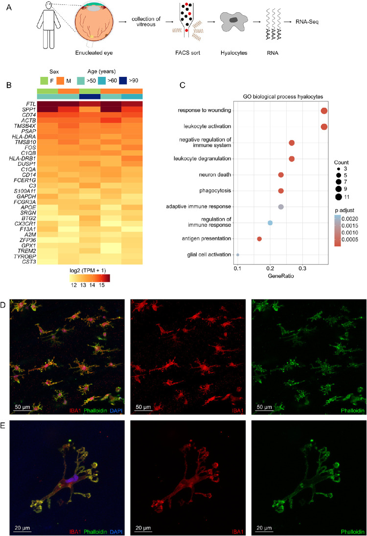
Hyalocytes are the tissue-resident innate immune cell population of the vitreous body with important functions in health and vitreoretinal disease. The purpose of this study is to gain new insights into the biology and function of human hyalocytes in comparison to other innate immune cells.

METHODS

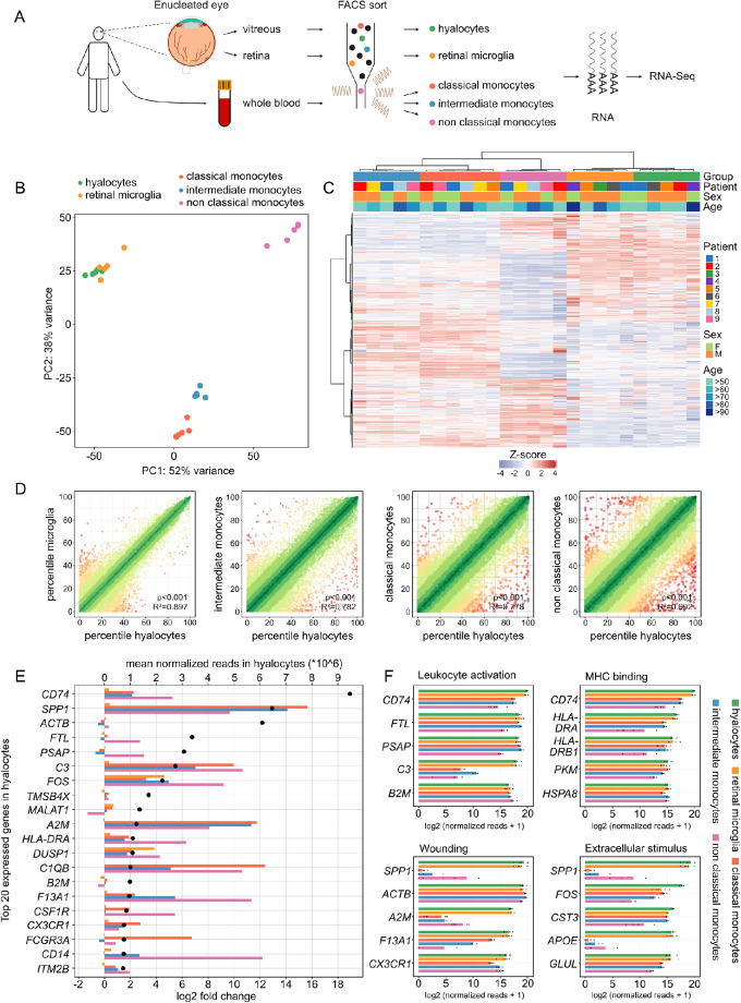
The present study applies fluorescence-activated cell sorting and RNA sequencing to compare the transcriptional profiles of human hyalocytes, retinal microglia (rMG) and classical, intermediate, and non-classical monocytes isolated from the same patients. Immunohistochemistry was applied for morphological characterization of human hyalocytes.

RESULTS

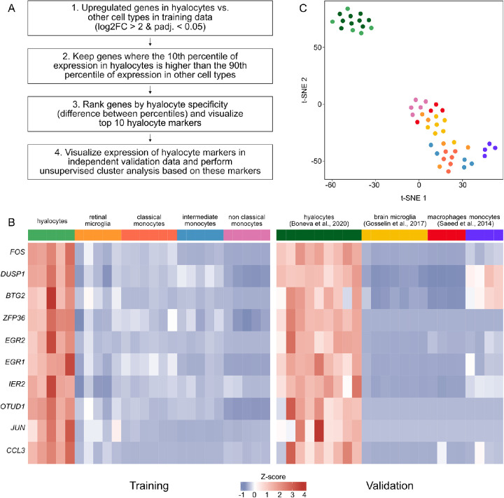
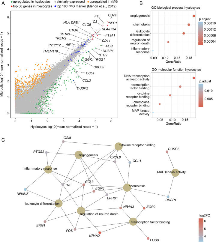
Pairwise analysis indicates distinct differences between hyalocytes and monocytes, whereas a high degree of similarity to rMG is apparent, with comparable expression levels of established microglia markers, such as TREM2, P2RY12, and TMEM119. Among the top expressed genes in hyalocytes, SPP1, CD74, and C3, were significantly upregulated when compared with monocytes. Despite the high level of similarity of hyalocytes and rMG, ten highly expressed genes in hyalocytes compared to microglia were identified, among them FOS, DUSP1, and EGR2.

CONCLUSIONS

This study reveals a high degree of similarity between hyalocytes and retinal microglia. Nevertheless, hyalocytes exhibit some expression differences that may adapt them to the specific needs of the vitreous and provide the basis for deciphering the multiple roles of this fascinating cell population in health and vitreoretinal diseases.

摘要

目的

玻璃体细胞是玻璃体组织驻留的先天免疫细胞群,在眼部健康和视网膜疾病中具有重要功能。本研究的目的是深入了解人类玻璃体细胞与其他先天免疫细胞的生物学和功能。

方法

本研究应用荧光激活细胞分选和 RNA 测序,比较从同一患者分离的人玻璃体细胞、视网膜小胶质细胞(rMG)以及经典、中间和非经典单核细胞的转录谱。免疫组织化学用于人玻璃体细胞的形态学特征分析。

结果

两两比较分析表明玻璃体细胞与单核细胞之间存在明显差异,而与 rMG 之间具有高度相似性,表现为类似的成熟小胶质细胞标志物,如 TREM2、P2RY12 和 TMEM119 的表达水平。在玻璃体细胞中表达最高的基因中,与单核细胞相比,SPP1、CD74 和 C3 显著上调。尽管玻璃体细胞与 rMG 具有高度相似性,但与小胶质细胞相比,在玻璃体细胞中鉴定出 10 个高表达基因,其中包括 FOS、DUSP1 和 EGR2。

结论

本研究揭示了玻璃体细胞与视网膜小胶质细胞之间具有高度相似性。然而,玻璃体细胞表现出一些表达差异,可能使其适应玻璃体的特殊需求,并为阐明该迷人细胞群在眼部健康和视网膜疾病中的多种作用提供基础。

https://cdn.ncbi.nlm.nih.gov/pmc/blobs/5cf5/8934546/9099609a0190/iovs-63-3-9-f004.jpg
https://cdn.ncbi.nlm.nih.gov/pmc/blobs/5cf5/8934546/15368c277f47/iovs-63-3-9-f001.jpg
https://cdn.ncbi.nlm.nih.gov/pmc/blobs/5cf5/8934546/e33d74be42e0/iovs-63-3-9-f002.jpg
https://cdn.ncbi.nlm.nih.gov/pmc/blobs/5cf5/8934546/10c6d769301e/iovs-63-3-9-f003.jpg
https://cdn.ncbi.nlm.nih.gov/pmc/blobs/5cf5/8934546/9099609a0190/iovs-63-3-9-f004.jpg

相似文献

1
Deciphering the Molecular Signature of Human Hyalocytes in Relation to Other Innate Immune Cell Populations.解析人玻璃体细胞与其他固有免疫细胞群体相关的分子特征。
Invest Ophthalmol Vis Sci. 2022 Mar 2;63(3):9. doi: 10.1167/iovs.63.3.9.
2
Transcriptional Profiling Uncovers Human Hyalocytes as a Unique Innate Immune Cell Population.转录谱分析揭示人玻璃体细胞是一种独特的固有免疫细胞群体。
Front Immunol. 2020 Sep 11;11:567274. doi: 10.3389/fimmu.2020.567274. eCollection 2020.
3
Hyalocytes-guardians of the vitreoretinal interface.玻璃体细胞——视网膜界面的守护者。
Graefes Arch Clin Exp Ophthalmol. 2024 Sep;262(9):2765-2784. doi: 10.1007/s00417-024-06448-3. Epub 2024 Apr 3.
4
Redefining the ontogeny of hyalocytes as yolk sac-derived tissue-resident macrophages of the vitreous body.重新定义玻璃体内的原纤维细胞为卵黄囊来源的组织驻留巨噬细胞。
J Neuroinflammation. 2024 Jul 3;21(1):168. doi: 10.1186/s12974-024-03110-x.
5
In-Depth Molecular Profiling Specifies Human Retinal Microglia Identity.深入的分子剖析明确了人视网膜小胶质细胞的身份。
Front Immunol. 2022 Mar 18;13:863158. doi: 10.3389/fimmu.2022.863158. eCollection 2022.
6
Hyalocyte origin, structure, and imaging.玻璃体细胞的起源、结构与成像。
Expert Rev Ophthalmol. 2022;17(4):233-248. doi: 10.1080/17469899.2022.2100762. Epub 2022 Sep 6.
7
Macrophages in close proximity to the vitreoretinal interface are potential biomarkers of inflammation during retinal vascular disease.紧邻玻璃体视网膜界面的巨噬细胞是视网膜血管疾病炎症过程中的潜在生物标志物。
J Neuroinflammation. 2022 Aug 8;19(1):203. doi: 10.1186/s12974-022-02562-3.
8
The characterisation of hyalocytes: the origin, phenotype, and turnover.玻璃体细胞的特征:起源、表型及更新
Br J Ophthalmol. 2005 Apr;89(4):513-7. doi: 10.1136/bjo.2004.050658.
9
[The effect of OPTC-shRNA on the bovine retinal pigment epithelial cells and hyalocytes co-culture collagen gel contraction system].[OPTC-shRNA对牛视网膜色素上皮细胞与玻璃体细胞共培养胶原凝胶收缩系统的影响]
Zhonghua Yan Ke Za Zhi. 2013 Feb;49(2):155-62.
10
Functional properties of hyalocytes under PDGF-rich conditions.富含血小板源性生长因子(PDGF)条件下玻璃体细胞的功能特性。
Invest Ophthalmol Vis Sci. 2004 Jul;45(7):2107-14. doi: 10.1167/iovs.03-1092.

引用本文的文献

1
Differentiation of human hyalocytes from induced pluripotent stem cells through ascorbic acid treatment.通过抗坏血酸处理从诱导多能干细胞分化出人类玻璃体细胞。
Hum Cell. 2025 Feb 12;38(2):52. doi: 10.1007/s13577-025-01182-2.
2
Annexin-V binds subpopulation of immune cells altering its interpretation as an biomarker for apoptosis in the retina.膜联蛋白V结合免疫细胞亚群,改变了其作为视网膜细胞凋亡生物标志物的解读。
Int J Biol Sci. 2024 Nov 11;20(15):6073-6089. doi: 10.7150/ijbs.102551. eCollection 2024.
3
Short-Term Culture of Human Hyalocytes Retains Their Initial Phenotype and Displays Their Contraction Abilities.

本文引用的文献

1
The Human Eye Transcriptome Atlas: A searchable comparative transcriptome database for healthy and diseased human eye tissue.《人类眼睛转录组图谱:一个可搜索的健康和患病人类眼组织比较转录组数据库》
Genomics. 2022 Mar;114(2):110286. doi: 10.1016/j.ygeno.2022.110286. Epub 2022 Feb 3.
2
Immunosenescence in Choroidal Neovascularization (CNV)-Transcriptional Profiling of Naïve and CNV-Associated Retinal Myeloid Cells during Aging.免疫衰老与脉络膜新生血管(CNV)- 衰老过程中幼稚和 CNV 相关视网膜髓样细胞的转录谱分析。
Int J Mol Sci. 2021 Dec 10;22(24):13318. doi: 10.3390/ijms222413318.
3
In-Depth Molecular Characterization of Neovascular Membranes Suggests a Role for Hyalocyte-to-Myofibroblast Transdifferentiation in Proliferative Diabetic Retinopathy.
人玻璃体细胞的短期培养保留了其初始表型,并显示出其收缩能力。
Cells. 2024 Nov 6;13(22):1837. doi: 10.3390/cells13221837.
4
The multifaceted role of vitreous hyalocytes: Orchestrating inflammation, angiomodulation and erythrophagocytosis in proliferative diabetic retinopathy.玻璃体细胞的多效作用:在增生性糖尿病视网膜病变中协调炎症、血管生成调节和红细胞吞噬作用。
J Neuroinflammation. 2024 Nov 14;21(1):297. doi: 10.1186/s12974-024-03291-5.
5
The application of artificial intelligence in diabetic retinopathy: progress and prospects.人工智能在糖尿病视网膜病变中的应用:进展与展望。
Front Cell Dev Biol. 2024 Oct 25;12:1473176. doi: 10.3389/fcell.2024.1473176. eCollection 2024.
6
Liquid Biopsy for Proliferative Diabetic Retinopathy: Single-Cell Transcriptomics of Human Vitreous Reveals Inflammatory T-Cell Signature.增殖性糖尿病视网膜病变的液体活检:人玻璃体的单细胞转录组学揭示炎症性T细胞特征
Ophthalmol Sci. 2024 Apr 26;4(6):100539. doi: 10.1016/j.xops.2024.100539. eCollection 2024 Nov-Dec.
7
Redefining the ontogeny of hyalocytes as yolk sac-derived tissue-resident macrophages of the vitreous body.重新定义玻璃体内的原纤维细胞为卵黄囊来源的组织驻留巨噬细胞。
J Neuroinflammation. 2024 Jul 3;21(1):168. doi: 10.1186/s12974-024-03110-x.
8
Impact of Equine Ocular Surface Squamous Neoplasia on Interactions between Ocular Transcriptome and Microbiome.马眼表面鳞状细胞瘤对眼转录组与微生物组之间相互作用的影响。
Vet Sci. 2024 Apr 7;11(4):167. doi: 10.3390/vetsci11040167.
9
Deciphering perivascular macrophages and microglia in the retinal ganglion cell layers.解析视网膜神经节细胞层中的血管周围巨噬细胞和小胶质细胞。
Front Cell Dev Biol. 2024 Mar 26;12:1368021. doi: 10.3389/fcell.2024.1368021. eCollection 2024.
10
Hyalocytes-guardians of the vitreoretinal interface.玻璃体细胞——视网膜界面的守护者。
Graefes Arch Clin Exp Ophthalmol. 2024 Sep;262(9):2765-2784. doi: 10.1007/s00417-024-06448-3. Epub 2024 Apr 3.
深入的分子特征分析表明,玻璃体细胞向肌成纤维细胞转分化在增生性糖尿病视网膜病变中起作用。
Front Immunol. 2021 Nov 2;12:757607. doi: 10.3389/fimmu.2021.757607. eCollection 2021.
4
Novel Insight Into Glycosaminoglycan Biosynthesis Based on Gene Expression Profiles.基于基因表达谱对糖胺聚糖生物合成的新见解。
Front Cell Dev Biol. 2021 Sep 6;9:709018. doi: 10.3389/fcell.2021.709018. eCollection 2021.
5
The role of interferon regulatory factor 8 for retinal tissue homeostasis and development of choroidal neovascularisation.干扰素调节因子 8 在视网膜组织稳态和脉络膜新生血管形成中的作用。
J Neuroinflammation. 2021 Sep 20;18(1):215. doi: 10.1186/s12974-021-02230-y.
6
Subretinal fibrosis in neovascular age-related macular degeneration: current concepts, therapeutic avenues, and future perspectives.新生血管性年龄相关性黄斑变性的视网膜下纤维化:当前概念、治疗途径和未来展望。
Cell Tissue Res. 2022 Mar;387(3):361-375. doi: 10.1007/s00441-021-03514-8. Epub 2021 Sep 3.
7
Complement System and Potential Therapeutics in Age-Related Macular Degeneration.补体系统与年龄相关性黄斑变性的潜在治疗方法。
Int J Mol Sci. 2021 Jun 25;22(13):6851. doi: 10.3390/ijms22136851.
8
Deep Learning Reveals Key Immunosuppression Genes and Distinct Immunotypes in Periodontitis.深度学习揭示牙周炎中的关键免疫抑制基因和不同免疫类型。
Front Genet. 2021 Mar 12;12:648329. doi: 10.3389/fgene.2021.648329. eCollection 2021.
9
Secreted Phosphoprotein 1 Expression in Retinal Mononuclear Phagocytes Links Murine to Human Choroidal Neovascularization.视网膜单核吞噬细胞中分泌型磷蛋白1的表达将小鼠与人脉络膜新生血管形成联系起来。
Front Cell Dev Biol. 2021 Jan 28;8:618598. doi: 10.3389/fcell.2020.618598. eCollection 2020.
10
Ensembl 2021.Ensembl 2021.
Nucleic Acids Res. 2021 Jan 8;49(D1):D884-D891. doi: 10.1093/nar/gkaa942.