van Buuren Nicholas, Ramirez Ricardo, Turner Scott, Chen Diana, Suri Vithika, Aggarwal Abhishek, Moon Christina, Kim Sam, Kornyeyev Dmytro, Bui Nam, Bhardwaj Neeru, Chan Henry Ly, Marcellin Patrick, Buti Maria, Wallin Jeffrey, Gaggar Anuj, Fletcher Simon P, Diehl Lauri, Li Li, Mo Hongmei, Feierbach Becket
Gilead Sciences Inc. 324 Lakeside Dr., Foster City, CA, 94404, United States.
Current address: Foundation Medicine, Cambridge, MA, 02141, United States.
JHEP Rep. 2021 Oct 24;4(1):100388. doi: 10.1016/j.jhepr.2021.100388. eCollection 2022 Jan.
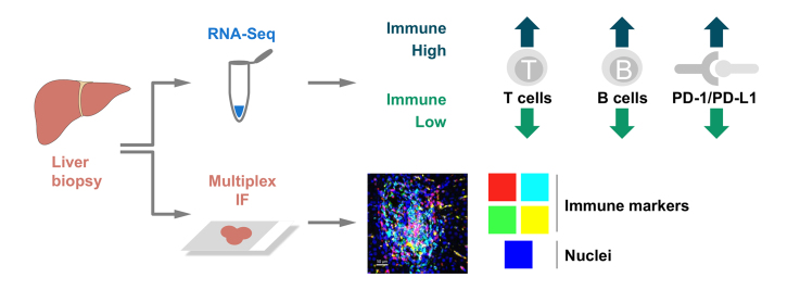
BACKGROUND & AIMS: We aim to describe the liver immune microenvironment by analyzing liver biopsies from patients with chronic HBV infection (CHB). Host immune cell signatures and their corresponding localization were characterized by analyzing the intrahepatic transcriptome in combination with a custom multiplex immunofluorescence panel.
Matching FFPE and fresh frozen liver biopsies were collected from immune active patients within the open-label phase IV study GS-US-174-0149. RNA-Seq was conducted on 53 CHB liver biopsies from 46 patients. Twenty-eight of the 53 samples had matched FFPE biopsies and were stained with a 12-plex panel including cell segmentation, immune and viral biomarkers. Corresponding serum samples were screened using the MSD Human V-plex Screen Service to identify peripheral correlates for the immune microenvironment.
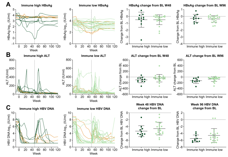
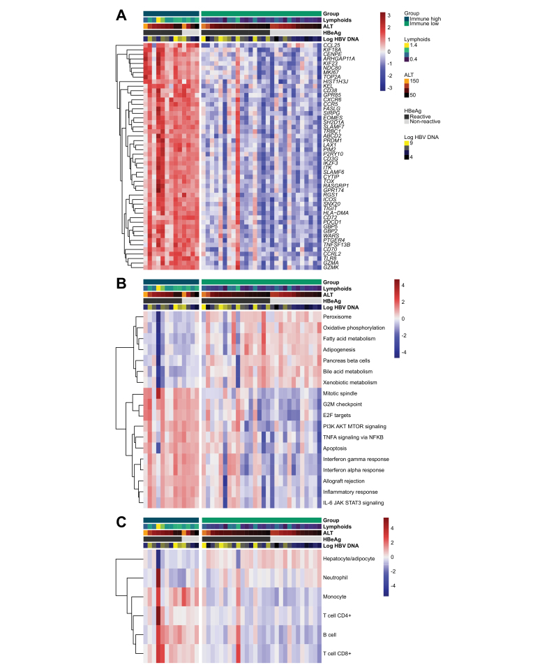
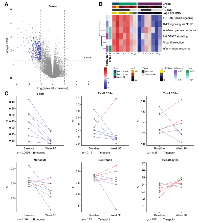
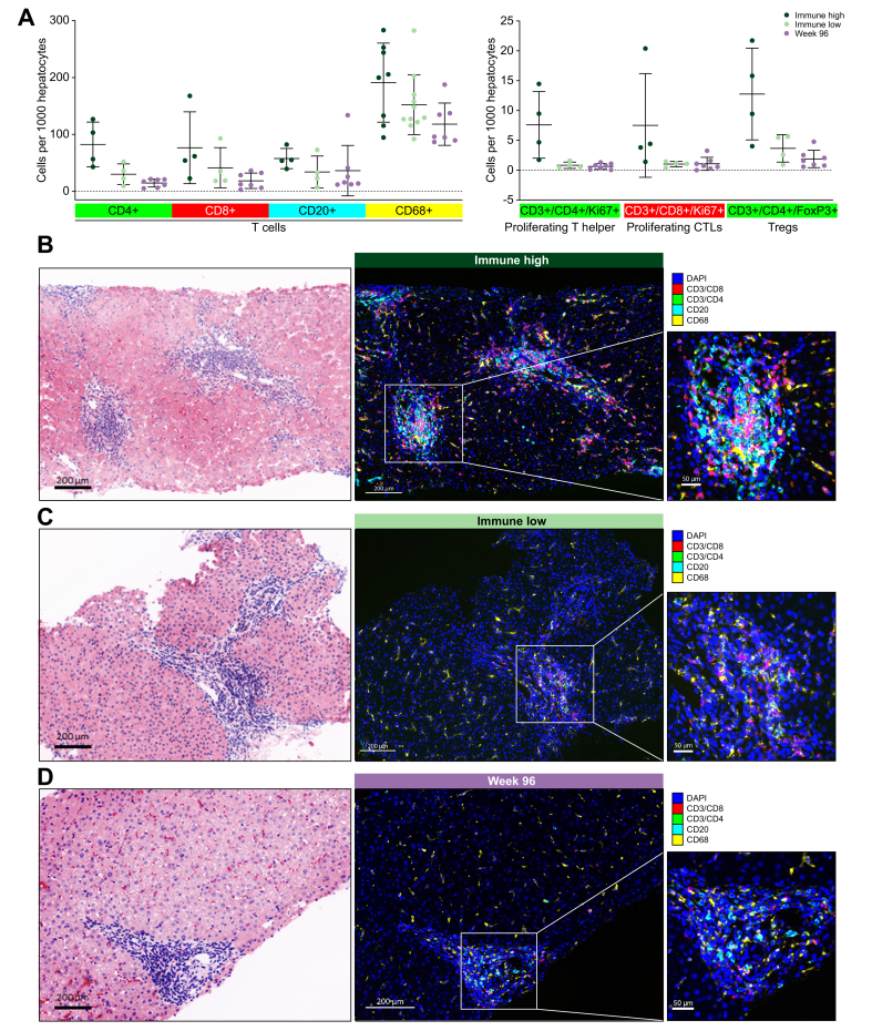
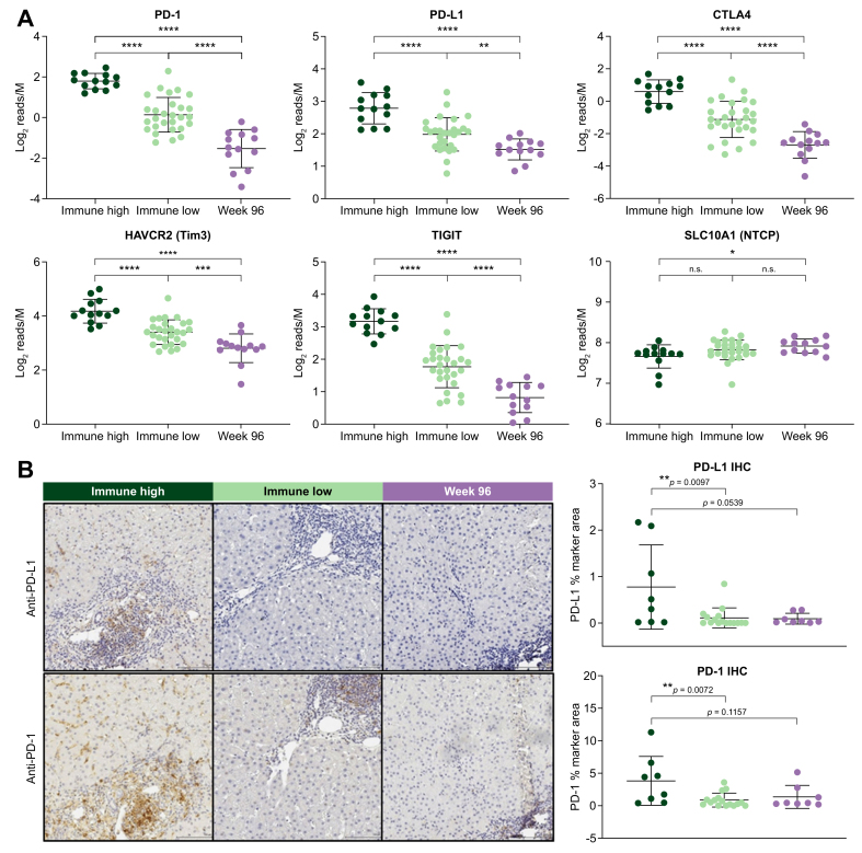
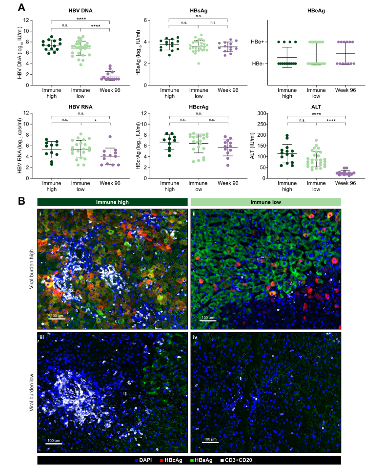
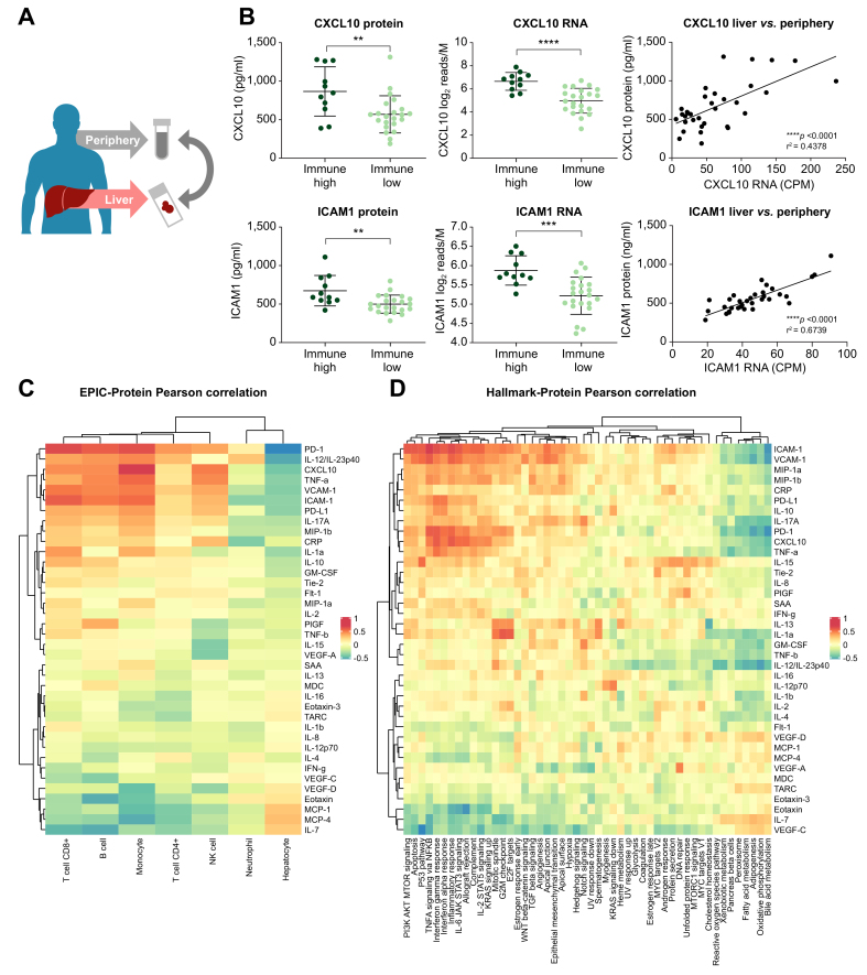
Using unsupervised clustering of the transcriptome, we reveal two unique liver immune signatures classified as immune high and immune low based on the quantification of the liver infiltrate gene signatures. Multiplex immunofluorescence analysis demonstrated large periportal lymphoid aggregates in immune high samples consisting of CD4 and CD8 T cells, B cells and macrophages. Differentiation of the high and low immune microenvironments was independent of HBeAg status and peripheral viral antigen levels. In addition, longitudinal analysis indicates that treatment and normalization of ALT correlates with a decrease in liver immune infiltrate and inflammation. Finally, we screened a panel of peripheral biomarkers and identified ICAM-1 and CXCL10 as biomarkers that strongly correlate with these unique immune microenvironments.
These data provide a description of immune phenotypes in patients with CHB and show that immune responses are downregulated in the liver following nucleotide analogue treatment. This may have important implications for both the safety and efficacy of immune modulator programs aimed at HBV cure.
Liver biopsies from patients with chronic hepatitis B were submitted to RNA-Seq and multiplex immunofluorescence and identified two different liver immune microenvironments: immune high and immune low. Immune high patients showed elevated immune pathways, including interferon signaling pathways, and increase presence of immune cells. Longitudinal analysis of biopsies from treatment experienced patients showed that treatment correlates with a marked decrease in inflammation and these findings may have important implications for both safety and efficacy of immune modulator programs for HBV cure.
我们旨在通过分析慢性乙型肝炎病毒感染(CHB)患者的肝脏活检样本,描述肝脏免疫微环境。通过结合定制的多重免疫荧光检测板分析肝内转录组,对宿主免疫细胞特征及其相应定位进行了表征。
在开放标签的IV期研究GS-US-174-0149中,从免疫活跃的患者中收集匹配的福尔马林固定石蜡包埋(FFPE)和新鲜冷冻肝脏活检样本。对来自46例患者的53份CHB肝脏活检样本进行了RNA测序。53份样本中的28份有匹配的FFPE活检样本,并用包括细胞分割、免疫和病毒生物标志物的12重检测板进行染色。使用MSD人V型检测板筛选服务对相应的血清样本进行检测,以确定免疫微环境的外周相关指标。
通过对转录组进行无监督聚类,我们基于肝脏浸润基因特征的量化,揭示了两种独特的肝脏免疫特征,分别归类为免疫高和免疫低。多重免疫荧光分析显示,免疫高样本中存在大量门静脉周围淋巴样聚集物,由CD4和CD8 T细胞、B细胞和巨噬细胞组成。高、低免疫微环境的分化与HBeAg状态和外周病毒抗原水平无关。此外,纵向分析表明,ALT的治疗和正常化与肝脏免疫浸润和炎症的减少相关。最后,我们筛选了一组外周生物标志物,确定细胞间黏附分子-1(ICAM-1)和CXC趋化因子配体10(CXCL10)为与这些独特免疫微环境密切相关的生物标志物。
这些数据描述了CHB患者的免疫表型,并表明核苷酸类似物治疗后肝脏中的免疫反应下调。这可能对旨在治愈HBV的免疫调节剂方案的安全性和有效性都具有重要意义。
对慢性乙型肝炎患者的肝脏活检样本进行RNA测序和多重免疫荧光检测,确定了两种不同的肝脏免疫微环境:免疫高和免疫低。免疫高的患者显示免疫途径升高,包括干扰素信号通路,并且免疫细胞存在增加。对接受过治疗的患者活检样本的纵向分析表明,治疗与炎症的显著减少相关,这些发现可能对HBV治愈的免疫调节剂方案的安全性和有效性都具有重要意义。