Zhang Ying, Zhang Zhixiang, Zhang Min, Cao Yin, Yun Wenwei
Department of Neurology, Changzhou Second People's Hospital Affiliated to Nanjing Medical University, Changzhou, China.
Front Neurosci. 2021 Dec 14;15:727998. doi: 10.3389/fnins.2021.727998. eCollection 2021.
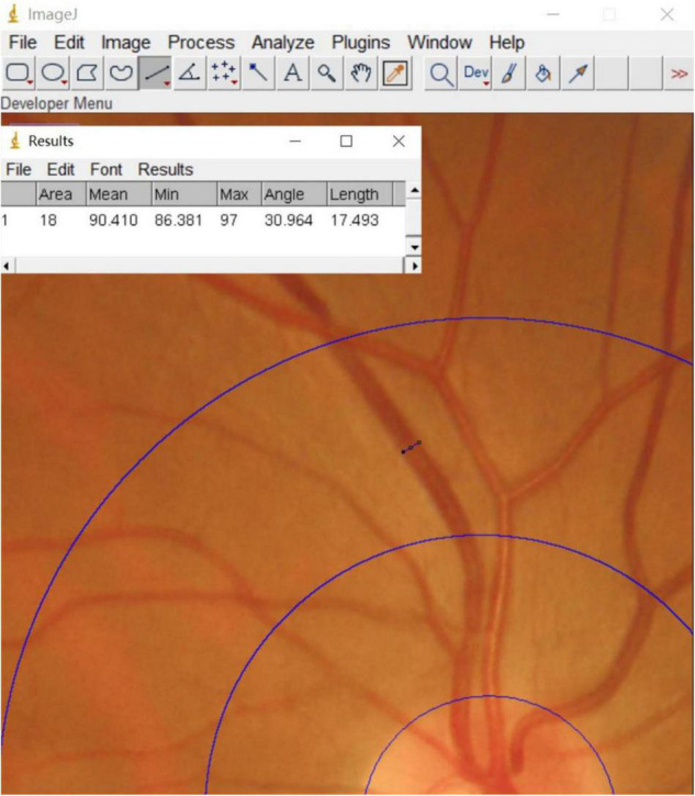
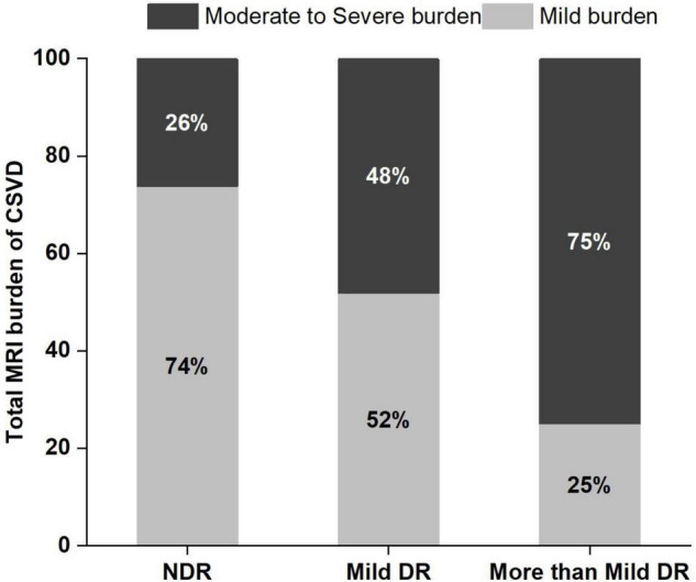
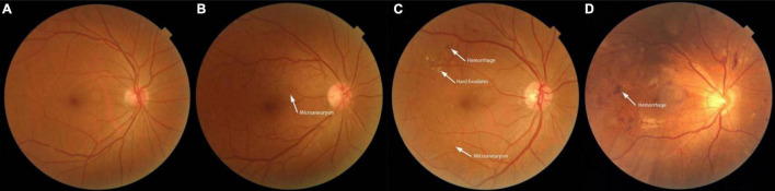
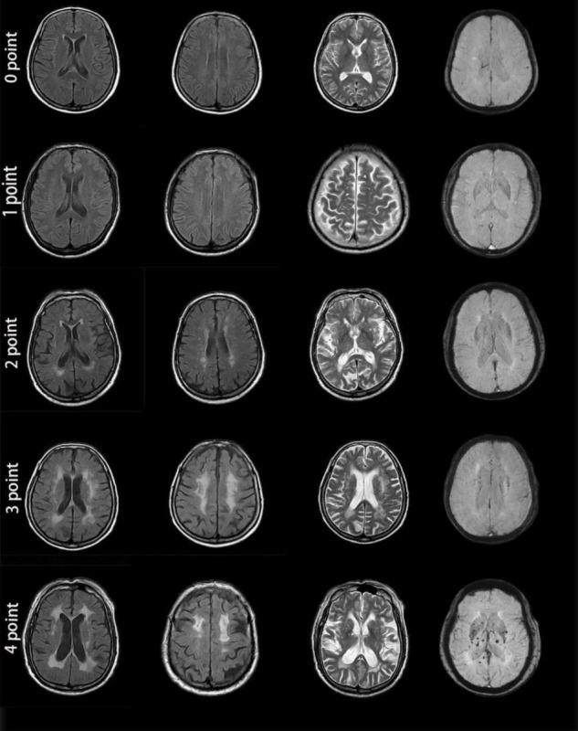
Diabetic retinopathy (DR) is one of the common microvascular complications in diabetes. The total magnetic resonance imaging (MRI) burden of cerebral small vessel disease (CSVD) tends to be increased in diabetic patients and is a marker of microvascular disease; however, the relationship between DR and CSVD is unclear. This study aimed to explore the relationship between retinal microvascular abnormalities and the total MRI burden of CSVD in patients with type 2 diabetes. Data were collected from patients with type 2 diabetes who were hospitalized between December 2019 and November 2020 in Changzhou Second People's Hospital affiliated to Nanjing Medical University. All patients underwent retinal photography and cerebral MRI. The central retinal artery equivalent (CRAE), the central retinal venous equivalent (CRVE), and arteriole-to-venule ratio (AVR) were calculated using Image J software to determine the retinal vascular calibers for each patient. The total MRI burden score for CSVD was determined, and the relationship between retinal microvascular abnormalities and the total MRI burden of CSVD was analyzed. Of the 151 diabetic patients included in the study, 84 (55.6%) had no diabetic retinopathy (NDR), 27 (17.9%) had mild DR, and 40 (26.5%) had moderate, or severe non-proliferative DR (grouped together for this study as "more than mild DR"). In patients with more than mild DR, the proportion of moderate to severe burden of CSVD was 75%, which was higher than in patients with mild DR (48.1%) or NDR (26.2%). Patients with moderate to severe burden of CSVD were more likely than those with mild burden of CSVD to have narrowed retinal arterioles (105.24 ± 8.42 μm vs. 109.45 ± 7.93 μm), widened retinal venules (201.67 ± 16.25 μm vs. 193.95 ± 13.54 μm), and lower arteriole-to-venule ratio (0.52 ± 0.05 vs. 0.57 ± 0.04) ( < 0.05 for all). The degree of DR ( = 0.465, < 0.001) and CRVE ( = 0.366, < 0.001) were positively correlated with the total MRI burden of CSVD. Multivariate logistic regression analysis indicated that, after adjustments were made for age, smoking, alcohol consumption, hypertension, and other factors, more than mild DR (OR, 4.383; = 0.028), CRAE (OR, 0.490; = 0.031), and CRVE (OR, 1.475; = 0.041) were independently associated with moderate to severe burden of CSVD. Retinal microvascular abnormalities in patients with type 2 diabetes are associated with the presence of cerebral small vessel lesions. The degree of DR and retinal vessel changes can be used as predictors of intracranial microcirculation lesions.
糖尿病视网膜病变(DR)是糖尿病常见的微血管并发症之一。糖尿病患者脑小血管病(CSVD)的总磁共振成像(MRI)负荷往往会增加,并且是微血管疾病的一个标志;然而,DR与CSVD之间的关系尚不清楚。本研究旨在探讨2型糖尿病患者视网膜微血管异常与CSVD的总MRI负荷之间的关系。数据收集自2019年12月至2020年11月在南京医科大学附属常州第二人民医院住院的2型糖尿病患者。所有患者均接受了视网膜照相和脑部MRI检查。使用Image J软件计算视网膜中央动脉等效直径(CRAE)、视网膜中央静脉等效直径(CRVE)和动静脉比(AVR),以确定每位患者的视网膜血管管径。确定CSVD的总MRI负荷评分,并分析视网膜微血管异常与CSVD的总MRI负荷之间的关系。在纳入研究的151例糖尿病患者中,84例(55.6%)无糖尿病视网膜病变(NDR),27例(17.9%)有轻度DR,40例(26.5%)有中度或重度非增殖性DR(本研究将其归为“轻度以上DR”)。在轻度以上DR患者中,CSVD中度至重度负荷的比例为75%,高于轻度DR患者(48.1%)或NDR患者(26.2%)。CSVD中度至重度负荷的患者比轻度负荷的患者更有可能出现视网膜小动脉狭窄(105.24±8.42μm对109.45±7.93μm)、视网膜小静脉增宽(201.67±16.25μm对193.95±13.54μm)以及动静脉比降低(0.52±0.05对0.57±0.04)(所有P均<0.05)。DR程度(r = 0.465,P < 0.001)和CRVE(r = 0.366,P < 0.001)与CSVD的总MRI负荷呈正相关。多因素逻辑回归分析表明,在对年龄、吸烟、饮酒、高血压等因素进行校正后,轻度以上DR(OR,4.383;P = 0.028)、CRAE(OR,0.490;P = 0.031)和CRVE(OR,1.475;P = 0.041)与CSVD中度至重度负荷独立相关。2型糖尿病患者的视网膜微血管异常与脑小血管病变的存在有关。DR程度和视网膜血管变化可作为颅内微循环病变的预测指标。