• 文献检索
  • 文档翻译
  • 深度研究
  • 学术资讯
  • Suppr Zotero 插件Zotero 插件
  • 邀请有礼
  • 套餐&价格
  • 历史记录
应用&插件
Suppr Zotero 插件Zotero 插件浏览器插件Mac 客户端Windows 客户端微信小程序
定价
高级版会员购买积分包购买API积分包
服务
文献检索文档翻译深度研究API 文档MCP 服务
关于我们
关于 Suppr公司介绍联系我们用户协议隐私条款
关注我们

Suppr 超能文献

核心技术专利:CN118964589B侵权必究
粤ICP备2023148730 号-1Suppr @ 2026

文献检索

告别复杂PubMed语法,用中文像聊天一样搜索,搜遍4000万医学文献。AI智能推荐,让科研检索更轻松。

立即免费搜索

文件翻译

保留排版,准确专业,支持PDF/Word/PPT等文件格式,支持 12+语言互译。

免费翻译文档

深度研究

AI帮你快速写综述,25分钟生成高质量综述,智能提取关键信息,辅助科研写作。

立即免费体验

脑小血管病中的视网膜结构和血管密度变化

Retinal structure and vessel density changes in cerebral small vessel disease.

作者信息

Wang Dandan, Wang Lina, Wang Jinjin, Du Yang, Wang Kaiyue, Wang Meizi, Yang Liu, Zhao Xingquan

机构信息

Department of Neurology, Beijing Tiantan Hospital, Capital Medical University, Beijing, China.

Department of Ophthalmology, Beijing Tiantan Hospital, Capital Medical University, Beijing, China.

出版信息

Front Neurosci. 2024 Feb 26;18:1288380. doi: 10.3389/fnins.2024.1288380. eCollection 2024.

DOI:10.3389/fnins.2024.1288380
PMID:38469574
原文链接:https://pmc.ncbi.nlm.nih.gov/articles/PMC10925719/
Abstract

BACKGROUND

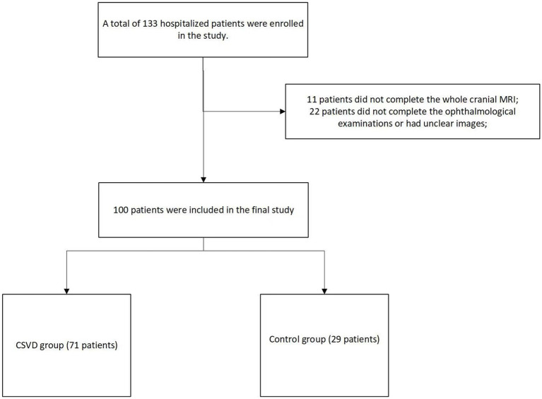
Cerebral small vessel disease (CSVD) attaches people's attention in recent years. In this study, we aim to explore retinal structure and vessel density changes in CSVD patients.

METHODS

We collected information on retinal metrics assessed by optical coherence tomography (OCT) and OCT angiography and CSVD characters. Logistic and liner regression was used to analyze the relationship between retinal metrics and CSVD.

RESULTS

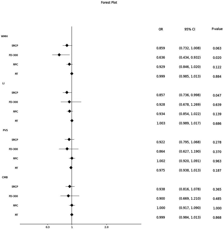
Vessel density of superficial retinal capillary plexus (SRCP), foveal density- 300 length (FD-300), radial peripapillary capillary (RPC) and thickness of retina were significantly lower in CSVD patients, the difference only existed in the thickness of retina after adjusted relevant risk factors (OR (95% CI): 0.954 (0.912, 0.997),  = 0.037). SRCP vessel density showed a significant downward trend with the increase of CSVD scores (β: -0.087, 95%CI: -0.166, -0.008,  = 0.031). SRCP and FD-300 were significantly lower in patients with lacunar infarctions and white matter hypertensions separately [OR (95% CI): 0.857 (0.736, 0.998), = 0.047 and OR (95% CI): 0.636 (0.434, 0.932), = 0.020, separately].

CONCLUSION

SRCP, FD-300 and thickness of retina were associated with the occurrence and severity of total CSVD scores and its different radiological manifestations. Exploring CSVD by observing alterations in retinal metrics has become an optional research direction in future.

摘要

背景

近年来,脑小血管病(CSVD)备受关注。在本研究中,我们旨在探讨CSVD患者的视网膜结构和血管密度变化。

方法

我们收集了通过光学相干断层扫描(OCT)和OCT血管造影评估的视网膜指标信息以及CSVD特征。采用逻辑回归和线性回归分析视网膜指标与CSVD之间的关系。

结果

CSVD患者的浅表视网膜毛细血管丛(SRCP)血管密度、黄斑中心凹密度-300长度(FD-300)、视乳头周围放射状毛细血管(RPC)和视网膜厚度显著降低,在调整相关危险因素后,仅视网膜厚度存在差异(OR(95%CI):0.954(0.912,0.997),P = 0.037)。SRCP血管密度随CSVD评分增加呈显著下降趋势(β:-0.087,95%CI:-0.166,-0.008,P = 0.031)。腔隙性脑梗死和白质高血压患者的SRCP和FD-300分别显著降低[OR(95%CI):0.857(0.736,0.998),P = 0.047和OR(95%CI):0.636(0.434,0.932),P = 0.020]。

结论

SRCP、FD-300和视网膜厚度与总CSVD评分及其不同影像学表现的发生和严重程度相关。通过观察视网膜指标的变化来探索CSVD已成为未来一个可选的研究方向。

https://cdn.ncbi.nlm.nih.gov/pmc/blobs/fc3a/10925719/5554695bf528/fnins-18-1288380-g002.jpg
https://cdn.ncbi.nlm.nih.gov/pmc/blobs/fc3a/10925719/8b8a9b63cc96/fnins-18-1288380-g001.jpg
https://cdn.ncbi.nlm.nih.gov/pmc/blobs/fc3a/10925719/5554695bf528/fnins-18-1288380-g002.jpg
https://cdn.ncbi.nlm.nih.gov/pmc/blobs/fc3a/10925719/8b8a9b63cc96/fnins-18-1288380-g001.jpg
https://cdn.ncbi.nlm.nih.gov/pmc/blobs/fc3a/10925719/5554695bf528/fnins-18-1288380-g002.jpg

相似文献

1
Retinal structure and vessel density changes in cerebral small vessel disease.脑小血管病中的视网膜结构和血管密度变化
Front Neurosci. 2024 Feb 26;18:1288380. doi: 10.3389/fnins.2024.1288380. eCollection 2024.
2
The vessel density of the superficial retinal capillary plexus as a new biomarker in cerebral small vessel disease: an optical coherence tomography angiography study.视网膜浅层毛细血管丛血管密度作为脑小血管病的一种新生物标志物:一项光相干断层扫描血管造影研究。
Neurol Sci. 2021 Sep;42(9):3615-3624. doi: 10.1007/s10072-021-05038-z. Epub 2021 Jan 11.
3
Fundus Changes Evaluated by OCTA in Patients With Cerebral Small Vessel Disease and Their Correlations: A Cross-Sectional Study.光学相干断层扫描血管造影评估脑小血管病患者的眼底改变及其相关性:一项横断面研究
Front Neurol. 2022 Apr 25;13:843198. doi: 10.3389/fneur.2022.843198. eCollection 2022.
4
Association between retinal vessel density and neuroimaging features and cognitive impairment in cerebral small vessel disease.大脑小血管病患者视网膜血管密度与神经影像学特征及认知障碍的相关性。
Clin Neurol Neurosurg. 2022 Oct;221:107407. doi: 10.1016/j.clineuro.2022.107407. Epub 2022 Aug 3.
5
Optical coherence tomography angiography as a potential screening tool for cerebral small vessel diseases.光学相干断层扫描血管造影术作为一种潜在的脑小血管疾病筛查工具。
Alzheimers Res Ther. 2020 Jun 11;12(1):73. doi: 10.1186/s13195-020-00638-x.
6
Peripapillary and parafoveal microvascular changes in eyes with optic neuritis and their fellow eyes measured by optical coherence tomography angiography: an Exploratory Study.应用光相干断层扫描血管造影术测量视神经炎及其对侧眼的视盘旁和中心凹旁微血管改变:一项探索性研究。
Acta Ophthalmol. 2021 May;99(3):288-298. doi: 10.1111/aos.14577. Epub 2020 Aug 24.
7
Changes in parafoveal and peripapillary perfusion after decompression surgery in chiasmal compression due to pituitary tumors.因垂体瘤导致的视交叉受压患者减压手术后旁中心区和视盘周围血流的变化。
Sci Rep. 2021 Feb 10;11(1):3464. doi: 10.1038/s41598-021-82151-1.
8
Differential patterns of parafoveal and peripapillary vessel density in multiple sclerosis and neuromyelitis optica spectrum disorder.多发性硬化症和视神经脊髓炎谱系障碍中黄斑旁和视乳头周围血管密度的差异模式。
Mult Scler Relat Disord. 2021 Apr;49:102780. doi: 10.1016/j.msard.2021.102780. Epub 2021 Jan 19.
9
Structural retinal changes in cerebral small vessel disease.脑小血管病的结构视网膜改变。
Sci Rep. 2022 Jun 3;12(1):9315. doi: 10.1038/s41598-022-13312-z.
10
[Correlation between total cerebral small vessel disease score and retinal vessel diameters in patients with mild stroke].[轻度卒中患者全脑小血管病评分与视网膜血管直径的相关性]
Zhonghua Yi Xue Za Zhi. 2021 Jan 5;101(1):62-67. doi: 10.3760/cma.j.cn112137-20200405-01088.

引用本文的文献

1
Retinal vessel density and cognitive function in healthy older adults.健康老年人的视网膜血管密度与认知功能
Exp Brain Res. 2025 Apr 15;243(5):114. doi: 10.1007/s00221-025-07076-x.

本文引用的文献

1
Associations of macular microvascular parameters with cerebral small vessel disease in rural older adults: A population-based OCT angiography study.农村老年人黄斑微血管参数与脑小血管疾病的关联:一项基于人群的光学相干断层扫描血管造影研究
Front Neurol. 2023 Mar 16;14:1133819. doi: 10.3389/fneur.2023.1133819. eCollection 2023.
2
Retinal microvascular signs and recurrent vascular events in patients with TIA or minor stroke.短暂性脑缺血发作或小卒中患者的视网膜微血管征象与再发血管事件。
Stroke Vasc Neurol. 2023 Oct;8(5):379-386. doi: 10.1136/svn-2022-001784. Epub 2023 Mar 1.
3
Correlation between Retinal Vascularization and Disease Aggressiveness in Amyotrophic Lateral Sclerosis.
肌萎缩侧索硬化症中视网膜血管生成与疾病侵袭性之间的相关性
Biomedicines. 2022 Sep 25;10(10):2390. doi: 10.3390/biomedicines10102390.
4
The global burden of cerebral small vessel disease in low- and middle-income countries: A systematic review and meta-analysis.低收入和中等收入国家脑小血管病的全球负担:一项系统评价和荟萃分析。
Int J Stroke. 2023 Jan;18(1):15-27. doi: 10.1177/17474930221137019. Epub 2022 Nov 25.
5
Association between retinal vessel density and neuroimaging features and cognitive impairment in cerebral small vessel disease.大脑小血管病患者视网膜血管密度与神经影像学特征及认知障碍的相关性。
Clin Neurol Neurosurg. 2022 Oct;221:107407. doi: 10.1016/j.clineuro.2022.107407. Epub 2022 Aug 3.
6
Structural retinal changes in cerebral small vessel disease.脑小血管病的结构视网膜改变。
Sci Rep. 2022 Jun 3;12(1):9315. doi: 10.1038/s41598-022-13312-z.
7
Prognostic value of radial peripapillary capillary density for visual field outcomes in pituitary adenoma: A case-control study.垂体腺瘤患者视盘周围放射状毛细血管密度对视野结果的预后价值:病例对照研究。
J Clin Neurosci. 2022 Jun;100:113-119. doi: 10.1016/j.jocn.2022.04.012. Epub 2022 Apr 18.
8
Age-related macular degeneration: Epidemiology, genetics, pathophysiology, diagnosis, and targeted therapy.年龄相关性黄斑变性:流行病学、遗传学、病理生理学、诊断及靶向治疗。
Genes Dis. 2021 Feb 27;9(1):62-79. doi: 10.1016/j.gendis.2021.02.009. eCollection 2022 Jan.
9
Correlation Between Retinal Microvascular Abnormalities and Total Magnetic Resonance Imaging Burden of Cerebral Small Vessel Disease in Patients With Type 2 Diabetes.2型糖尿病患者视网膜微血管异常与脑小血管病磁共振成像总负担的相关性
Front Neurosci. 2021 Dec 14;15:727998. doi: 10.3389/fnins.2021.727998. eCollection 2021.
10
Retinopathy in Malignant Hypertension.恶性高血压性视网膜病变
N Engl J Med. 2021 Nov 18;385(21):1994. doi: 10.1056/NEJMicm2109500. Epub 2021 Nov 13.