• 文献检索
  • 文档翻译
  • 深度研究
  • 学术资讯
  • Suppr Zotero 插件Zotero 插件
  • 邀请有礼
  • 套餐&价格
  • 历史记录
应用&插件
Suppr Zotero 插件Zotero 插件浏览器插件Mac 客户端Windows 客户端微信小程序
定价
高级版会员购买积分包购买API积分包
服务
文献检索文档翻译深度研究API 文档MCP 服务
关于我们
关于 Suppr公司介绍联系我们用户协议隐私条款
关注我们

Suppr 超能文献

核心技术专利:CN118964589B侵权必究
粤ICP备2023148730 号-1Suppr @ 2026

文献检索

告别复杂PubMed语法,用中文像聊天一样搜索,搜遍4000万医学文献。AI智能推荐,让科研检索更轻松。

立即免费搜索

文件翻译

保留排版,准确专业,支持PDF/Word/PPT等文件格式,支持 12+语言互译。

免费翻译文档

深度研究

AI帮你快速写综述,25分钟生成高质量综述,智能提取关键信息,辅助科研写作。

立即免费体验

管状上皮细胞与巨噬细胞的通讯通过细胞外囊泡转移形成负反馈回路,从而促进糖尿病肾病中的肾脏炎症和细胞凋亡。

Tubular epithelial cell-to-macrophage communication forms a negative feedback loop via extracellular vesicle transfer to promote renal inflammation and apoptosis in diabetic nephropathy.

机构信息

Inflammation and Immune Mediated Diseases Laboratory of Anhui Province, Anhui Institute of Innovative Drugs, School of Pharmacy, Anhui Medical University, Hefei, 230032, China.

The Center for Scientific Research of Anhui Medical University, Hefei, 230032, China.

出版信息

Theranostics. 2022 Jan 1;12(1):324-339. doi: 10.7150/thno.63735. eCollection 2022.

DOI:10.7150/thno.63735
PMID:34987648
原文链接:https://pmc.ncbi.nlm.nih.gov/articles/PMC8690920/
Abstract

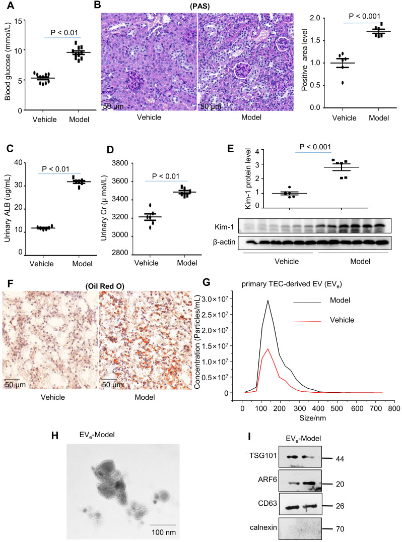
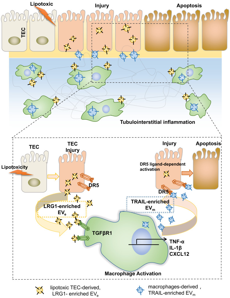
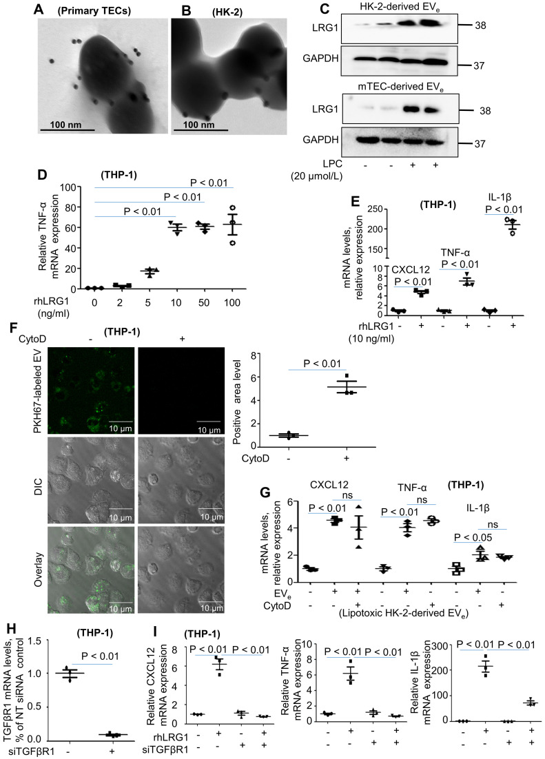
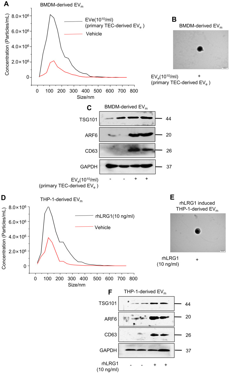
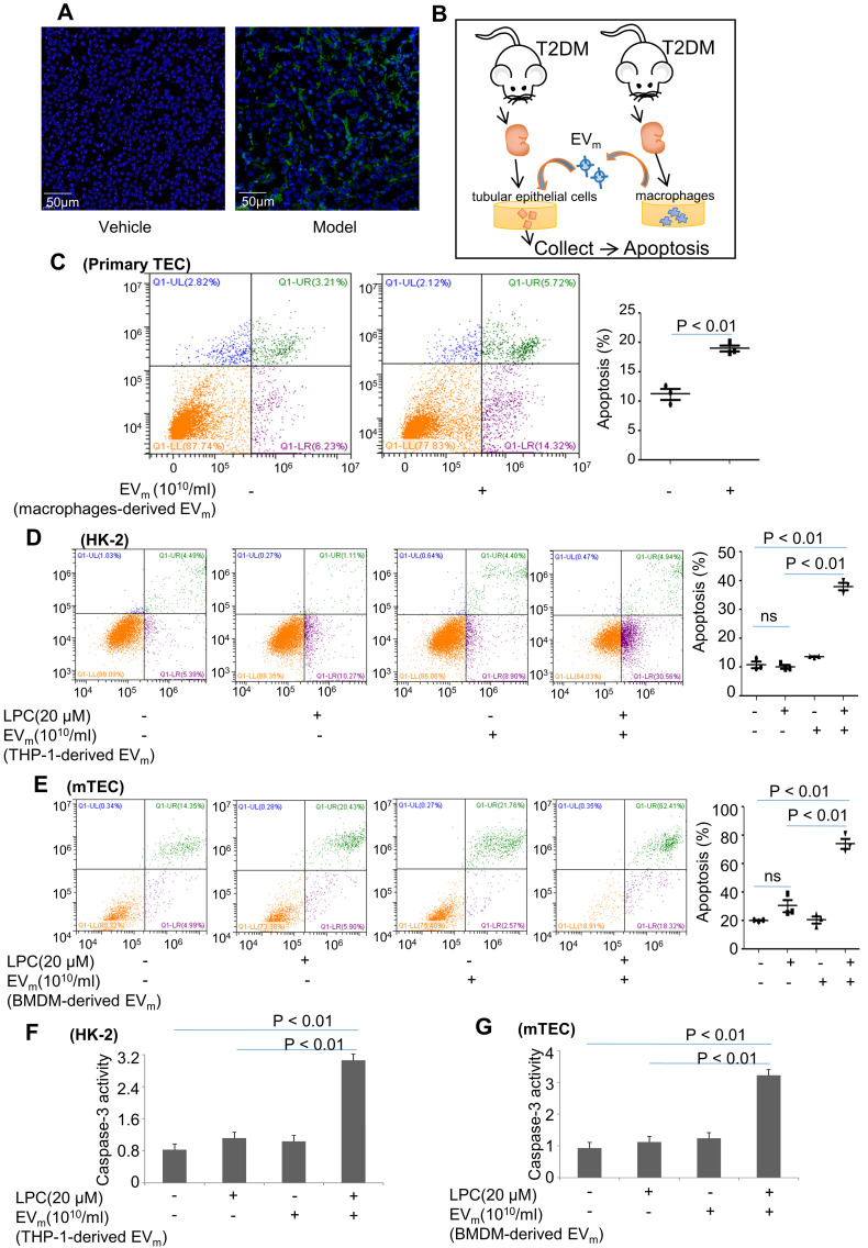
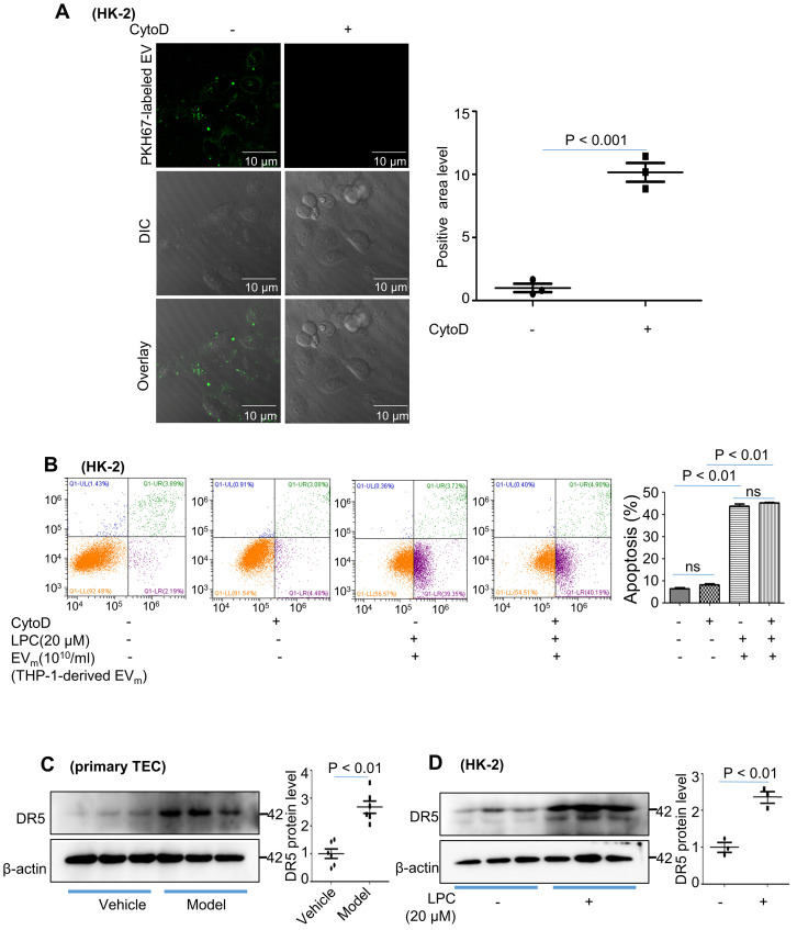
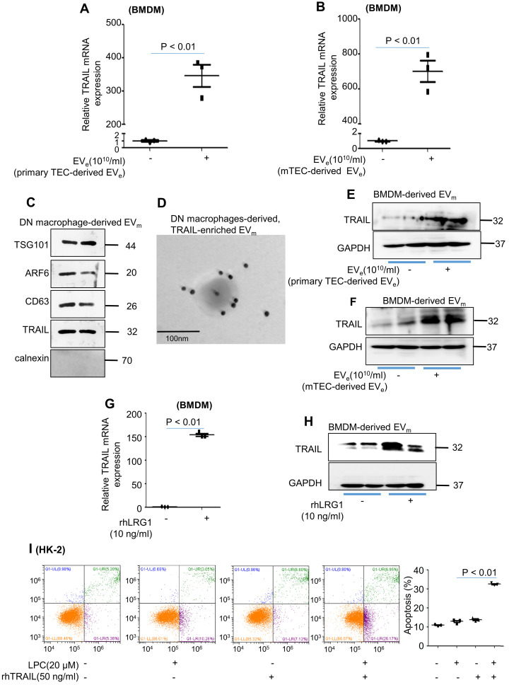
Macrophage infiltration around lipotoxic tubular epithelial cells (TECs) is a hallmark of diabetic nephropathy (DN). However, how these two types of cells communicate remains obscure. We previously demonstrated that LRG1 was elevated in the process of kidney injury. Here, we demonstrated that macrophage-derived, LRG1-enriched extracellular vesicles (EVs) exacerbated DN. We induced an experimental T2DM mouse model with a HFD diet for four months. Renal primary epithelial cells and macrophage-derived EVs were isolated from T2D mice by differential ultracentrifugation. To investigate whether lipotoxic TEC-derived EV (EV) activate macrophages, mouse bone marrow-derived macrophages (BMDMs) were incubated with EV. To investigate whether activated macrophage-derived EVs (EV) induce lipotoxic TEC apoptosis, EV were cocultured with primary renal tubular epithelial cells. Subsequently, we evaluated the effect of LRG1 in EV by investigating the apoptosis mechanism. We demonstrated that incubation of primary TECs of DN or HK-2 mTECs with lysophosphatidyl choline (LPC) increased the release of EV. Interestingly, TEC-derived EV activated an inflammatory phenotype in macrophages and induced the release of macrophage-derived EV. Furthermore, EV could induce apoptosis in TECs injured by LPC. Importantly, we found that leucine-rich α-2-glycoprotein 1 (LRG1)-enriched EV activated macrophages via a TGFβR1-dependent process and that tumor necrosis factor-related apoptosis-inducing ligand (TRAIL)-enriched EV induced apoptosis in injured TECs via a death receptor 5 (DR5)-dependent process. Our findings indicated a novel cell communication mechanism between tubular epithelial cells and macrophages in DN, which could be a potential therapeutic target.

摘要

脂肪毒性管状上皮细胞(TEC)周围的巨噬细胞浸润是糖尿病肾病(DN)的一个标志。然而,这两种细胞如何相互交流仍然不清楚。我们之前的研究表明,LRG1 在肾脏损伤过程中升高。在这里,我们证明了巨噬细胞衍生的富含 LRG1 的细胞外囊泡(EV)加剧了 DN。我们通过高脂肪饮食诱导了一个实验性 T2DM 小鼠模型,持续四个月。通过差速超速离心从 T2D 小鼠中分离出肾脏原代上皮细胞和巨噬细胞衍生的 EV。为了研究脂毒性 TEC 衍生的 EV(EV)是否激活巨噬细胞,用 EV 孵育小鼠骨髓来源的巨噬细胞(BMDM)。为了研究激活的巨噬细胞衍生的 EV(EV)是否诱导脂毒性 TEC 凋亡,将 EV 与原代肾小管上皮细胞共培养。随后,我们通过研究凋亡机制来评估 EV 中 LRG1 的作用。我们证明,用溶血磷脂酰胆碱(LPC)孵育 DN 或 HK-2 mTEC 的原代 TEC 会增加 EV 的释放。有趣的是,TEC 衍生的 EV 激活了巨噬细胞中的炎症表型,并诱导了巨噬细胞衍生的 EV 的释放。此外,EV 可诱导 LPC 损伤的 TEC 凋亡。重要的是,我们发现富含亮氨酸的α-2-糖蛋白 1(LRG1)的 EV 通过 TGFβR1 依赖性过程激活巨噬细胞,而富含肿瘤坏死因子相关凋亡诱导配体(TRAIL)的 EV 通过死亡受体 5(DR5)依赖性过程诱导损伤的 TEC 凋亡。我们的研究结果表明,DN 中肾小管上皮细胞和巨噬细胞之间存在一种新的细胞通讯机制,这可能是一个潜在的治疗靶点。

https://cdn.ncbi.nlm.nih.gov/pmc/blobs/9637/8690920/eac4d98636e9/thnov12p0324g008.jpg
https://cdn.ncbi.nlm.nih.gov/pmc/blobs/9637/8690920/7523c1834ca7/thnov12p0324g001.jpg
https://cdn.ncbi.nlm.nih.gov/pmc/blobs/9637/8690920/3ecc8850f3b2/thnov12p0324g002.jpg
https://cdn.ncbi.nlm.nih.gov/pmc/blobs/9637/8690920/1c895415e081/thnov12p0324g003.jpg
https://cdn.ncbi.nlm.nih.gov/pmc/blobs/9637/8690920/031569bd06e9/thnov12p0324g004.jpg
https://cdn.ncbi.nlm.nih.gov/pmc/blobs/9637/8690920/5b8ec8788340/thnov12p0324g005.jpg
https://cdn.ncbi.nlm.nih.gov/pmc/blobs/9637/8690920/6944f22afae6/thnov12p0324g006.jpg
https://cdn.ncbi.nlm.nih.gov/pmc/blobs/9637/8690920/6e83a95f1925/thnov12p0324g007.jpg
https://cdn.ncbi.nlm.nih.gov/pmc/blobs/9637/8690920/eac4d98636e9/thnov12p0324g008.jpg

相似文献

1
Tubular epithelial cell-to-macrophage communication forms a negative feedback loop via extracellular vesicle transfer to promote renal inflammation and apoptosis in diabetic nephropathy.管状上皮细胞与巨噬细胞的通讯通过细胞外囊泡转移形成负反馈回路,从而促进糖尿病肾病中的肾脏炎症和细胞凋亡。
Theranostics. 2022 Jan 1;12(1):324-339. doi: 10.7150/thno.63735. eCollection 2022.
2
Macrophage-derived, LRG1-enriched extracellular vesicles exacerbate aristolochic acid nephropathy in a TGFβR1-dependent manner.巨噬细胞衍生的富含 LRG1 的细胞外囊泡以 TGFβR1 依赖的方式加重马兜铃酸肾病。
Cell Biol Toxicol. 2022 Aug;38(4):629-648. doi: 10.1007/s10565-021-09666-1. Epub 2021 Oct 22.
3
Tubular epithelial cell-derived extracellular vesicles induce macrophage glycolysis by stabilizing HIF-1α in diabetic kidney disease.管状上皮细胞衍生的细胞外囊泡通过稳定糖尿病肾病中的 HIF-1α 诱导巨噬细胞糖酵解。
Mol Med. 2022 Aug 12;28(1):95. doi: 10.1186/s10020-022-00525-1.
4
Epsin1-mediated exosomal sorting of Dll4 modulates the tubular-macrophage crosstalk in diabetic nephropathy.Epsin1 介导的 Dll4 外泌体分拣调节糖尿病肾病中的管状细胞-巨噬细胞串扰。
Mol Ther. 2023 May 3;31(5):1451-1467. doi: 10.1016/j.ymthe.2023.03.027. Epub 2023 Apr 3.
5
Keratinocyte-to-macrophage communication exacerbate psoriasiform dermatitis via LRG1-enriched extracellular vesicles.角质形成细胞与巨噬细胞的通讯通过富含 LRG1 的细胞外囊泡加剧银屑病样皮炎。
Theranostics. 2024 Jan 1;14(3):1049-1064. doi: 10.7150/thno.89180. eCollection 2024.
6
Lipid-Induced Signaling Causes Release of Inflammatory Extracellular Vesicles From Hepatocytes.脂质诱导的信号传导导致肝细胞释放炎性细胞外囊泡。
Gastroenterology. 2016 Apr;150(4):956-67. doi: 10.1053/j.gastro.2015.12.037. Epub 2016 Jan 5.
7
Tim-3 aggravates podocyte injury in diabetic nephropathy by promoting macrophage activation via the NF-κB/TNF-α pathway.Tim-3 通过 NF-κB/TNF-α 通路促进巨噬细胞活化,加重糖尿病肾病足细胞损伤。
Mol Metab. 2019 May;23:24-36. doi: 10.1016/j.molmet.2019.02.007. Epub 2019 Feb 26.
8
Exosomal miRNA-19b-3p of tubular epithelial cells promotes M1 macrophage activation in kidney injury.肾小管上皮细胞来源的外泌体 miR-19b-3p 促进肾损伤中 M1 巨噬细胞的活化。
Cell Death Differ. 2020 Jan;27(1):210-226. doi: 10.1038/s41418-019-0349-y. Epub 2019 May 16.
9
Mesenchymal stem cells prevent the progression of diabetic nephropathy by improving mitochondrial function in tubular epithelial cells.间充质干细胞通过改善肾小管上皮细胞的线粒体功能来阻止糖尿病肾病的进展。
Exp Mol Med. 2019 Jul 9;51(7):1-14. doi: 10.1038/s12276-019-0268-5.
10
Macrophage-derived exosomes promote telomere fragility and senescence in tubular epithelial cells by delivering miR-155.巨噬细胞衍生的外泌体通过递送 miR-155 促进管状上皮细胞端粒脆弱和衰老。
Cell Commun Signal. 2024 Jul 10;22(1):357. doi: 10.1186/s12964-024-01708-5.

引用本文的文献

1
Research progress on non-coding RNA regulatory networks and targeted therapy in diabetic nephropathy.糖尿病肾病中非编码RNA调控网络与靶向治疗的研究进展
Front Endocrinol (Lausanne). 2025 Jul 29;16:1625307. doi: 10.3389/fendo.2025.1625307. eCollection 2025.
2
A Network Pharmacology-Based Investigation into the Mechanism of Quercetin Combined with Rosuvastatin in Delaying Diabetic Nephropathy via Inhibiting NRK-52E Cell Ferroptosis.基于网络药理学探究槲皮素联合瑞舒伐他汀通过抑制NRK-52E细胞铁死亡延缓糖尿病肾病的机制
Diabetes Metab Syndr Obes. 2025 Aug 5;18:2681-2694. doi: 10.2147/DMSO.S524983. eCollection 2025.
3

本文引用的文献

1
Schisandrin C attenuates renal damage in diabetic nephropathy by regulating macrophage polarization.五味子丙素通过调节巨噬细胞极化减轻糖尿病肾病中的肾损伤。
Am J Transl Res. 2021 Jan 15;13(1):210-222. eCollection 2021.
2
2. Classification and Diagnosis of Diabetes: .2. 糖尿病的分类和诊断: 。
Diabetes Care. 2021 Jan;44(Suppl 1):S15-S33. doi: 10.2337/dc21-S002.
3
Intestinal Epithelium-Derived Luminally Released Extracellular Vesicles in Sepsis Exhibit the Ability to Suppress TNF-a and IL-17A Expression in Mucosal Inflammation.
The role of macrophages in renal fibrosis and therapeutic prospects.
巨噬细胞在肾纤维化中的作用及治疗前景。
PeerJ. 2025 Jul 23;13:e19769. doi: 10.7717/peerj.19769. eCollection 2025.
4
Caffeic acid phenethyl ester protects renal tubular epithelial cells against ferroptosis in diabetic kidney disease via restoring PINK1-mediated mitophagy.咖啡酸苯乙酯通过恢复PINK1介导的线粒体自噬保护糖尿病肾病中的肾小管上皮细胞免受铁死亡。
Mol Med. 2025 Jul 24;31(1):264. doi: 10.1186/s10020-025-01318-y.
5
Plasma and Urine Metabolites Associated With Nondiabetic Chronic Kidney Disease: The HELIUS Study.与非糖尿病慢性肾脏病相关的血浆和尿液代谢物:HELIUS研究
Kidney Med. 2025 Apr 17;7(7):101009. doi: 10.1016/j.xkme.2025.101009. eCollection 2025 Jul.
6
Targeting programmed cell death pathways: emerging therapeutic strategies for diabetic kidney disease.靶向程序性细胞死亡途径:糖尿病肾病的新兴治疗策略
Front Endocrinol (Lausanne). 2025 Jun 11;16:1513895. doi: 10.3389/fendo.2025.1513895. eCollection 2025.
7
Hemoglobin-associated in proximal tubule cells can be used as a biomarker for idiopathic membranous nephropathy.近端肾小管细胞中与血红蛋白相关的物质可作为特发性膜性肾病的生物标志物。
Front Med (Lausanne). 2025 Jun 11;12:1574852. doi: 10.3389/fmed.2025.1574852. eCollection 2025.
8
Exosomes in inflammation and cancer: from bench to bedside applications.炎症与癌症中的外泌体:从实验台到临床应用
Mol Biomed. 2025 Jun 10;6(1):41. doi: 10.1186/s43556-025-00280-9.
9
Cuproptosis-related gene signatures define the immune microenvironment in diabetic nephropathy.铜死亡相关基因特征定义糖尿病肾病中的免疫微环境。
PLoS One. 2025 Jun 3;20(6):e0321636. doi: 10.1371/journal.pone.0321636. eCollection 2025.
10
PRPF19 mediates the proteasomal degradation of VDR to exacerbate ferroptosis in diabetic nephropathy.PRPF19介导维生素D受体的蛋白酶体降解,以加剧糖尿病肾病中的铁死亡。
Cell Commun Signal. 2025 May 25;23(1):242. doi: 10.1186/s12964-025-02253-5.
肠道上皮细胞衍生的腔内分泌细胞外囊泡在脓毒症中表现出抑制黏膜炎症中 TNF-a 和 IL-17A 表达的能力。
Int J Mol Sci. 2020 Nov 10;21(22):8445. doi: 10.3390/ijms21228445.
4
A Multifunctional Role of Leucine-Rich α-2-Glycoprotein 1 in Cutaneous Wound Healing Under Normal and Diabetic Conditions.富含亮氨酸α-2-糖蛋白 1 在正常和糖尿病条件下的皮肤创伤愈合中的多功能作用。
Diabetes. 2020 Nov;69(11):2467-2480. doi: 10.2337/db20-0585. Epub 2020 Sep 4.
5
Structural cells are key regulators of organ-specific immune responses.结构细胞是器官特异性免疫反应的关键调节者。
Nature. 2020 Jul;583(7815):296-302. doi: 10.1038/s41586-020-2424-4. Epub 2020 Jul 1.
6
Extracellular Vesicle Delivery of TRAIL Eradicates Resistant Tumor Growth in Combination with CDK Inhibition by Dinaciclib.通过细胞外囊泡递送TRAIL与地西他滨抑制CDK联合使用可根除耐药肿瘤生长。
Cancers (Basel). 2020 May 4;12(5):1157. doi: 10.3390/cancers12051157.
7
Lipotoxicity and Diabetic Nephropathy: Novel Mechanistic Insights and Therapeutic Opportunities.脂毒性与糖尿病肾病:新的发病机制见解与治疗机遇。
Int J Mol Sci. 2020 Apr 10;21(7):2632. doi: 10.3390/ijms21072632.
8
Extracellular Vesicles as Mediators of Cellular Crosstalk Between Immune System and Kidney Graft.细胞外囊泡作为免疫系统和肾脏移植物之间细胞串扰的介质。
Front Immunol. 2020 Feb 27;11:74. doi: 10.3389/fimmu.2020.00074. eCollection 2020.
9
Clinical course and risk factors for mortality of adult inpatients with COVID-19 in Wuhan, China: a retrospective cohort study.中国武汉成人 COVID-19 住院患者的临床病程和死亡危险因素:一项回顾性队列研究。
Lancet. 2020 Mar 28;395(10229):1054-1062. doi: 10.1016/S0140-6736(20)30566-3. Epub 2020 Mar 11.
10
Renal Lipid Metabolism and Lipotoxicity in Diabetes.糖尿病中的肾脏脂质代谢和脂毒性
Am J Med Sci. 2020 Feb;359(2):84-99. doi: 10.1016/j.amjms.2019.11.004. Epub 2019 Nov 25.