• 文献检索
  • 文档翻译
  • 深度研究
  • 学术资讯
  • Suppr Zotero 插件Zotero 插件
  • 邀请有礼
  • 套餐&价格
  • 历史记录
应用&插件
Suppr Zotero 插件Zotero 插件浏览器插件Mac 客户端Windows 客户端微信小程序
定价
高级版会员购买积分包购买API积分包
服务
文献检索文档翻译深度研究API 文档MCP 服务
关于我们
关于 Suppr公司介绍联系我们用户协议隐私条款
关注我们

Suppr 超能文献

核心技术专利:CN118964589B侵权必究
粤ICP备2023148730 号-1Suppr @ 2026

文献检索

告别复杂PubMed语法,用中文像聊天一样搜索,搜遍4000万医学文献。AI智能推荐,让科研检索更轻松。

立即免费搜索

文件翻译

保留排版,准确专业,支持PDF/Word/PPT等文件格式,支持 12+语言互译。

免费翻译文档

深度研究

AI帮你快速写综述,25分钟生成高质量综述,智能提取关键信息,辅助科研写作。

立即免费体验

Epsin1 介导的 Dll4 外泌体分拣调节糖尿病肾病中的管状细胞-巨噬细胞串扰。

Epsin1-mediated exosomal sorting of Dll4 modulates the tubular-macrophage crosstalk in diabetic nephropathy.

机构信息

Department of Nephrology, The Second Xiangya Hospital, Central South University, Changsha, Hunan, China.

Department of Nephrology, The Second Xiangya Hospital, Central South University, Changsha, Hunan, China; Department of Cellular Biology and Anatomy, Medical College of Georgia at Augusta University, Augusta, GA, USA; Charlie Norwood VA Medical Center, Augusta, GA, USA.

出版信息

Mol Ther. 2023 May 3;31(5):1451-1467. doi: 10.1016/j.ymthe.2023.03.027. Epub 2023 Apr 3.

DOI:10.1016/j.ymthe.2023.03.027
PMID:37016580
原文链接:https://pmc.ncbi.nlm.nih.gov/articles/PMC10188907/
Abstract

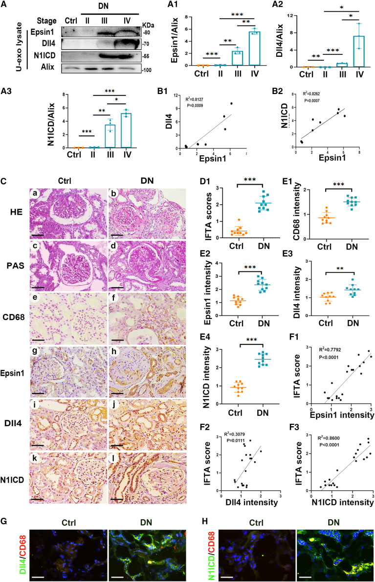
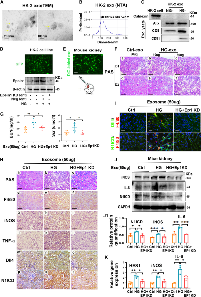
Tubular epithelial cells (TECs) play critical roles in the development of diabetic nephropathy (DN), and can activate macrophages through the secretion of exosomes. However, the mechanism(s) of TEC-exosomes in macrophage activation under DN remains unknown. By mass spectrometry, 1,644 differentially expressed proteins, especially Dll4, were detected in the urine exosomes of DN patients compared with controls, which was confirmed by western blot assay. Elevated Epsin1 and Dll4/N1ICD expression was observed in kidney tissues in both DN patients and db/db mice and was positively associated with tubulointerstitial damage. Exosomes from high glucose (HG)-treated tubular cells (HK-2) with Epsin1 knockdown (KD) ameliorated macrophage activation, TNF-α, and IL-6 expression, and tubulointerstitial damage in C57BL/6 mice in vivo. In an in vitro study, enriched Dll4 was confirmed in HK-2 cells stimulated with HG, which was captured by THP-1 cells and promoted M1 macrophage activation. In addition, Epsin1 modulated the content of Dll4 in TEC-exosomes stimulated with HG. TEC-exosomes with Epsin1-KD significantly inhibited N1ICD activation and iNOS expression in THP-1 cells compared with incubation with HG alone. These findings suggested that Epsin1 could modulate tubular-macrophage crosstalk in DN by mediating exosomal sorting of Dll4 and Notch1 activation.

摘要

管状上皮细胞 (TECs) 在糖尿病肾病 (DN) 的发展中起着关键作用,并且可以通过外泌体的分泌激活巨噬细胞。然而,DN 条件下 TEC 外泌体在巨噬细胞激活中的机制尚不清楚。通过质谱分析,与对照组相比,DN 患者的尿液外泌体中检测到 1644 种差异表达蛋白,尤其是 Dll4,Western blot 检测也证实了这一点。在 DN 患者和 db/db 小鼠的肾脏组织中均观察到 Epsin1 和 Dll4/N1ICD 表达升高,并且与肾小管间质损伤呈正相关。用 Epsin1 敲低 (KD) 的高糖 (HG) 处理的管状细胞 (HK-2) 的外泌体处理 C57BL/6 小鼠,可改善巨噬细胞激活、TNF-α 和 IL-6 表达以及肾小管间质损伤。在体外研究中,用 HG 刺激的 HK-2 细胞中证实了富含 Dll4 的外泌体,该外泌体被 THP-1 细胞捕获并促进 M1 巨噬细胞激活。此外,Epsin1 调节了 HG 刺激的 TEC 外泌体中 Dll4 的含量。与单独用 HG 孵育相比,用 Epsin1-KD 的 TEC 外泌体明显抑制了 THP-1 细胞中 N1ICD 的激活和 iNOS 的表达。这些发现表明,Epsin1 可以通过调节 Dll4 和 Notch1 激活的外泌体分拣来调节 DN 中的管状细胞-巨噬细胞串扰。

https://cdn.ncbi.nlm.nih.gov/pmc/blobs/f0f1/10188907/ea2801b7ac32/gr7.jpg
https://cdn.ncbi.nlm.nih.gov/pmc/blobs/f0f1/10188907/f4fcf2175efd/fx1.jpg
https://cdn.ncbi.nlm.nih.gov/pmc/blobs/f0f1/10188907/8e901590ec08/gr1.jpg
https://cdn.ncbi.nlm.nih.gov/pmc/blobs/f0f1/10188907/cb3500f1aa25/gr2.jpg
https://cdn.ncbi.nlm.nih.gov/pmc/blobs/f0f1/10188907/5f1de3e96277/gr3.jpg
https://cdn.ncbi.nlm.nih.gov/pmc/blobs/f0f1/10188907/20b206e7c0eb/gr4.jpg
https://cdn.ncbi.nlm.nih.gov/pmc/blobs/f0f1/10188907/a8f9fa32ab0b/gr5.jpg
https://cdn.ncbi.nlm.nih.gov/pmc/blobs/f0f1/10188907/fda3c74b855c/gr6.jpg
https://cdn.ncbi.nlm.nih.gov/pmc/blobs/f0f1/10188907/ea2801b7ac32/gr7.jpg

相似文献

1
Epsin1-mediated exosomal sorting of Dll4 modulates the tubular-macrophage crosstalk in diabetic nephropathy.Epsin1 介导的 Dll4 外泌体分拣调节糖尿病肾病中的管状细胞-巨噬细胞串扰。
Mol Ther. 2023 May 3;31(5):1451-1467. doi: 10.1016/j.ymthe.2023.03.027. Epub 2023 Apr 3.
2
Exosomal miRNA-19b-3p of tubular epithelial cells promotes M1 macrophage activation in kidney injury.肾小管上皮细胞来源的外泌体 miR-19b-3p 促进肾损伤中 M1 巨噬细胞的活化。
Cell Death Differ. 2020 Jan;27(1):210-226. doi: 10.1038/s41418-019-0349-y. Epub 2019 May 16.
3
Tubular epithelial cell-to-macrophage communication forms a negative feedback loop via extracellular vesicle transfer to promote renal inflammation and apoptosis in diabetic nephropathy.管状上皮细胞与巨噬细胞的通讯通过细胞外囊泡转移形成负反馈回路,从而促进糖尿病肾病中的肾脏炎症和细胞凋亡。
Theranostics. 2022 Jan 1;12(1):324-339. doi: 10.7150/thno.63735. eCollection 2022.
4
Renal Tissue-Derived Exosomal miRNA-34a in Diabetic Nephropathy Induces Renal Tubular Cell Fibrosis by Promoting the Polarization of M1 Macrophages.肾组织来源的外泌体 miRNA-34a 通过促进 M1 巨噬细胞极化诱导糖尿病肾病肾小管细胞纤维化。
IET Nanobiotechnol. 2024 Apr 17;2024:5702517. doi: 10.1049/2024/5702517. eCollection 2024.
5
Proximal tubule-derived exosomes contribute to mesangial cell injury in diabetic nephropathy via miR-92a-1-5p transfer.近端肾小管来源的外泌体通过 miR-92a-1-5p 转移促进糖尿病肾病系膜细胞损伤。
Cell Commun Signal. 2023 Jan 13;21(1):10. doi: 10.1186/s12964-022-00997-y.
6
Epac activation ameliorates tubulointerstitial inflammation in diabetic nephropathy.Epac 的激活可改善糖尿病肾病的肾小管间质炎症。
Acta Pharmacol Sin. 2022 Mar;43(3):659-671. doi: 10.1038/s41401-021-00689-2. Epub 2021 Jun 8.
7
HNRNPA1-mediated exosomal sorting of miR-483-5p out of renal tubular epithelial cells promotes the progression of diabetic nephropathy-induced renal interstitial fibrosis.HNRNPA1 介导的 miR-483-5p 外泌体分选从肾小管上皮细胞中排出,促进糖尿病肾病诱导的肾间质纤维化的进展。
Cell Death Dis. 2021 Mar 10;12(3):255. doi: 10.1038/s41419-021-03460-x.
8
Exosomes from high glucose-treated macrophages promote epithelial-mesenchymal transition of renal tubular epithelial cells via long non-coding RNAs.高糖处理的巨噬细胞来源的外泌体通过长非编码 RNA 促进肾小管上皮细胞的上皮-间充质转化。
BMC Nephrol. 2023 Jan 30;24(1):24. doi: 10.1186/s12882-023-03065-w.
9
Exosomal CCL2 from Tubular Epithelial Cells Is Critical for Albumin-Induced Tubulointerstitial Inflammation.管状上皮细胞来源的外泌体 CCL2 对于白蛋白诱导的肾小管间质炎症至关重要。
J Am Soc Nephrol. 2018 Mar;29(3):919-935. doi: 10.1681/ASN.2017050523. Epub 2018 Jan 2.
10
Alpha-kinase1 promotes tubular injury and interstitial inflammation in diabetic nephropathy by canonical pyroptosis pathway.α-激酶 1 通过经典的焦亡途径促进糖尿病肾病的肾小管损伤和间质炎症。
Biol Res. 2023 Feb 2;56(1):5. doi: 10.1186/s40659-023-00416-7.

引用本文的文献

1
Exo-hydrogel therapy: a revolutionary approach to managing diabetic complications.外泌体水凝胶疗法:一种治疗糖尿病并发症的革命性方法。
J Nanobiotechnology. 2025 Aug 11;23(1):558. doi: 10.1186/s12951-025-03621-6.
2
Exosomes applications in kidney diseases.外泌体在肾脏疾病中的应用。
Mol Biol Rep. 2025 Jun 21;52(1):622. doi: 10.1007/s11033-025-10727-5.
3
Extracellular Vesicles in Renal Inflammatory Diseases: Revealing Mechanisms of Extracellular Vesicle-Mediated Macrophage Regulation.肾脏炎症性疾病中的细胞外囊泡:揭示细胞外囊泡介导的巨噬细胞调节机制

本文引用的文献

1
Proximal tubule-derived exosomes contribute to mesangial cell injury in diabetic nephropathy via miR-92a-1-5p transfer.近端肾小管来源的外泌体通过 miR-92a-1-5p 转移促进糖尿病肾病系膜细胞损伤。
Cell Commun Signal. 2023 Jan 13;21(1):10. doi: 10.1186/s12964-022-00997-y.
2
Exosomes as Carriers for Notch Molecules.外泌体作为 Notch 分子的载体。
Methods Mol Biol. 2022;2472:197-208. doi: 10.1007/978-1-0716-2201-8_16.
3
Inhibition of Notch activity suppresses hyperglycemia-augmented polarization of macrophages to the M1 phenotype and alleviates acute pancreatitis.
Int J Mol Sci. 2025 Apr 12;26(8):3646. doi: 10.3390/ijms26083646.
4
Paeoniflorin: a review of its pharmacology, pharmacokinetics and toxicity in diabetes.芍药苷:糖尿病药理学、药代动力学及毒性研究综述
Front Pharmacol. 2025 Apr 7;16:1551368. doi: 10.3389/fphar.2025.1551368. eCollection 2025.
5
Urinary exosomes as promising biomarkers for early kidney disease detection.尿外泌体作为早期肾病检测的有前景的生物标志物。
Am J Clin Exp Urol. 2025 Feb 15;13(1):1-19. doi: 10.62347/DAKE5842. eCollection 2025.
6
Research progress on small extracellular vesicles in diabetic nephropathy.糖尿病肾病中小细胞外囊泡的研究进展
Front Cell Dev Biol. 2025 Mar 5;13:1535249. doi: 10.3389/fcell.2025.1535249. eCollection 2025.
7
Epigenetic regulation-mediated disorders in dopamine transporter endocytosis: A novel mechanism for the pathogenesis of Parkinson's disease.多巴胺转运体胞吞作用中表观遗传调控介导的紊乱:帕金森病发病机制的一种新机制。
Theranostics. 2025 Jan 13;15(6):2250-2278. doi: 10.7150/thno.107436. eCollection 2025.
8
Integration of transcriptome and Mendelian randomization analyses in exploring the extracellular vesicle-related biomarkers of diabetic kidney disease.转录组与孟德尔随机化分析相结合以探索糖尿病肾病细胞外囊泡相关生物标志物
Ren Fail. 2025 Dec;47(1):2458767. doi: 10.1080/0886022X.2025.2458767. Epub 2025 Feb 17.
9
m6A demethylase Fto inhibited macrophage activation and glycolysis in diabetic nephropathy via m6A/Npas2/Hif-1α axis.m6A去甲基化酶Fto通过m6A/Npas2/Hif-1α轴抑制糖尿病肾病中的巨噬细胞活化和糖酵解。
FASEB J. 2025 Jan 31;39(2):e70332. doi: 10.1096/fj.202403014R.
10
Tigulixostat Alleviates Hyperuricemic Nephropathy by Promoting M2 Macrophage Polarization.替古利昔他通过促进M2巨噬细胞极化减轻高尿酸血症肾病。
J Inflamm Res. 2025 Jan 3;18:17-30. doi: 10.2147/JIR.S500101. eCollection 2025.
抑制 Notch 活性可抑制高血糖增强的巨噬细胞向 M1 表型的极化,并减轻急性胰腺炎。
Clin Sci (Lond). 2022 Apr 14;136(7):455-471. doi: 10.1042/CS20211031.
4
Excessive Activation of Notch Signaling in Macrophages Promote Kidney Inflammation, Fibrosis, and Necroptosis.过度激活巨噬细胞中的 Notch 信号通路可促进肾脏炎症、纤维化和坏死性凋亡。
Front Immunol. 2022 Feb 25;13:835879. doi: 10.3389/fimmu.2022.835879. eCollection 2022.
5
Effects of Exosomes Derived from Kidney Tubular Cells on Diabetic Nephropathy in Rats.肾小管细胞来源的外泌体对大鼠糖尿病肾病的影响
Cell J. 2022 Jan;24(1):28-35. doi: 10.22074/cellj.2022.7591.
6
Tubular epithelial cell-to-macrophage communication forms a negative feedback loop via extracellular vesicle transfer to promote renal inflammation and apoptosis in diabetic nephropathy.管状上皮细胞与巨噬细胞的通讯通过细胞外囊泡转移形成负反馈回路,从而促进糖尿病肾病中的肾脏炎症和细胞凋亡。
Theranostics. 2022 Jan 1;12(1):324-339. doi: 10.7150/thno.63735. eCollection 2022.
7
EHD2 modulates Dll4 endocytosis during blood vessel development.EHD2在血管发育过程中调节Dll4的内吞作用。
Microcirculation. 2022 Jan;29(1):e12740. doi: 10.1111/micc.12740. Epub 2021 Dec 5.
8
Epsins Negatively Regulate Aortic Endothelial Cell Function by Augmenting Inflammatory Signaling.Epsins 通过增强炎症信号负调控主动脉内皮细胞功能。
Cells. 2021 Jul 29;10(8):1918. doi: 10.3390/cells10081918.
9
Urinary small extracellular vesicles derived CCL21 mRNA as biomarker linked with pathogenesis for diabetic nephropathy.尿中小细胞外囊泡衍生的 CCL21 mRNA 作为与糖尿病肾病发病机制相关的生物标志物。
J Transl Med. 2021 Aug 17;19(1):355. doi: 10.1186/s12967-021-03030-x.
10
Exosomes: Advances, development and potential therapeutic strategies in diabetic nephropathy.外泌体:糖尿病肾病的研究进展、发展及潜在治疗策略
Metabolism. 2021 Sep;122:154834. doi: 10.1016/j.metabol.2021.154834. Epub 2021 Jul 2.