Scherer Katharina M, Mascheroni Luca, Carnell George W, Wunderlich Lucia C S, Makarchuk Stanislaw, Brockhoff Marius, Mela Ioanna, Fernandez-Villegas Ana, Barysevich Max, Stewart Hazel, Suau Sans Maria, George Charlotte L, Lamb Jacob R, Kaminski-Schierle Gabriele S, Heeney Jonathan L, Kaminski Clemens F
Department of Chemical Engineering and Biotechnology, University of Cambridge, Cambridge, UK.
Department of Veterinary Medicine, University of Cambridge, Cambridge, UK.
Sci Adv. 2022 Jan 7;8(1):eabl4895. doi: 10.1126/sciadv.abl4895.
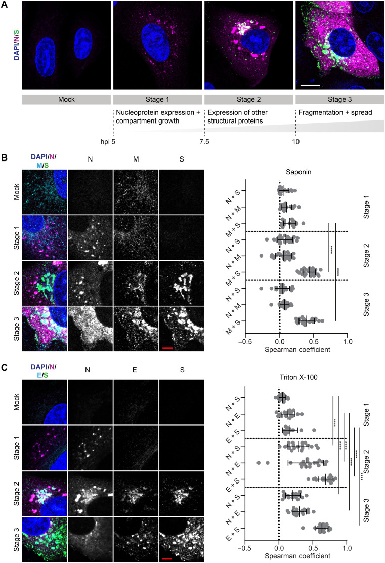
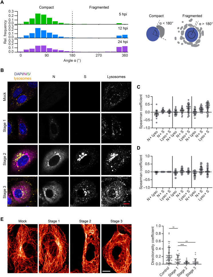
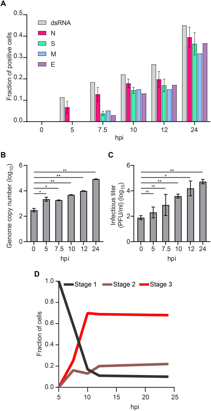
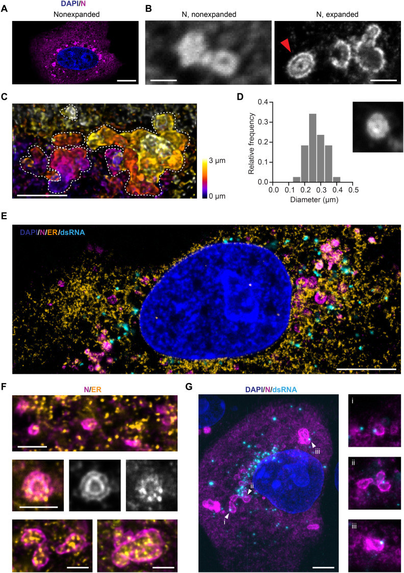
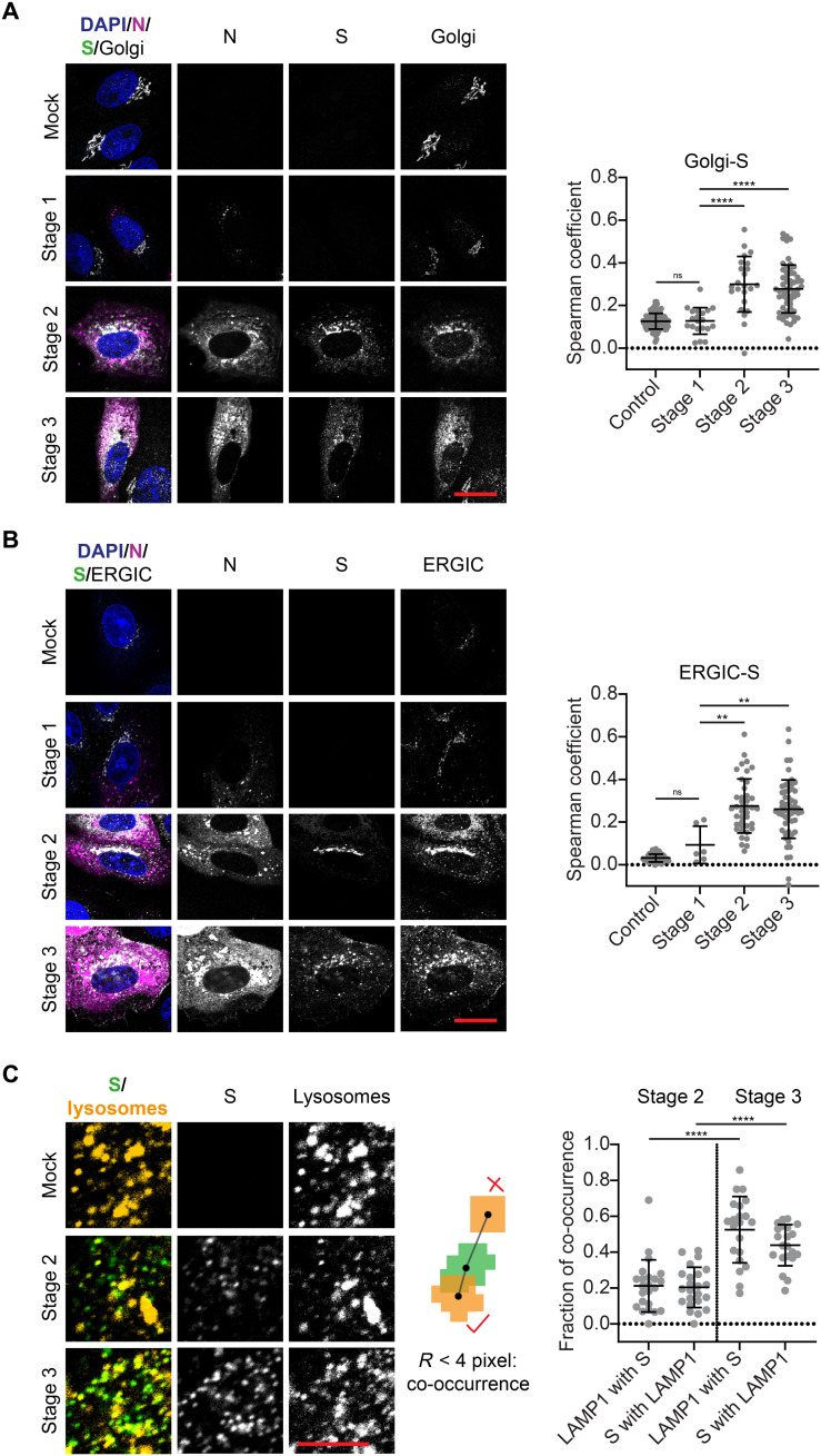
Despite being the target of extensive research efforts due to the COVID-19 (coronavirus disease 2019) pandemic, relatively little is known about the dynamics of severe acute respiratory syndrome coronavirus 2 (SARS-CoV-2) replication within cells. We investigate and characterize the tightly orchestrated virus assembly by visualizing the spatiotemporal dynamics of the four structural SARS-CoV-2 proteins at high resolution. The nucleoprotein is expressed first and accumulates around folded endoplasmic reticulum (ER) membranes in convoluted layers that contain viral RNA replication foci. We find that, of the three transmembrane proteins, the membrane protein appears at the Golgi apparatus/ER-to-Golgi intermediate compartment before the spike and envelope proteins. Relocation of a lysosome marker toward the assembly compartment and its detection in transport vesicles of viral proteins confirm an important role of lysosomes in SARS-CoV-2 egress. These data provide insights into the spatiotemporal regulation of SARS-CoV-2 assembly and refine the current understanding of SARS-CoV-2 replication.
尽管由于2019年冠状病毒病(COVID-19)大流行,严重急性呼吸综合征冠状病毒2(SARS-CoV-2)成为广泛研究的对象,但人们对其在细胞内的复制动态了解相对较少。我们通过高分辨率可视化SARS-CoV-2四种结构蛋白的时空动态,对其紧密协调的病毒组装过程进行了研究和表征。核蛋白首先表达,并在含有病毒RNA复制位点的卷曲层中围绕折叠的内质网(ER)膜积累。我们发现,在三种跨膜蛋白中,膜蛋白在刺突蛋白和包膜蛋白之前出现在高尔基体/内质网到高尔基体中间区室。溶酶体标记物向组装区室的重新定位及其在病毒蛋白运输囊泡中的检测证实了溶酶体在SARS-CoV-2释放中的重要作用。这些数据为SARS-CoV-2组装的时空调控提供了见解,并完善了目前对SARS-CoV-2复制的理解。