Department of Burn and Plastic Surgery, The First Affiliated Hospital of Nanjing Medical University, Nanjing, China.
Department of General Surgery, Fuyang Hospital Affiliated to Anhui Medical University, Fuyang, China.
Front Immunol. 2021 Dec 22;12:783907. doi: 10.3389/fimmu.2021.783907. eCollection 2021.
The pathophysiology of keloid formation is not yet understood, so the identification of biomarkers for kelod can be one step towards designing new targeting therapies which will improve outcomes for patients with keloids or at risk of developing keloids.
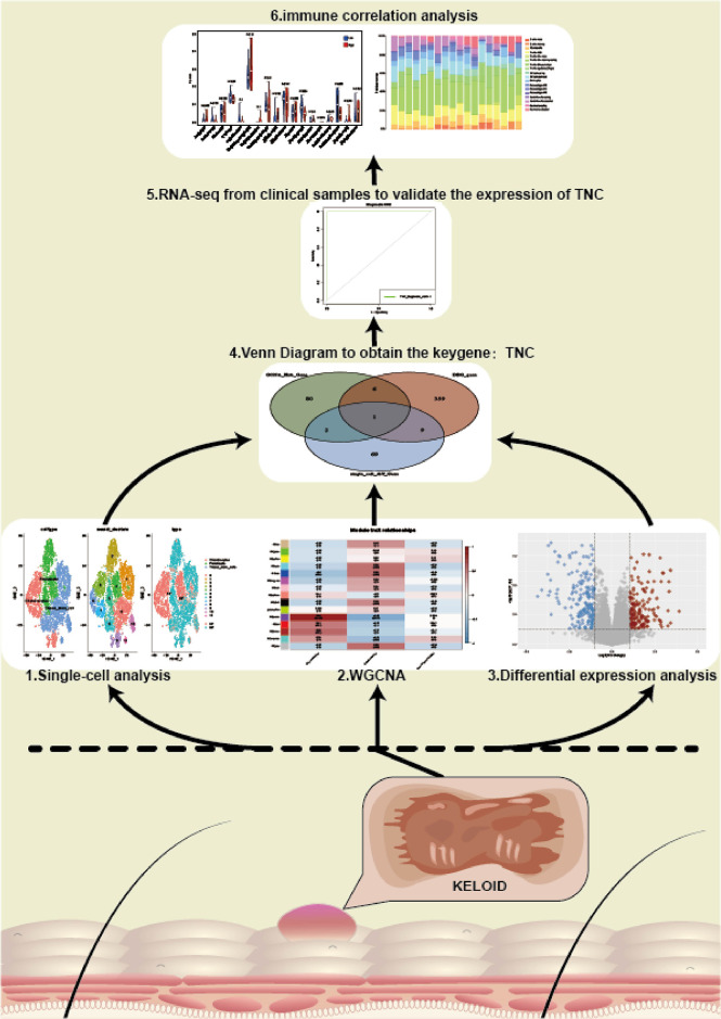
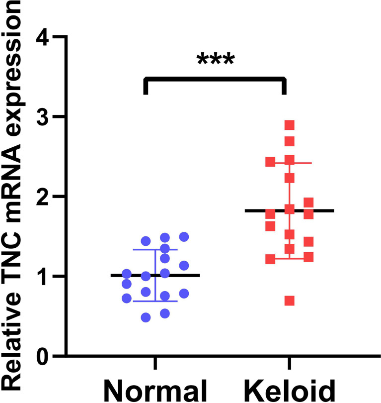
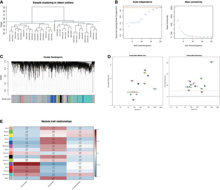
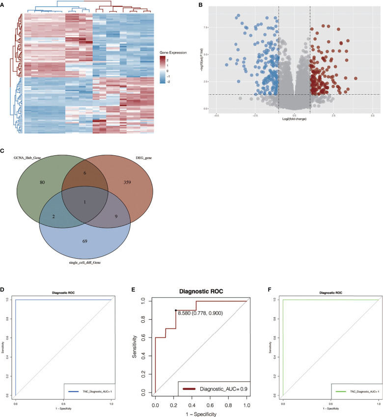
In this study, we performed single-cell RNA sequencing analysis, weighted co-expression network analysis, and differential expression analysis of keloids based on public databases. And 3 RNA sequencing data from keloid patients in our center were used for validation. Besides, we performed QRT-PCR on keloid tissue and adjacent normal tissues from 16 patients for further verification.
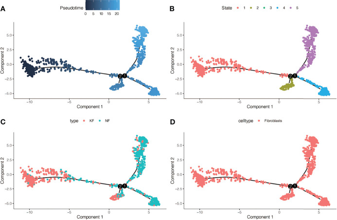
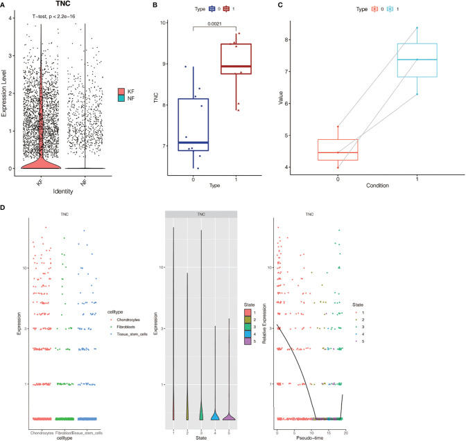
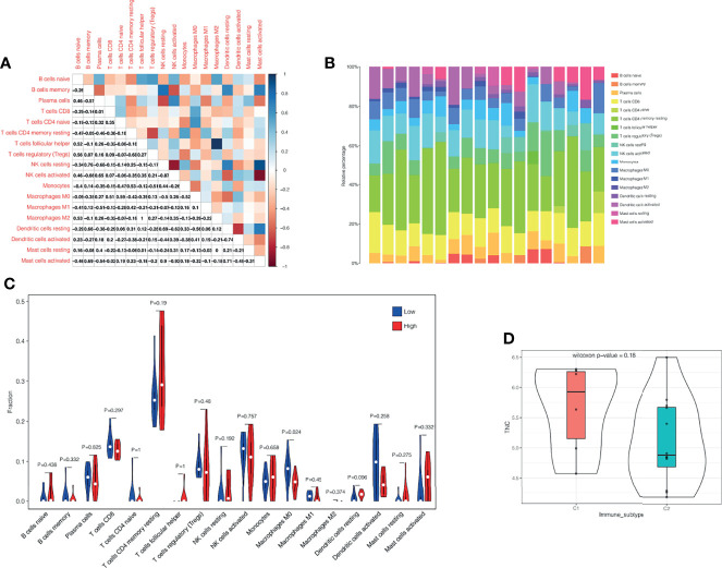
We identified the sensitive biomarker of keloid: Tenascin-C (TNC). Then, Pseudotime analysis found that the expression level of TNC decreased first, then stabilized and finally increased with fibroblast differentiation, suggesting that TNC may play an potential role in fibroblast differentiation. In addition, there were differences in the infiltration level of macrophages M0 between the TNC-high group and the TNC-low group. Macrophages M0 had a higher infiltration level in low TNC- group (P<0.05).
Our results can provide a new idea for the diagnosis and treatment of keloid.
瘢痕疙瘩形成的病理生理学尚不清楚,因此鉴定瘢痕疙瘩的生物标志物可以是设计新的靶向治疗方法的一步,这将改善瘢痕疙瘩患者或有发生瘢痕疙瘩风险的患者的治疗效果。
本研究我们基于公共数据库进行了瘢痕疙瘩的单细胞 RNA 测序分析、加权共表达网络分析和差异表达分析。并使用来自我们中心的 3 例瘢痕疙瘩患者的 RNA 测序数据进行验证。此外,我们对 16 例患者的瘢痕疙瘩组织和相邻正常组织进行了 QRT-PCR 检测,以进一步验证。
我们鉴定出了瘢痕疙瘩的敏感生物标志物:Tenascin-C(TNC)。然后,拟时分析发现 TNC 的表达水平随着成纤维细胞分化先降低,然后稳定,最后升高,提示 TNC 可能在成纤维细胞分化中发挥潜在作用。此外,TNC 高表达组和 TNC 低表达组之间 M0 型巨噬细胞的浸润水平存在差异。在 TNC 低表达组中,M0 型巨噬细胞的浸润水平更高(P<0.05)。
我们的研究结果为瘢痕疙瘩的诊断和治疗提供了新的思路。