Department of Medicine, Division of Diabetes, Metabolism and Endocrinology, Showa University School of Medicine, Shinagawa, Tokyo, Japan.
Department of Medicine, Division of Diabetes, Metabolism and Endocrinology, Anti-glycation Research Section, Showa University School of Medicine, Shinagawa, Tokyo, Japan.
Diab Vasc Dis Res. 2021 Mar-Apr;18(2):1479164121999034. doi: 10.1177/1479164121999034.
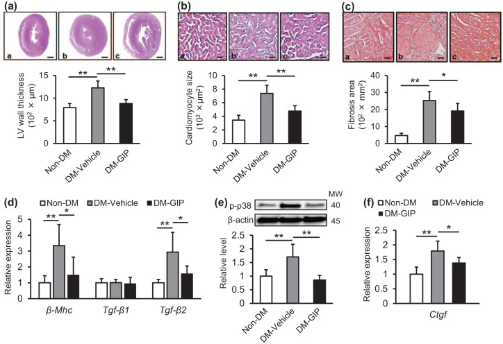
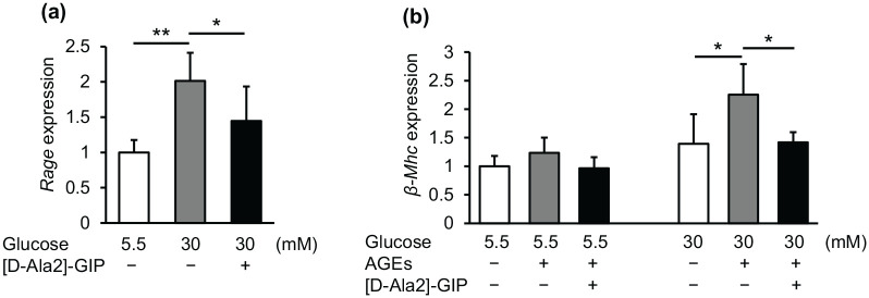
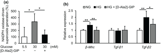
Diabetic cardiomyopathy is associated with an increased risk for heart failure and death in patients with diabetes. We investigated here whether and how GIP attenuated cardiac hypertrophy and fibrosis in diabetic mice with obesity. Diabetic db/db mice at 7 weeks old were infused with vehicle or GIP (50 nmol/kg/day) for 6 weeks, and hearts were collected for histological and RT-PCR analyzes. Cardiomyocytes isolated from neonatal mice were incubated with or without 300 nM [D-Ala2]-GIP, 30 mM glucose, or 100 μg/mL advanced glycation end products (AGEs) for RT-PCR and lucigenin assays. Compared with non-diabetic mice, diabetic mice exhibited larger left ventricle wall thickness and cardiomyocyte sizes and more fibrotic areas in association with up-regulation of myosin heavy chain β (β-Mhc) and transforming growth factor-beta2 (Tgf-β2) mRNA levels, all of which were inhibited by GIP infusion. High glucose increased NADPH oxidase-driven superoxide generation and up-regulated β-Mhc, Tgf-β2, and receptor for AGEs mRNA levels in cardiomyocytes, and augmented the AGE-induced β-Mhc gene expression. [D-Ala2]-GIP attenuated all of the deleterious effects of high glucose and/or AGEs on cardiomyocytes. Our present findings suggest that GIP could inhibit cardiac hypertrophy and fibrosis in diabetic mice via suppression of TGF-β2.
糖尿病心肌病与糖尿病患者心力衰竭和死亡风险增加有关。我们在此研究了 GIP 是否以及如何减轻肥胖糖尿病 db/db 小鼠的心脏肥大和纤维化。7 周龄的糖尿病 db/db 小鼠接受载体或 GIP(50nmol/kg/天)输注 6 周,收集心脏进行组织学和 RT-PCR 分析。将新生小鼠的心肌细胞与或不与 300nM [D-Ala2]-GIP、30mM 葡萄糖或 100μg/mL 晚期糖基化终产物(AGEs)孵育进行 RT-PCR 和荧光素酶测定。与非糖尿病小鼠相比,糖尿病小鼠的左心室壁厚度和心肌细胞大小更大,纤维化区域更多,与肌球蛋白重链 β(β-Mhc)和转化生长因子-β2(Tgf-β2)mRNA 水平上调有关,所有这些都被 GIP 输注抑制。高葡萄糖增加了 NADPH 氧化酶驱动的超氧化物生成,并上调了心肌细胞中的β-Mhc、Tgf-β2 和 AGE 受体 mRNA 水平,并增强了 AGE 诱导的β-Mhc 基因表达。[D-Ala2]-GIP 减弱了高葡萄糖和/或 AGEs 对心肌细胞的所有有害影响。我们目前的研究结果表明,GIP 通过抑制 TGF-β2 可抑制糖尿病小鼠的心脏肥大和纤维化。