• 文献检索
  • 文档翻译
  • 深度研究
  • 学术资讯
  • Suppr Zotero 插件Zotero 插件
  • 邀请有礼
  • 套餐&价格
  • 历史记录
应用&插件
Suppr Zotero 插件Zotero 插件浏览器插件Mac 客户端Windows 客户端微信小程序
定价
高级版会员购买积分包购买API积分包
服务
文献检索文档翻译深度研究API 文档MCP 服务
关于我们
关于 Suppr公司介绍联系我们用户协议隐私条款
关注我们

Suppr 超能文献

核心技术专利:CN118964589B侵权必究
粤ICP备2023148730 号-1Suppr @ 2026

文献检索

告别复杂PubMed语法,用中文像聊天一样搜索,搜遍4000万医学文献。AI智能推荐,让科研检索更轻松。

立即免费搜索

文件翻译

保留排版,准确专业,支持PDF/Word/PPT等文件格式,支持 12+语言互译。

免费翻译文档

深度研究

AI帮你快速写综述,25分钟生成高质量综述,智能提取关键信息,辅助科研写作。

立即免费体验

使用性能灵敏度分析研究用于语义分割的卷积神经网络中的偏差。

Investigation of biases in convolutional neural networks for semantic segmentation using performance sensitivity analysis.

机构信息

Medical Physics Group, Institute of Diagnostic and Interventional Radiology, Jena University Hospital - Friedrich Schiller University Jena, Germany.

Medical Physics Group, Institute of Diagnostic and Interventional Radiology, Jena University Hospital - Friedrich Schiller University Jena, Germany.

出版信息

Z Med Phys. 2022 Aug;32(3):346-360. doi: 10.1016/j.zemedi.2021.11.004. Epub 2022 Jan 10.

DOI:10.1016/j.zemedi.2021.11.004
PMID:35016819
原文链接:https://pmc.ncbi.nlm.nih.gov/articles/PMC9948839/
Abstract

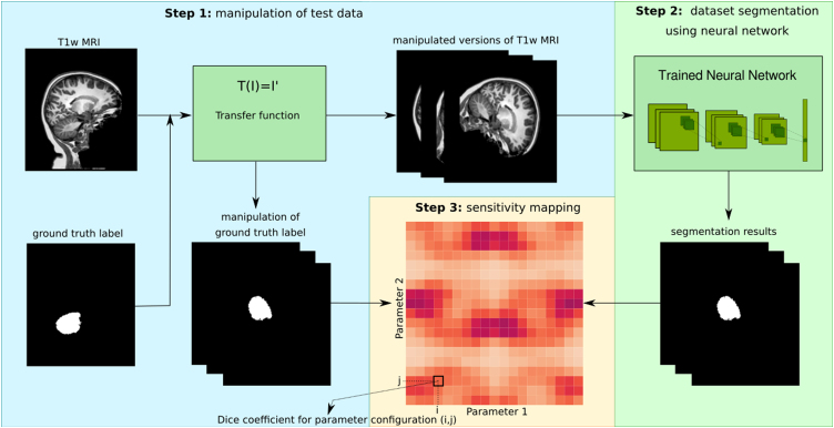
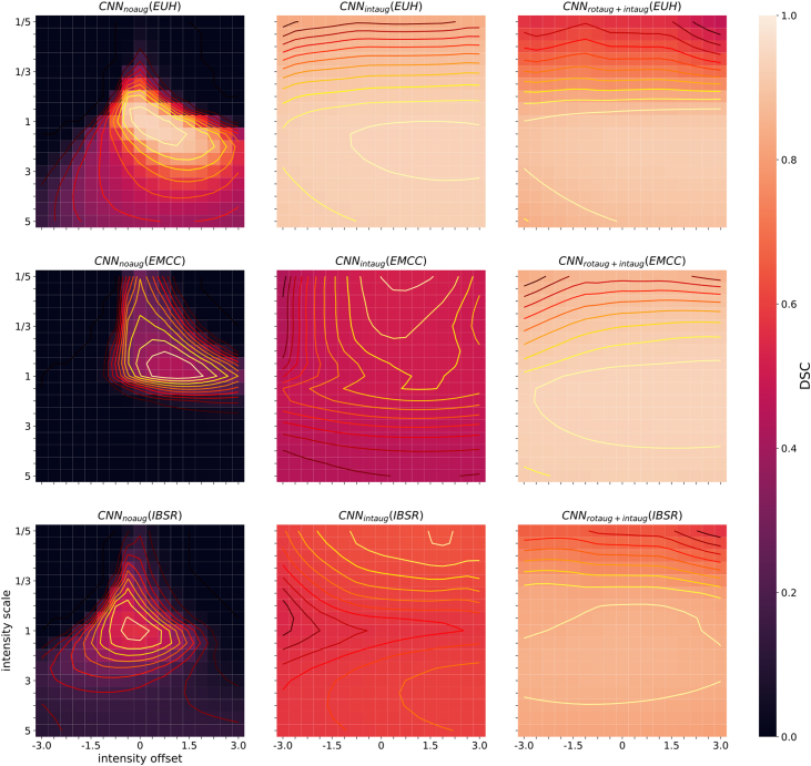
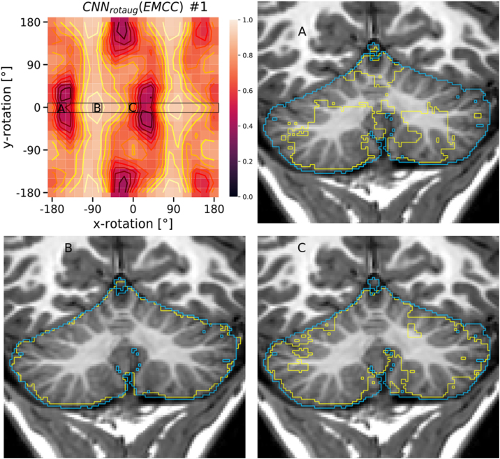
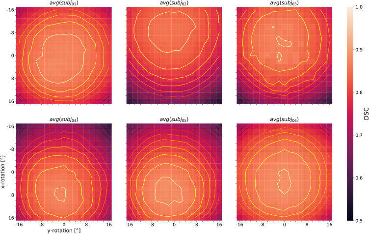
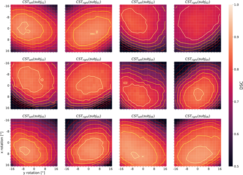
The application of deep neural networks for segmentation in medical imaging has gained substantial interest in recent years. In many cases, this variant of machine learning has been shown to outperform other conventional segmentation approaches. However, little is known about its general applicability. Especially the robustness against image modifications (e.g., intensity variations, contrast variations, spatial alignment) has hardly been investigated. Data augmentation is often used to compensate for sensitivity to such changes, although its effectiveness has not yet been studied. Therefore, the goal of this study was to systematically investigate the sensitivity to variations in input data with respect to segmentation of medical images using deep learning. This approach was tested with two publicly available segmentation frameworks (DeepMedic and TractSeg). In the case of DeepMedic, the performance was tested using ground truth data, while in the case of TractSeg, the STAPLE technique was employed. In both cases, sensitivity analysis revealed significant dependence of the segmentation performance on input variations. The effects of different data augmentation strategies were also shown, making this type of analysis a useful tool for selecting the right parameters for augmentation. The proposed analysis should be applied to any deep learning image segmentation approach, unless the assessment of sensitivity to input variations can be directly derived from the network.

摘要

近年来,深度学习网络在医学图像分割中的应用引起了广泛关注。在许多情况下,这种机器学习变体已被证明优于其他传统的分割方法。然而,其普遍适用性知之甚少。特别是针对图像修改(例如,强度变化、对比度变化、空间配准)的鲁棒性几乎没有被研究过。数据增强通常用于补偿对这些变化的敏感性,尽管其有效性尚未得到研究。因此,本研究的目的是系统地研究使用深度学习对医学图像分割的输入数据变化的敏感性。该方法使用两个公开的分割框架(DeepMedic 和 TractSeg)进行了测试。在 DeepMedic 的情况下,使用真实数据测试了性能,而在 TractSeg 的情况下,使用了 STAPLE 技术。在这两种情况下,敏感性分析都表明分割性能对输入变化有很大的依赖性。还展示了不同的数据增强策略的效果,这使得这种类型的分析成为选择正确增强参数的有用工具。除非可以直接从网络中得出对输入变化的敏感性评估,否则应将所提出的分析应用于任何深度学习图像分割方法。

https://cdn.ncbi.nlm.nih.gov/pmc/blobs/44ce/9948839/b89109ea4841/gr9.jpg
https://cdn.ncbi.nlm.nih.gov/pmc/blobs/44ce/9948839/90654bfe6bb6/gr1.jpg
https://cdn.ncbi.nlm.nih.gov/pmc/blobs/44ce/9948839/0f1ee3c4f773/gr2.jpg
https://cdn.ncbi.nlm.nih.gov/pmc/blobs/44ce/9948839/e0960165a9dd/gr3.jpg
https://cdn.ncbi.nlm.nih.gov/pmc/blobs/44ce/9948839/63b49177abd7/gr4.jpg
https://cdn.ncbi.nlm.nih.gov/pmc/blobs/44ce/9948839/ef0b17fbf593/gr5.jpg
https://cdn.ncbi.nlm.nih.gov/pmc/blobs/44ce/9948839/450b6dfeff92/gr6.jpg
https://cdn.ncbi.nlm.nih.gov/pmc/blobs/44ce/9948839/ff075390358a/gr7.jpg
https://cdn.ncbi.nlm.nih.gov/pmc/blobs/44ce/9948839/2b3266843ecd/gr8.jpg
https://cdn.ncbi.nlm.nih.gov/pmc/blobs/44ce/9948839/b89109ea4841/gr9.jpg

相似文献

1
Investigation of biases in convolutional neural networks for semantic segmentation using performance sensitivity analysis.使用性能灵敏度分析研究用于语义分割的卷积神经网络中的偏差。
Z Med Phys. 2022 Aug;32(3):346-360. doi: 10.1016/j.zemedi.2021.11.004. Epub 2022 Jan 10.
2
[Application of semantic segmentation based on convolutional neural network in medical images].基于卷积神经网络的语义分割在医学图像中的应用
Sheng Wu Yi Xue Gong Cheng Xue Za Zhi. 2020 Jun 25;37(3):533-540. doi: 10.7507/1001-5515.201906067.
3
A comparison between two semantic deep learning frameworks for the autosomal dominant polycystic kidney disease segmentation based on magnetic resonance images.基于磁共振图像的常染色体显性遗传性多囊肾病分割的两种语义深度学习框架的比较。
BMC Med Inform Decis Mak. 2019 Dec 12;19(Suppl 9):244. doi: 10.1186/s12911-019-0988-4.
4
Analysis of intensity normalization for optimal segmentation performance of a fully convolutional neural network.分析强度归一化对全卷积神经网络最佳分割性能的影响。
Z Med Phys. 2019 May;29(2):128-138. doi: 10.1016/j.zemedi.2018.11.004. Epub 2018 Dec 20.
5
Combined Transfer Learning and Test-Time Augmentation Improves Convolutional Neural Network-Based Semantic Segmentation of Prostate Cancer from Multi-Parametric MR Images.联合迁移学习和测试时增强提高了基于卷积神经网络的多参数磁共振图像前列腺癌的语义分割。
Comput Methods Programs Biomed. 2021 Oct;210:106375. doi: 10.1016/j.cmpb.2021.106375. Epub 2021 Aug 28.
6
Convolutional neural network for automated mass segmentation in mammography.卷积神经网络在乳腺 X 线摄影中用于自动肿块分割。
BMC Bioinformatics. 2020 Dec 9;21(Suppl 1):192. doi: 10.1186/s12859-020-3521-y.
7
Machine learning and image analysis in vascular surgery.机器学习和血管外科学中的图像分析。
Semin Vasc Surg. 2023 Sep;36(3):413-418. doi: 10.1053/j.semvascsurg.2023.07.001. Epub 2023 Jul 7.
8
Semantic segmentation of cerebrospinal fluid and brain volume with a convolutional neural network in pediatric hydrocephalus-transfer learning from existing algorithms.基于卷积神经网络的小儿脑积水脑积水分割及脑容量计算——从现有算法进行迁移学习。
Acta Neurochir (Wien). 2020 Oct;162(10):2463-2474. doi: 10.1007/s00701-020-04447-x. Epub 2020 Jun 25.
9
A Lightweight Semantic Segmentation Algorithm Based on Deep Convolutional Neural Networks.基于深度卷积神经网络的轻量级语义分割算法。
Comput Intell Neurosci. 2022 Sep 6;2022:5339664. doi: 10.1155/2022/5339664. eCollection 2022.
10
Segment-then-Segment: Context-Preserving Crop-Based Segmentation for Large Biomedical Images.分段再分段:基于上下文保留的作物分割在大型生物医学图像中的应用。
Sensors (Basel). 2023 Jan 5;23(2):633. doi: 10.3390/s23020633.

引用本文的文献

1
Sensitivity of Diffusion MRI to White Matter Pathology: Influence of Diffusion Protocol, Magnetic Field Strength, and Processing Pipeline in Systemic Lupus Erythematosus.扩散加权磁共振成像对脑白质病变的敏感性:扩散协议、磁场强度及处理流程在系统性红斑狼疮中的影响
Front Neurol. 2022 Apr 26;13:837385. doi: 10.3389/fneur.2022.837385. eCollection 2022.

本文引用的文献

1
GANs for medical image analysis.生成对抗网络在医学图像分析中的应用。
Artif Intell Med. 2020 Sep;109:101938. doi: 10.1016/j.artmed.2020.101938. Epub 2020 Aug 9.
2
Generation of annotated multimodal ground truth datasets for abdominal medical image registration.生成带注释的多模态腹部医学图像配准地面实况数据集。
Int J Comput Assist Radiol Surg. 2021 Aug;16(8):1277-1285. doi: 10.1007/s11548-021-02372-7. Epub 2021 May 2.
3
Investigation of Deep-Learning-Driven Identification of Multiple Sclerosis Patients Based on Susceptibility-Weighted Images Using Relevance Analysis.
基于相关性分析的深度学习驱动的利用磁化率加权成像识别多发性硬化症患者的研究。
Front Neurosci. 2020 Dec 18;14:609468. doi: 10.3389/fnins.2020.609468. eCollection 2020.
4
Convolutional Neural Network Ensemble Segmentation With Ratio-Based Sampling for the Arteries and Veins in Abdominal CT Scans.基于比值采样的卷积神经网络集成分割在腹部 CT 扫描中的动脉和静脉。
IEEE Trans Biomed Eng. 2021 May;68(5):1518-1526. doi: 10.1109/TBME.2020.3042640. Epub 2021 Apr 21.
5
Unravelling the effect of data augmentation transformations in polyp segmentation.揭示数据增强变换在息肉分割中的作用。
Int J Comput Assist Radiol Surg. 2020 Dec;15(12):1975-1988. doi: 10.1007/s11548-020-02262-4. Epub 2020 Sep 28.
6
Latent space manipulation for high-resolution medical image synthesis via the StyleGAN.基于 StyleGAN 的高分辨率医学图像合成的潜在空间处理。
Z Med Phys. 2020 Nov;30(4):305-314. doi: 10.1016/j.zemedi.2020.05.001. Epub 2020 Jun 18.
7
Resolving challenges in deep learning-based analyses of histopathological images using explanation methods.运用解释方法解决基于深度学习的组织病理学图像分析中的挑战。
Sci Rep. 2020 Apr 14;10(1):6423. doi: 10.1038/s41598-020-62724-2.
8
Deep Learning-Based Gleason Grading of Prostate Cancer From Histopathology Images-Role of Multiscale Decision Aggregation and Data Augmentation.基于深度学习的前列腺癌组织病理图像 Gleason 分级——多尺度决策聚合和数据增强的作用。
IEEE J Biomed Health Inform. 2020 May;24(5):1413-1426. doi: 10.1109/JBHI.2019.2944643. Epub 2019 Sep 30.
9
Synthesis of CT images from digital body phantoms using CycleGAN.使用 CycleGAN 从数字体模型生成 CT 图像。
Int J Comput Assist Radiol Surg. 2019 Oct;14(10):1741-1750. doi: 10.1007/s11548-019-02042-9. Epub 2019 Aug 5.
10
A gentle introduction to deep learning in medical image processing.深度学习在医学图像处理中的应用简介。
Z Med Phys. 2019 May;29(2):86-101. doi: 10.1016/j.zemedi.2018.12.003. Epub 2019 Jan 25.