Department of Zoology, Faculty of Science, Cairo University, Giza, 12613, Egypt.
Pharmaceutical Research Institute, Albany College of Pharmacy and Health Sciences, Albany, NY, USA.
Biol Trace Elem Res. 2022 Dec;200(12):5145-5158. doi: 10.1007/s12011-022-03102-z. Epub 2022 Jan 15.
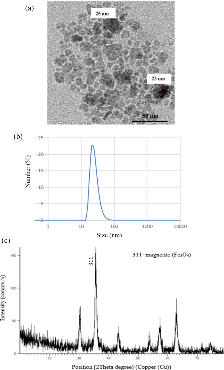
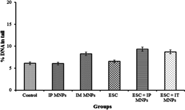
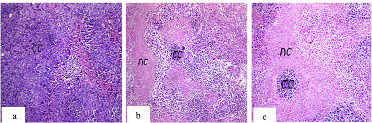
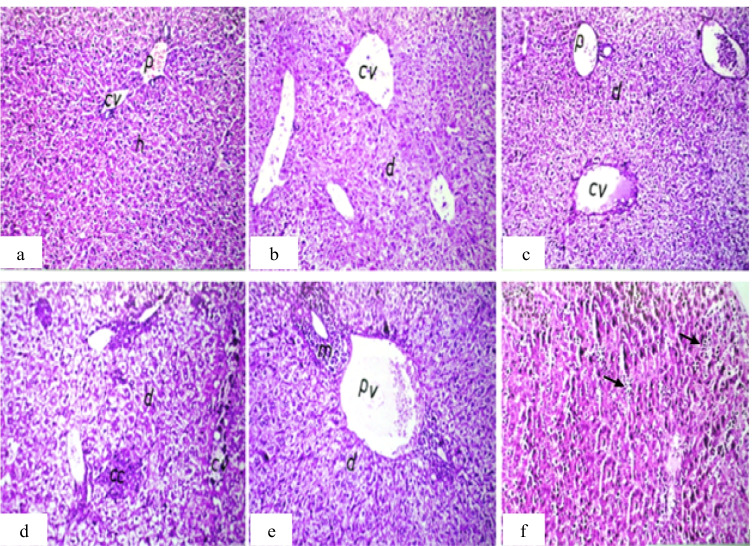
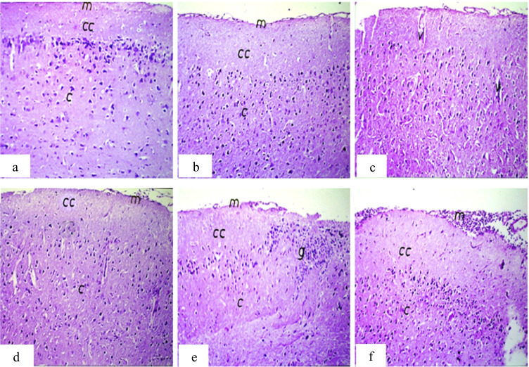
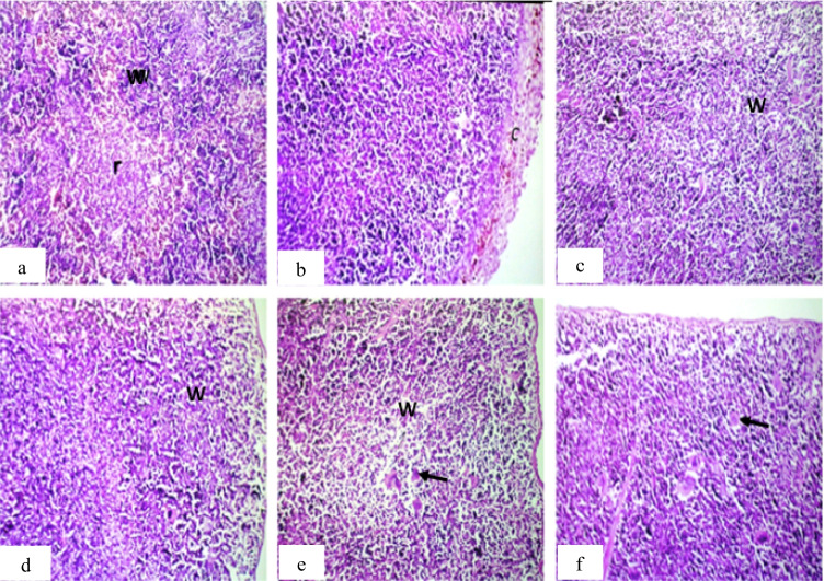
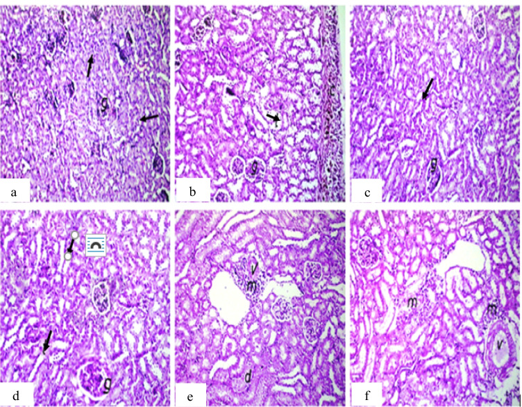
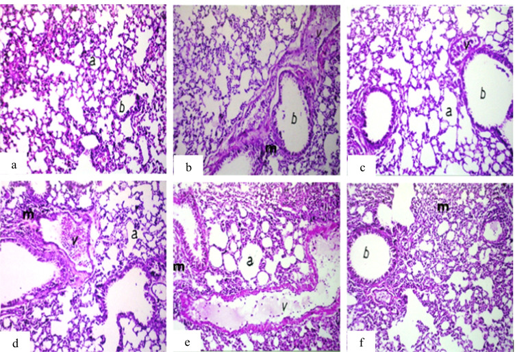
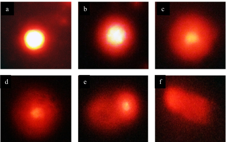
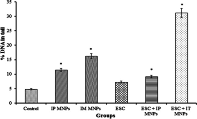
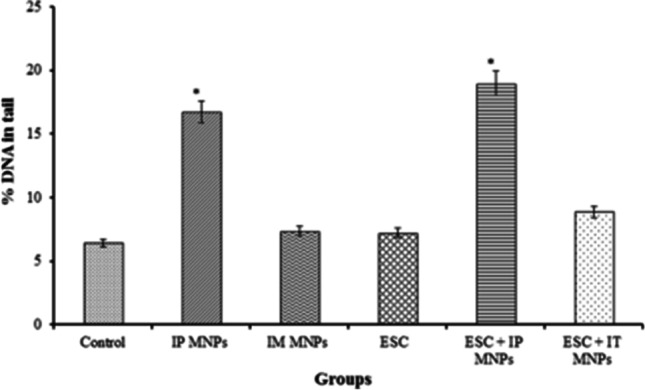
Nanoparticles can potentially cause adverse effects on cellular and molecular level. The present study aimed to investigate the histopathological changes and DNA damage effects of magnetite nanoparticles (MNPs) on female albino mice model with Ehrlich solid carcinoma (ESC). Magnetite nanoparticles coated with L-ascorbic acid (size ~ 25.0 nm) were synthesized and characterized. Mice were treated with MNPs day by day, intraperitoneally (IP), intramuscularly (IM), or intratumorally (IT). Autopsy samples were taken from the solid tumor, thigh muscle, liver, kidney, lung, spleen, and brain for assessment of iron content, histopathological examination, and genotoxicity using comet assay. The liver, spleen, lung, and heart had significantly higher iron content in groups treated IP. On the other hand, tumor, muscles, and the liver had significantly higher iron content in groups treated IT. MNPs induced a significant DNA damage in IT treated ESC. While a significant DNA damage was detected in the liver of the IP treated group, but no significant DNA damage could be detected in the brain. Histopathological findings in ESC revealed a marked tumor necrosis, 50% in group injected IT but 40% in group injected IP and 20% only in untreated tumors. Other findings include inflammatory cell infiltration, dilatation, and congestion of blood vessels of different organs of treated groups in addition to appearance of metastatic cancer cells in the liver of non-treated tumor group. MNPs could have an antitumor effect but it is recommended to be injected intratumorally to be directed to the tumor tissues and reduce its adverse effects on healthy tissues.
纳米粒子可能会对细胞和分子水平造成不良影响。本研究旨在探讨磁铁矿纳米粒子(MNPs)对艾氏腹水癌(ESC)雌性白化小鼠模型的组织病理学变化和 DNA 损伤效应。合成并表征了用 L-抗坏血酸(大小约 25.0nm)涂覆的磁铁矿纳米粒子。每天通过腹腔内(IP)、肌肉内(IM)或肿瘤内(IT)途径向小鼠给药 MNPs。从实体瘤、大腿肌肉、肝、肾、肺、脾和脑采集尸检样本,以评估铁含量、进行组织病理学检查和使用彗星试验评估遗传毒性。肝、脾、肺和心脏在 IP 治疗组中具有明显更高的铁含量。另一方面,在 IT 治疗组中,肿瘤、肌肉和肝脏具有明显更高的铁含量。MNPs 在 IT 治疗的 ESC 中诱导了明显的 DNA 损伤。虽然在 IP 治疗组的肝脏中检测到明显的 DNA 损伤,但在大脑中未检测到明显的 DNA 损伤。ESC 的组织病理学发现显示出明显的肿瘤坏死,在 IT 注射组中为 50%,在 IP 注射组中为 40%,而未治疗的肿瘤组中仅为 20%。其他发现包括处理组的不同器官的血管扩张和充血以及非处理肿瘤组的肝脏中出现转移性癌细胞。MNPs 可能具有抗肿瘤作用,但建议将其注入肿瘤内,以靶向肿瘤组织并减少其对健康组织的不良反应。