Laboratory of Neurogenesis, Carlos Chagas Filho Institute of Biophysics, Universidade Federal do Rio de Janeiro, Rio de Janeiro, Brazil.
Laboratory of Neurobiology, Department of Morphology, Institute of Biological Sciences, Universidade Federal de Minas Gerais, Belo Horizonte, Brazil.
Invest Ophthalmol Vis Sci. 2022 Feb 1;63(2):5. doi: 10.1167/iovs.63.2.5.
Based on our preview evidence that reduced nuclear content of the transcription factor Myc-associated protein X (MAX) is an early event associated with degeneration of retinal ganglion cells (RGCs), in the present study, our purpose was to test whether the overexpression of human MAX had a neuroprotective effect against RGC injury.
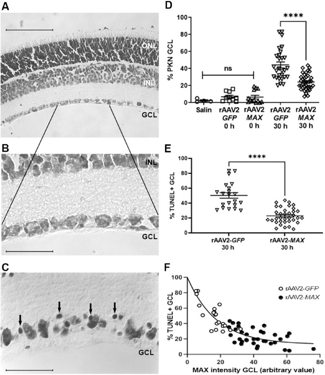
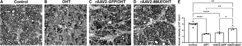
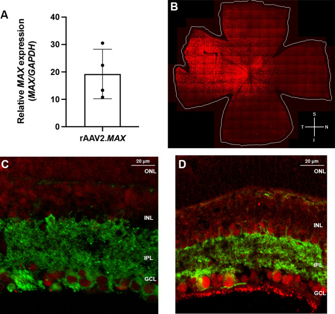
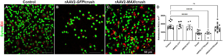
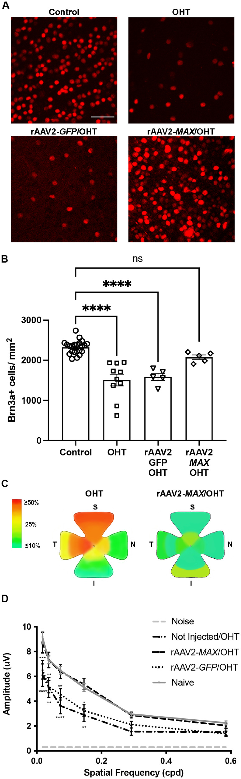
Overexpression of either MAX or green fluorescent protein (GFP) in the retina was achieved by intravitreal injections of recombinant adenovirus-associated viruses (rAAVs). Lister Hooded rats were used in three models of RGC degeneration: (1) cultures of retinal explants for 30 hours ex vivo from the eyes of 14-day-old rats that had received intravitreal injections of rAAV2-MAX or the control vector rAAV2-GFP at birth; (2) an optic nerve crush model, in which 1-month-old rats received intravitreal injection of either rAAV2-MAX or rAAV2-GFP and, 4 weeks later, were operated on; and (3) an ocular hypertension (OHT) glaucoma model, in which 1-month-old rats received intravitreal injection of either rAAV2-MAX or rAAV2-GFP and, 4 weeks later, were subject to cauterization of the limbal plexus. Cell death was estimated by detection of pyknotic nuclei and TUNEL technique and correlated with MAX immunocontent in an ex vivo model of retinal explants. MAX expression was detected by quantitative RT-PCR. In the OHT model, survival of RGCs was quantified by retrograde labeling with DiI or immunostaining for BRN3a at 14 days after in vivo injury. Functional integrity of RGCs was analyzed through pattern electroretinography, and damage to the optic nerve was examined in semithin sections.
In all three models of RGC insult, gene therapy by overexpression of MAX prevented RGC death. Also, ON degeneration and electrophysiologic deficits were prevented in the OHT model.
Our experiments offer proof of concept for a novel neuroprotective gene therapy for glaucomatous neurodegeneration based on overexpression of MAX.
基于我们的前期研究证据,即转录因子 Myc 相关蛋白 X(MAX)核内含量减少是与视网膜神经节细胞(RGC)变性相关的早期事件,本研究旨在检测人 MAX 的过表达是否对 RGC 损伤具有神经保护作用。
通过玻璃体内注射重组腺相关病毒(rAAV)实现 MAX 或绿色荧光蛋白(GFP)在视网膜中的过表达。使用李斯特褐鼠进行三种 RGC 变性模型的实验:(1)从出生时接受 rAAV2-MAX 或对照载体 rAAV2-GFP 玻璃体内注射的 14 日龄大鼠眼睛中分离出视网膜外植体,在体外培养 30 小时;(2)视神经挤压模型,1 月龄大鼠接受 rAAV2-MAX 或 rAAV2-GFP 的玻璃体内注射,4 周后进行手术;(3)眼高压(OHT)青光眼模型,1 月龄大鼠接受 rAAV2-MAX 或 rAAV2-GFP 的玻璃体内注射,4 周后进行角膜缘丛烧灼。通过检测固缩核和 TUNEL 技术来估计细胞死亡,并与体外视网膜外植体模型中的 MAX 免疫含量相关联。通过定量 RT-PCR 检测 MAX 表达。在 OHT 模型中,通过 DiI 逆行标记或 BRN3a 免疫染色在体内损伤后 14 天定量 RGC 存活。通过图形视网膜电图分析 RGC 功能完整性,并通过半薄切片检查视神经损伤。
在所有三种 RGC 损伤模型中,过表达 MAX 的基因治疗均可预防 RGC 死亡。此外,OHT 模型中还预防了视神经变性和电生理缺陷。
我们的实验为基于 MAX 过表达的新型青光眼神经退行性变神经保护基因治疗提供了概念验证。