Cong Ping, Qiu Qingtao, Li Xingchao, Sun Qian, Yu Xiaoming, Yin Yong
Department of Oncology, The Second Hospital, Cheeloo College of Medicine, Shandong University, Jinan, China.
Department of Radiation Oncology, Shandong Cancer Hospital and Institute, Shandong First Medical University and Shandong Academy of Medical Sciences, Jinan, China.
Transl Cancer Res. 2021 Oct;10(10):4375-4386. doi: 10.21037/tcr-21-702.
To develop and validate a radiomics model using computed tomography (CT) images acquired from the first diagnosis to estimate the status of occult brain metastases (BM) in patients with stage IV lung adenocarcinoma (LADC).
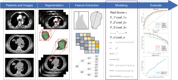
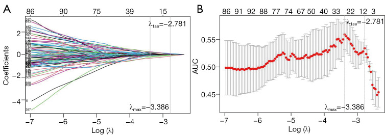
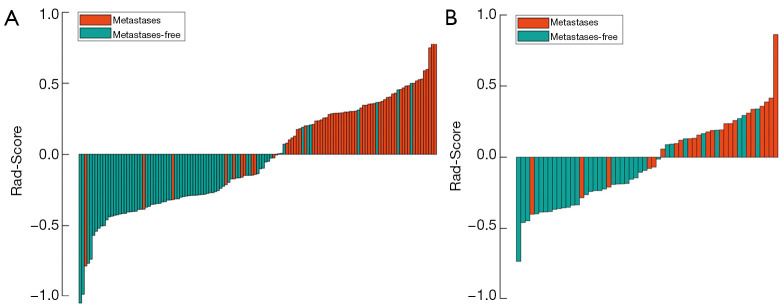
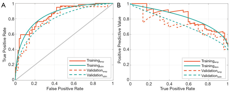
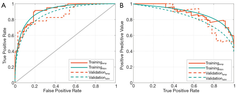
One hundred and ninety-three patients who were first diagnosed with stage IV LADC were enrolled and divided into a training cohort (n=135) and a validation cohort (n=58). Then, 725 radiomic features were extracted from contoured primary tumor volumes of LADCs. Intra- and interobserver reliabilities were calculated, and the least absolute shrinkage and selection operator (LASSO) was applied for feature selection. Subsequently, a radiomics signature (Rad-Score) was built. To improve performance, a nomogram incorporating a radiomics signature and an independent clinical predictor was developed. Finally, the established signature and nomogram were assessed using receiver operating characteristic (ROC) curves and precision-recall curves (PRC). Both empirical and α-binomial model-based ROCs and PRCs were plotted, and the area under the curve (AUC) and average precision (AP) of ROCs and PRCs were calculated and compared.
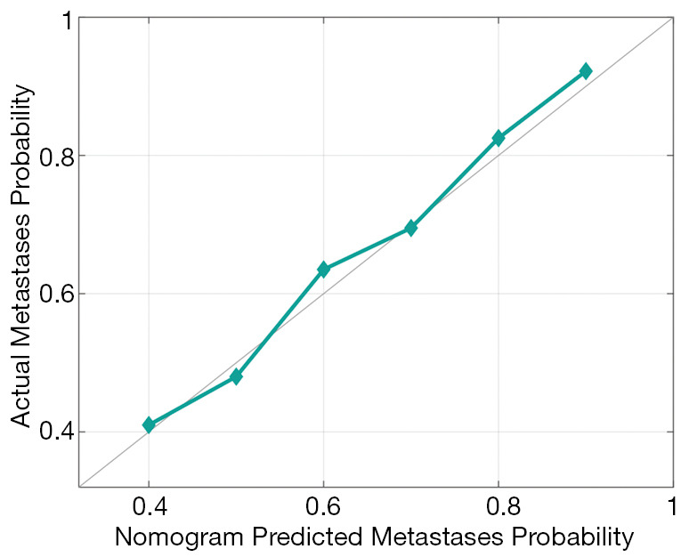
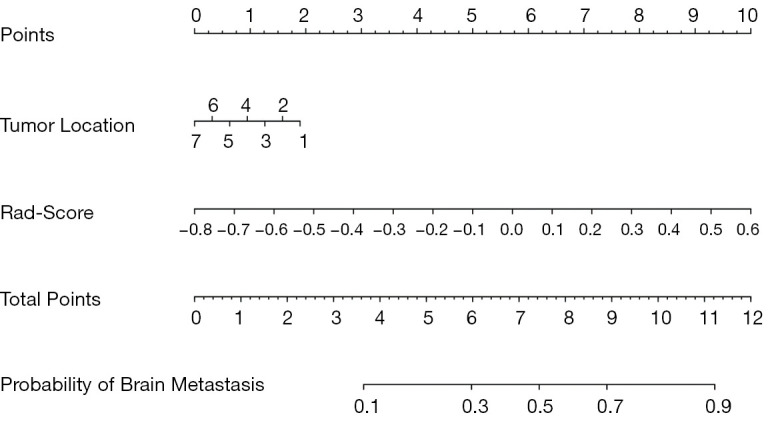
A radiomics signature and Rad-Score were constructed using eight radiomic features, and these had significant correlations with occult BM status. A nomogram was developed by incorporating a Rad-Score and the primary tumor location. The nomogram yielded an optimal AUC of 0.911 [95% confidence interval (CI): 0.903-0.919] and an AP of 0.885 (95% CI: 0.876-0.894) in the training cohort, and an AUC of 0.873 (95% CI: 0.866-0.80) and an AP of 0.827 (95% CI: 0.820-0.834) in the validation cohort using α-binomial model-based method. The calibration curve demonstrated that the nomogram showed high agreement between the actual occult BM probability and predicted by the nomogram (P=0.427).
The nomogram incorporating a radiomics signature and a clinical risk factor achieved optimal performance after holistic assessment using unbiased indexes for diagnosing occult BM of patients who were first diagnosed with stage IV LADC.
利用首次诊断时获取的计算机断层扫描(CT)图像开发并验证一种放射组学模型,以评估IV期肺腺癌(LADC)患者隐匿性脑转移(BM)的状态。
纳入193例首次诊断为IV期LADC的患者,分为训练队列(n = 135)和验证队列(n = 58)。然后,从LADC的轮廓化原发肿瘤体积中提取725个放射组学特征。计算观察者内和观察者间的可靠性,并应用最小绝对收缩和选择算子(LASSO)进行特征选择。随后,构建放射组学特征(Rad-Score)。为提高性能,开发了一种结合放射组学特征和独立临床预测指标的列线图。最后,使用受试者工作特征(ROC)曲线和精确召回率曲线(PRC)评估所建立的特征和列线图。绘制基于经验和α-二项式模型的ROC曲线和PRC,并计算和比较ROC曲线和PRC的曲线下面积(AUC)和平均精度(AP)。
利用8个放射组学特征构建了放射组学特征和Rad-Score,这些特征与隐匿性BM状态具有显著相关性。通过纳入Rad-Score和原发肿瘤位置开发了列线图。在训练队列中,列线图的最佳AUC为0.911[95%置信区间(CI):0.903 - 0.919],AP为0.885(95%CI:0.