Bristol Heart Institute and Translational Biomedical Research Centre, Faculty of Health Science, University of Bristol, Bristol, BS2 8HW, UK.
Centre for Nanosciences & Molecular Medicine, Amrita Vishwa Vidyapeetham, Kochi, 682 041, India.
J Nanobiotechnology. 2022 Feb 8;20(1):71. doi: 10.1186/s12951-022-01268-1.
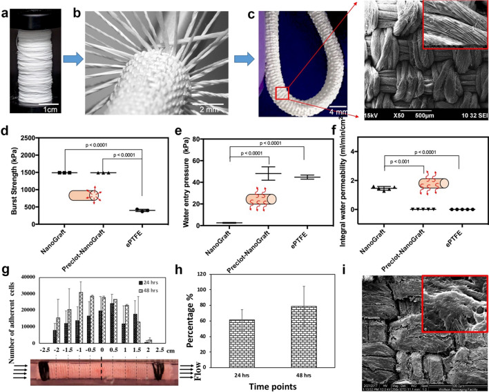
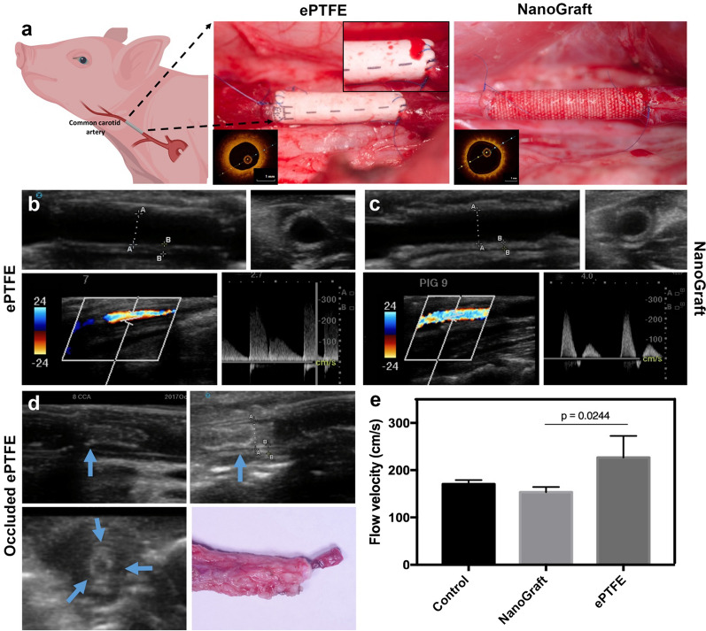
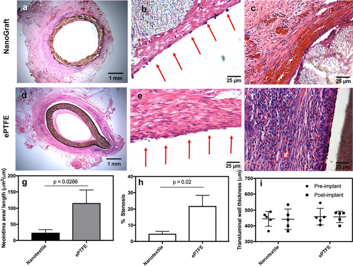
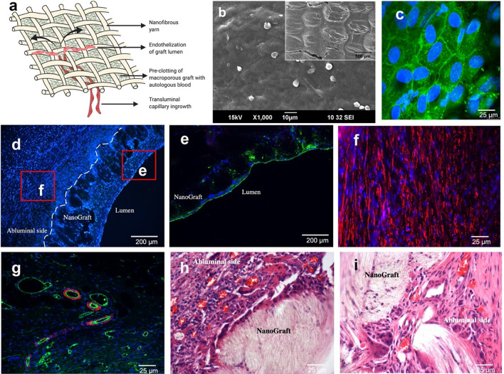
Globally, millions of patients are affected by myocardial infarction or lower limb gangrene/amputation due to atherosclerosis. Available surgical treatment based on vein and synthetic grafts provides sub-optimal benefits. We engineered a highly flexible and mechanically robust nanotextile-based vascular graft (NanoGraft) by interweaving nanofibrous threads of poly-L-lactic acid to address the unmet need. The NanoGrafts were rendered impervious with selective fibrin deposition in the micropores by pre-clotting. The pre-clotted NanoGrafts (4 mm diameter) and ePTFE were implanted in a porcine carotid artery replacement model. The fibrin-laden porous milieu facilitated rapid endothelization by the transmural angiogenesis in the NanoGraft. In-vivo patency of NanoGrafts was 100% at 2- and 4-weeks, with no changes over time in lumen size, flow velocities, and minimal foreign-body inflammatory reaction. However, the patency of ePTFE at 2-week was 66% and showed marked infiltration, neointimal thickening, and poor host tissue integration. The study demonstrates the in-vivo feasibility and safety of a thin-layered vascular prosthesis, viz., NanoGraft, and its potential superiority over the commercial ePTFE.
全球有数百万的患者因动脉粥样硬化而患有心肌梗塞或下肢坏疽/截肢。现有的基于静脉和合成移植物的手术治疗提供的效果并不理想。我们通过编织聚乳酸的纳米纤维线,开发出一种高度灵活且机械强度高的纳米纤维血管移植物(NanoGraft),以满足未满足的需求。通过预凝在微孔中选择性地沉积纤维蛋白,使 NanoGrafts 不透水。将预凝的 NanoGrafts(直径 4 毫米)和 ePTFE 植入猪颈动脉替代模型中。纤维蛋白填充的多孔环境促进了 NanoGraft 中壁内血管生成的快速内皮化。在 2 周和 4 周时,NanoGrafts 的通畅率为 100%,管腔大小、流速没有随时间变化,异物反应最小。然而,ePTFE 在 2 周时的通畅率为 66%,并显示出明显的浸润、新生内膜增厚和不良的宿主组织整合。该研究证明了薄层血管假体,即 NanoGraft 的体内可行性和安全性,及其相对于商业 ePTFE 的潜在优势。