Oulu Functional Neuroimaging, Department of Diagnostic Radiology, Oulu University Hospital, 90220 Oulu, Finland
Medical Imaging, Physics and Technology, Faculty of Medicine, University of Oulu, 90220 Oulu, Finland.
J Neurosci. 2022 Mar 23;42(12):2503-2515. doi: 10.1523/JNEUROSCI.0934-21.2022. Epub 2022 Feb 8.
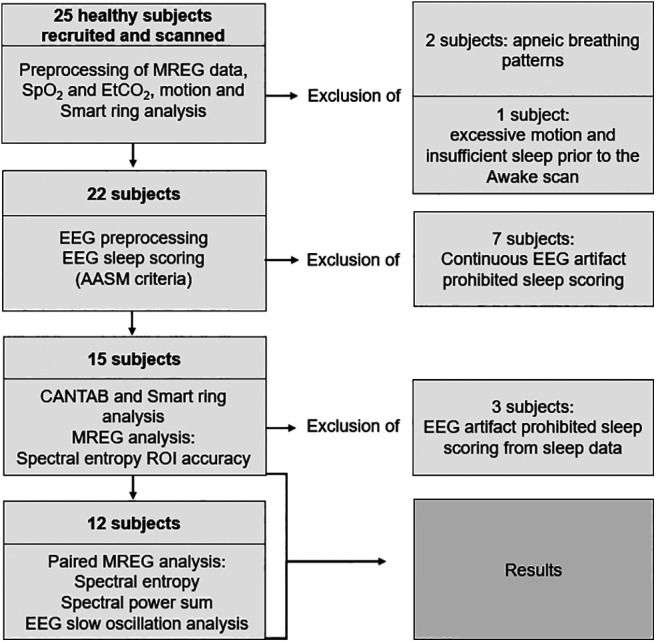
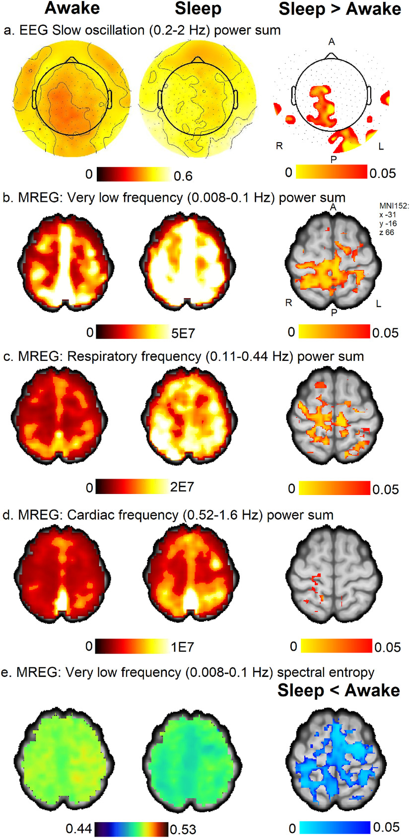
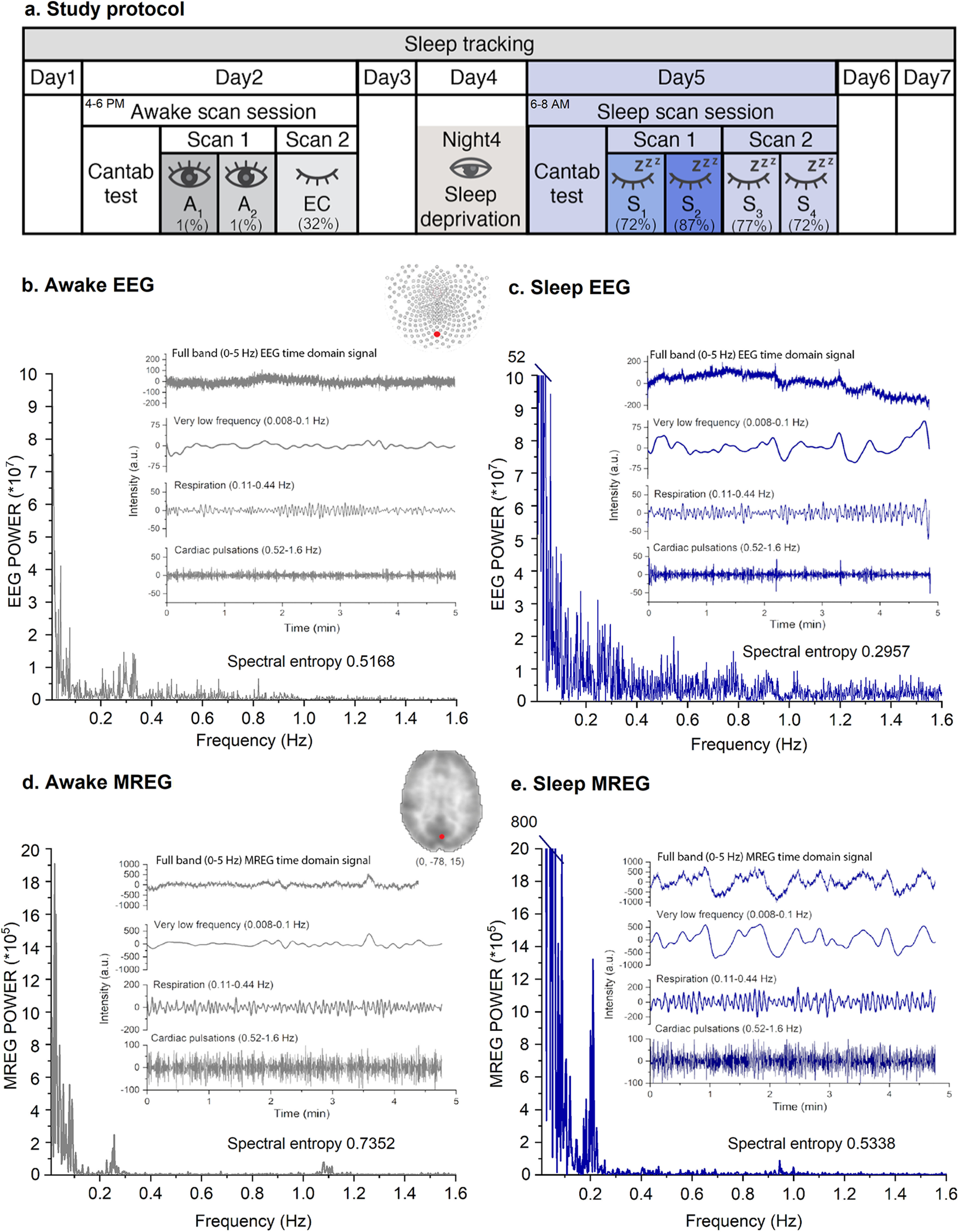
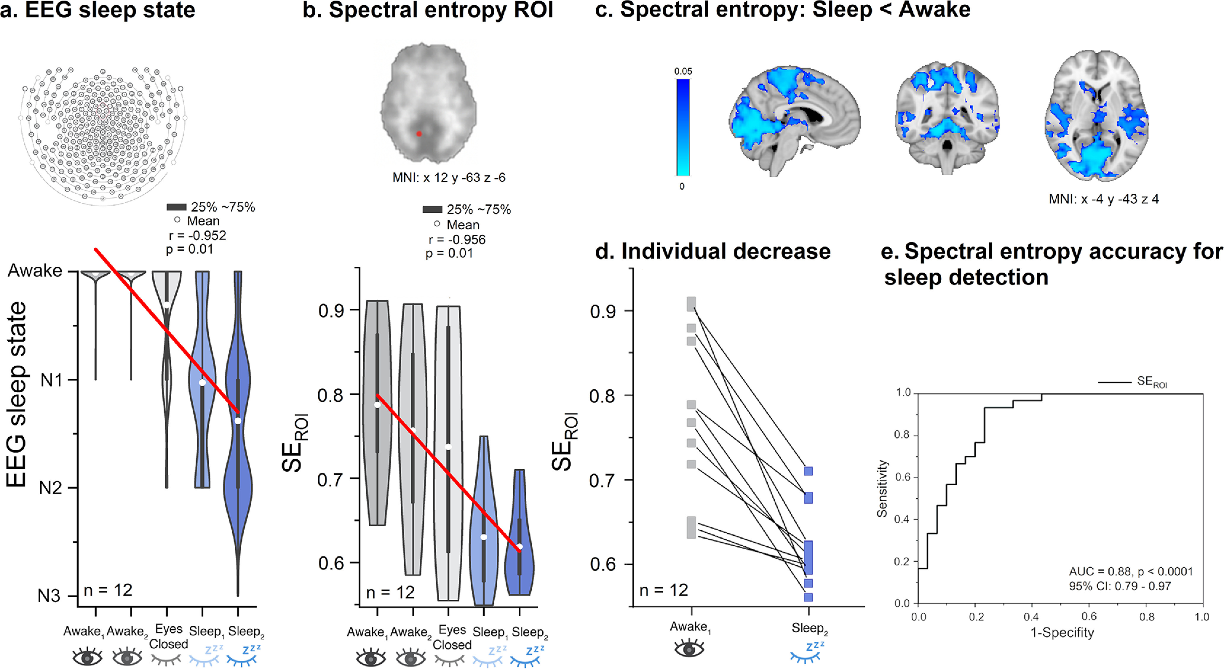
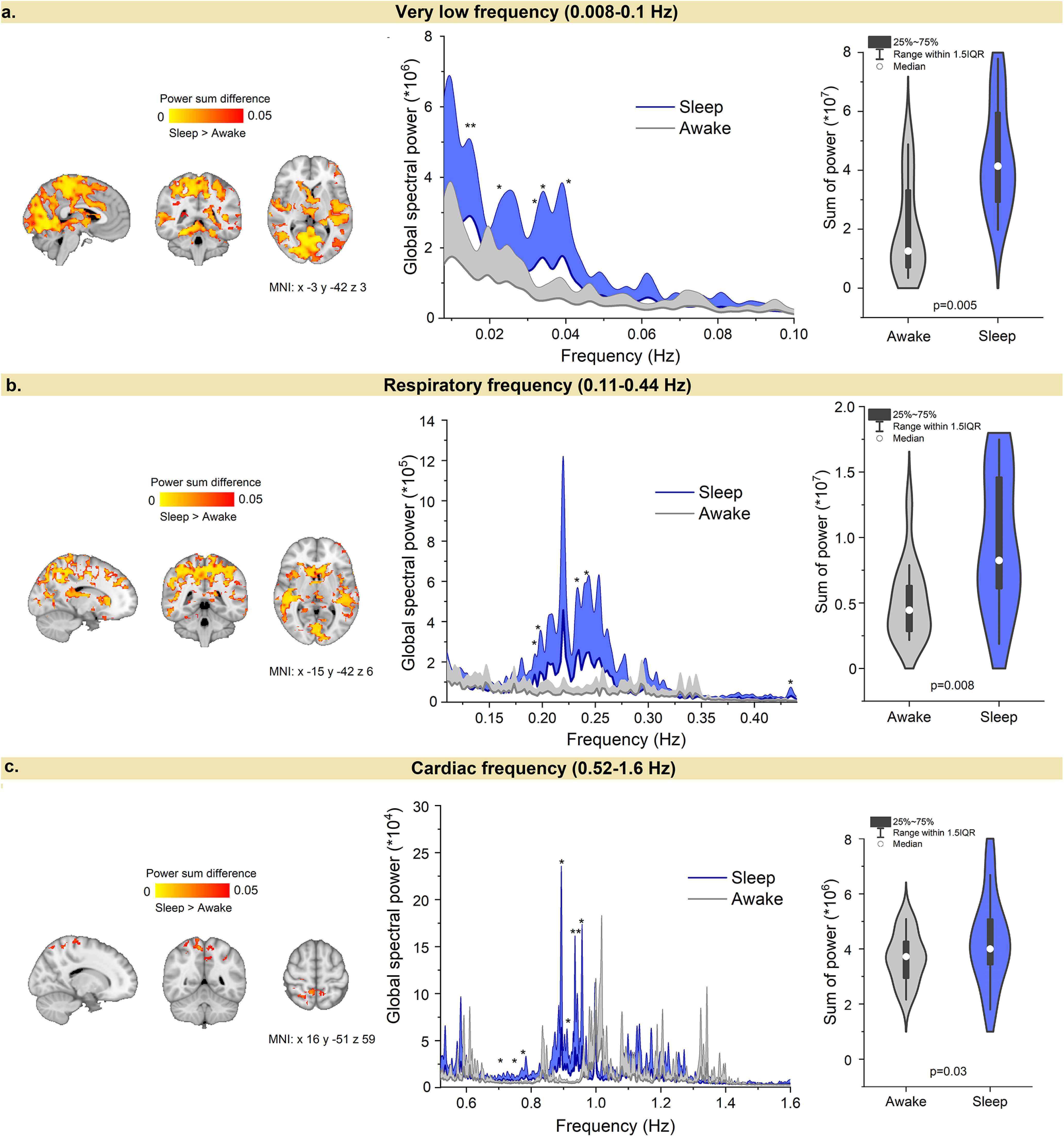
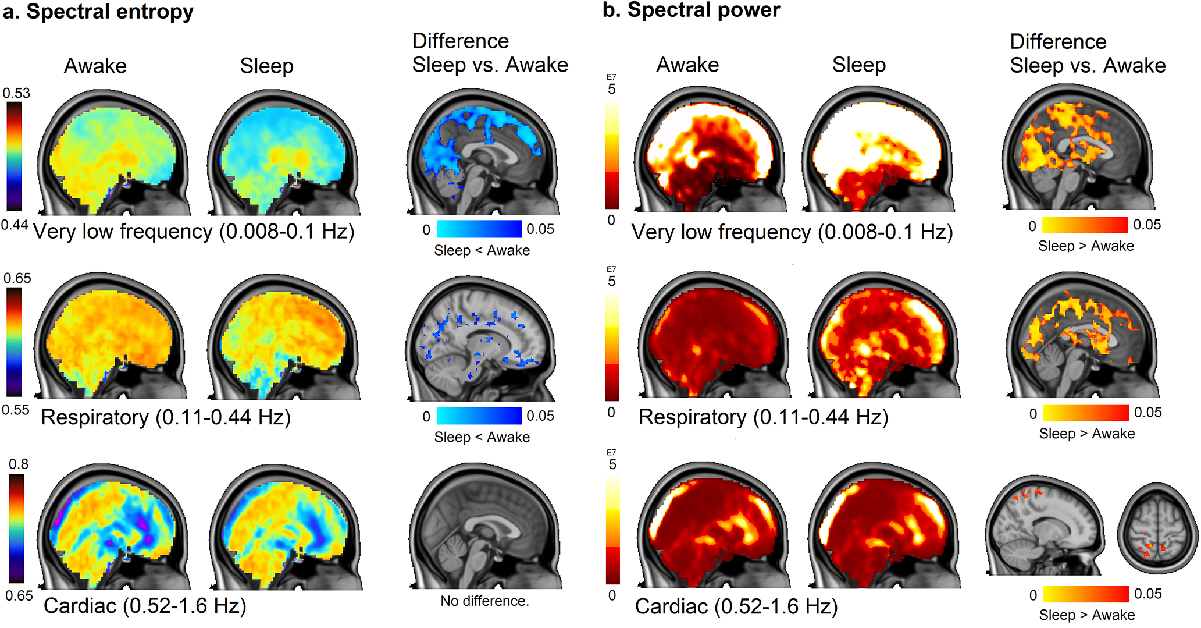
The physiological underpinnings of the necessity of sleep remain uncertain. Recent evidence suggests that sleep increases the convection of cerebrospinal fluid (CSF) and promotes the export of interstitial solutes, thus providing a framework to explain why all vertebrate species require sleep. Cardiovascular, respiratory and vasomotor brain pulsations have each been shown to drive CSF flow along perivascular spaces, yet it is unknown how such pulsations may change during sleep in humans. To investigate these pulsation phenomena in relation to sleep, we simultaneously recorded fast fMRI, magnetic resonance encephalography (MREG), and electroencephalography (EEG) signals in a group of healthy volunteers. We quantified sleep-related changes in the signal frequency distributions by spectral entropy analysis and calculated the strength of the physiological (vasomotor, respiratory, and cardiac) brain pulsations by power sum analysis in 15 subjects (age 26.5 ± 4.2 years, 6 females). Finally, we identified spatial similarities between EEG slow oscillation (0.2-2 Hz) power and MREG pulsations. Compared with wakefulness, nonrapid eye movement (NREM) sleep was characterized by reduced spectral entropy and increased brain pulsation intensity. These effects were most pronounced in posterior brain areas for very low-frequency (≤0.1 Hz) vasomotor pulsations but were also evident brain-wide for respiratory pulsations, and to a lesser extent for cardiac brain pulsations. There was increased EEG slow oscillation power in brain regions spatially overlapping with those showing sleep-related MREG pulsation changes. We suggest that reduced spectral entropy and enhanced pulsation intensity are characteristic of NREM sleep. With our findings of increased power of slow oscillation, the present results support the proposition that sleep promotes fluid transport in human brain. We report that the spectral power of physiological brain pulsation mechanisms driven by vasomotor, respiration, and cardiac rhythms in human brain increase during sleep, extending previous observations of their association with glymphatic brain clearance during sleep in rodents. The magnitudes of increased pulsations follow the rank order of vasomotor greater than respiratory greater than cardiac pulsations, with correspondingly declining spatial extents. Spectral entropy, previously known as vigilance and as an anesthesia metric, decreased during NREM sleep compared with the awake state in very low and respiratory frequencies, indicating reduced signal complexity. An EEG slow oscillation power increase occurring in the early sleep phase (NREM 1-2) spatially overlapped with pulsation changes, indicating reciprocal mechanisms between those measures.
睡眠的必要性的生理基础仍不确定。最近的证据表明,睡眠会增加脑脊液 (CSF) 的对流,并促进间质溶质的排出,从而为解释为什么所有脊椎动物都需要睡眠提供了一个框架。心血管、呼吸和血管搏动已被证明可驱动 CSF 沿血管周围空间流动,但尚不清楚在人类睡眠期间这些搏动如何变化。为了研究这些与睡眠相关的搏动现象,我们在一组健康志愿者中同时记录了快速 fMRI、磁共振脑图 (MREG) 和脑电图 (EEG) 信号。我们通过频谱熵分析量化了睡眠相关的信号频率分布变化,并通过功率和分析在 15 名受试者(年龄 26.5±4.2 岁,6 名女性)中计算了生理(血管搏动、呼吸和心脏)脑搏动的强度。最后,我们确定了 EEG 慢波 (0.2-2 Hz) 功率和 MREG 搏动之间的空间相似性。与清醒状态相比,非快速眼动 (NREM) 睡眠的特征是频谱熵降低和脑搏动强度增加。这些效应在极低频率 (≤0.1 Hz) 血管搏动的后脑部最为明显,但在呼吸搏动中也很明显,在心脏脑搏动中则不那么明显。在与睡眠相关的 MREG 搏动变化的脑区存在 EEG 慢波功率增加。我们认为,频谱熵降低和搏动强度增强是 NREM 睡眠的特征。我们发现慢波功率增加,这一结果支持了睡眠促进人脑内液体转运的假说。我们报告说,在人类大脑中由血管搏动、呼吸和心脏节律驱动的生理脑搏动机制的频谱功率在睡眠期间增加,扩展了先前在啮齿动物中观察到的它们与睡眠期间糖质运输的关联。搏动增加的幅度遵循血管搏动大于呼吸大于心脏搏动的顺序,相应的空间范围减小。频谱熵,以前称为警觉和麻醉指标,在 NREM 睡眠期间与清醒状态相比,在极低和呼吸频率下降低,表明信号复杂性降低。在早期睡眠阶段 (NREM1-2) 出现的 EEG 慢波功率增加与搏动变化在空间上重叠,表明这些测量之间存在相互作用机制。