• 文献检索
  • 文档翻译
  • 深度研究
  • 学术资讯
  • Suppr Zotero 插件Zotero 插件
  • 邀请有礼
  • 套餐&价格
  • 历史记录
应用&插件
Suppr Zotero 插件Zotero 插件浏览器插件Mac 客户端Windows 客户端微信小程序
定价
高级版会员购买积分包购买API积分包
服务
文献检索文档翻译深度研究API 文档MCP 服务
关于我们
关于 Suppr公司介绍联系我们用户协议隐私条款
关注我们

Suppr 超能文献

核心技术专利:CN118964589B侵权必究
粤ICP备2023148730 号-1Suppr @ 2026

文献检索

告别复杂PubMed语法,用中文像聊天一样搜索,搜遍4000万医学文献。AI智能推荐,让科研检索更轻松。

立即免费搜索

文件翻译

保留排版,准确专业,支持PDF/Word/PPT等文件格式,支持 12+语言互译。

免费翻译文档

深度研究

AI帮你快速写综述,25分钟生成高质量综述,智能提取关键信息,辅助科研写作。

立即免费体验

磁共振成像在肩袖损伤评估中的作用

Role of Magnetic Resonance Imaging in the Evaluation of Rotator Cuff Tears.

作者信息

Koganti Deepak V, Lamghare Purnachandra, Parripati Vinay Kumar, Khandelwal Rachit, Reddy Ayapaneni Dileep

机构信息

Radiology, Dr. D.Y. Patil Medical College, Hospital and Research Centre, Pune, IND.

出版信息

Cureus. 2022 Jan 8;14(1):e21025. doi: 10.7759/cureus.21025. eCollection 2022 Jan.

DOI:10.7759/cureus.21025
PMID:35154995
原文链接:https://pmc.ncbi.nlm.nih.gov/articles/PMC8819335/
Abstract

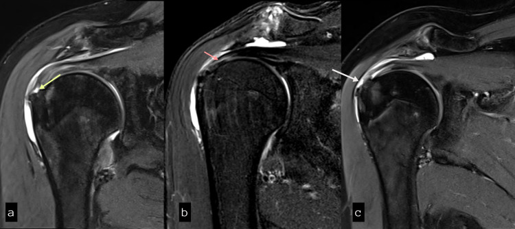
Background Magnetic resonance imaging (MRI), with the advent of surface coils, is becoming the modality of choice for imaging soft tissues around the shoulder joint. Good knowledge regarding the MR characteristics of rotator cuff tendons, acromion, and the abnormalities in these tendons is necessary for appropriate diagnosis. Methods This was a hospital-based descriptive, analytical and prospective study conducted at our tertiary care hospital. The study was performed on 50 patients with rotator cuff lesions detected on MRI of the shoulder joint. Results The age distribution found in the study is between 19 and 66 years with mean being 43 ± 14.8 years. The peak incidence was found in the fifth and sixth decades of life. Gender-wise distribution of rotator cuff pathologies has shown no significant gender variation. The pain was the most common presenting complaint. An abnormal supraspinatus tendon was seen in 82% of the 50 study patients, making it the most commonly affected tendons, followed by subscapularis and infraspinatus tendons. No apparent teres minor pathology was identified in the study patients. The most common pathology affecting the supraspinatus tendon was tendinosis (38%) closely followed by a partial tear (36%). Among the partial tears, the articular surface type of tear was the most common. About 52% patients had type II (curved) acromion; making it the most common type of acromion followed by type III (hook), supraspinatus tendinopathy was more common in type II acromion. A reduction in the acromiohumeral distance can cause supraspinatus tendinosis and also makes it more susceptible to tear. About 45.5% showed supraspinatus tendon tears when the acromiohumeral distance was less than 8mm as compared to 13.6% when more than 10mm. Only 4.2% had normal supraspinatus tendon in patients with this distance less than 7mm. Conclusion MRI provides valuable information to the orthopaedic surgeon regarding the status of tendons, bones, and joints. In order to choose the appropriate course of action, it is crucial first to identify the issue and report relevant data from rotator cuff imaging. A full grasp of the rotator cuff's architecture and function, as well as the repercussions of rotator cuff diseases, is required.

摘要

背景 随着表面线圈的出现,磁共振成像(MRI)正成为肩关节周围软组织成像的首选方式。了解肩袖肌腱、肩峰的磁共振特征以及这些肌腱的异常情况对于准确诊断至关重要。方法 这是一项在我们的三级护理医院进行的基于医院的描述性、分析性和前瞻性研究。该研究对50例肩关节MRI检查发现肩袖损伤的患者进行。结果 研究发现年龄分布在19至66岁之间,平均年龄为43±14.8岁。发病高峰出现在50至60岁。肩袖病变的性别分布未显示出明显的性别差异。疼痛是最常见的主诉。在50例研究患者中,82%可见冈上肌腱异常,使其成为最常受累的肌腱,其次是肩胛下肌腱和冈下肌腱。研究患者中未发现明显的小圆肌病变。影响冈上肌腱最常见的病变是肌腱病(38%),其次是部分撕裂(36%)。在部分撕裂中,关节面型撕裂最为常见。约52%的患者为II型(弧形)肩峰,使其成为最常见的肩峰类型,其次是III型(钩形),II型肩峰中冈上肌腱病更为常见。肩峰下间隙减小可导致冈上肌腱病,也使其更容易撕裂。当肩峰下间隙小于8mm时,约45.5%的患者出现冈上肌腱撕裂,而间隙大于10mm时为13.6%。当该间隙小于7mm时,只有4.2%的患者冈上肌腱正常。结论 MRI为骨科医生提供了有关肌腱、骨骼和关节状况的有价值信息。为了选择合适的治疗方案,首先确定问题并报告肩袖成像的相关数据至关重要。需要全面掌握肩袖的结构和功能,以及肩袖疾病的影响。

https://cdn.ncbi.nlm.nih.gov/pmc/blobs/f4c2/8819335/ff5a314c71bf/cureus-0014-00000021025-i03.jpg
https://cdn.ncbi.nlm.nih.gov/pmc/blobs/f4c2/8819335/1eec2d03b627/cureus-0014-00000021025-i01.jpg
https://cdn.ncbi.nlm.nih.gov/pmc/blobs/f4c2/8819335/7eefc7323a71/cureus-0014-00000021025-i02.jpg
https://cdn.ncbi.nlm.nih.gov/pmc/blobs/f4c2/8819335/ff5a314c71bf/cureus-0014-00000021025-i03.jpg
https://cdn.ncbi.nlm.nih.gov/pmc/blobs/f4c2/8819335/1eec2d03b627/cureus-0014-00000021025-i01.jpg
https://cdn.ncbi.nlm.nih.gov/pmc/blobs/f4c2/8819335/7eefc7323a71/cureus-0014-00000021025-i02.jpg
https://cdn.ncbi.nlm.nih.gov/pmc/blobs/f4c2/8819335/ff5a314c71bf/cureus-0014-00000021025-i03.jpg

相似文献

1
Role of Magnetic Resonance Imaging in the Evaluation of Rotator Cuff Tears.磁共振成像在肩袖损伤评估中的作用
Cureus. 2022 Jan 8;14(1):e21025. doi: 10.7759/cureus.21025. eCollection 2022 Jan.
2
Interobserver agreement in the classification of rotator cuff tears using magnetic resonance imaging.使用磁共振成像对肩袖撕裂进行分类时的观察者间一致性。
Am J Sports Med. 2008 Jan;36(1):99-103. doi: 10.1177/0363546507307504. Epub 2007 Oct 11.
3
High Acromial Slope and Low Acromiohumeral Distance Increase the Risk of Retear of the Supraspinatus Tendon After Repair.肩峰高斜率和肩峰肱骨头间距低会增加修复后冈上肌腱再次撕裂的风险。
Clin Orthop Relat Res. 2023 Jun 1;481(6):1158-1170. doi: 10.1097/CORR.0000000000002520. Epub 2022 Dec 20.
4
Comparison of shoulder muscle strength, cross-sectional area, acromiohumeral distance, and thickness of the supraspinatus tendon between symptomatic and asymptomatic patients with rotator cuff tears.比较肩袖撕裂症患者与无症状患者的肩部肌肉力量、横截面积、肩峰肱骨关节距离和冈上肌腱厚度。
J Shoulder Elbow Surg. 2020 Oct;29(10):2043-2050. doi: 10.1016/j.jse.2020.02.017. Epub 2020 May 26.
5
Association between rotator cuff abnormalities and reduced acromiohumeral distance.肩袖异常与肩峰下间隙减小之间的关联。
AJR Am J Roentgenol. 2006 Aug;187(2):376-82. doi: 10.2214/AJR.05.0435.
6
[Clinical application of acromion radiological classification in diagnosis and treatment of rotator cuff injury].肩峰放射学分类在肩袖损伤诊疗中的临床应用
Zhongguo Gu Shang. 2022 Aug 25;35(8):757-62. doi: 10.12200/j.issn.1003-0034.2022.08.011.
7
Partial-Thickness Rotator Cuff Tear by Itself Does Not Cause Shoulder Pain or Muscle Weakness in Baseball Players.单纯部分厚度肩袖撕裂不会导致棒球运动员肩部疼痛或肌肉无力。
Am J Sports Med. 2019 Dec;47(14):3476-3482. doi: 10.1177/0363546519878141. Epub 2019 Oct 14.
8
The acromiohumeral and coracohumeral intervals are abnormal in rotator cuff tears with muscular fatty degeneration.在伴有肌肉脂肪变性的肩袖撕裂中,肩峰肱骨间隙和喙突肱骨间隙异常。
Clin Orthop Relat Res. 2005 Apr(433):90-6. doi: 10.1097/01.blo.0000151441.05180.0e.
9
Association Between Rotator Cuff Tears and Superior Migration of the Humeral Head: An MRI-Based Anatomic Study.肩袖撕裂与肱骨头向上移位之间的关联:一项基于MRI的解剖学研究。
Orthop J Sports Med. 2021 Jun 10;9(6):23259671211009846. doi: 10.1177/23259671211009846. eCollection 2021 Jun.
10
Combined tears of the subscapularis and supraspinatus tendon: clinical outcome, rotator cuff strength and structural integrity following open repair.肩胛下肌和冈上肌腱联合撕裂:开放修复后的临床结果、肩袖力量和结构完整性。
Arch Orthop Trauma Surg. 2012 Jan;132(1):41-50. doi: 10.1007/s00402-011-1400-8. Epub 2011 Oct 12.

引用本文的文献

1
Shear wave elastography of the supraspinatus tendon with early degeneration in asymptomatic type II diabetes mellitus patients: a multicenter study.无症状II型糖尿病患者早期退变的冈上肌腱剪切波弹性成像:一项多中心研究。
BMC Musculoskelet Disord. 2025 Jul 4;26(1):637. doi: 10.1186/s12891-025-08864-w.
2
The Use of Magnetic Resonance Imaging in Patients With Shoulder Pain at King Abdulaziz University Hospital.磁共振成像在阿卜杜勒阿齐兹国王大学医院肩痛患者中的应用
Cureus. 2025 Jan 25;17(1):e77983. doi: 10.7759/cureus.77983. eCollection 2025 Jan.
3
Cross-cultural adaptation and validation of the shoulder rating questionnaire in Spanish patients with chronic nonsurgical shoulder pain.

本文引用的文献

1
Accuracy of MRI, MR arthrography, and ultrasound in the diagnosis of rotator cuff tears: a meta-analysis.磁共振成像(MRI)、磁共振关节造影和超声在诊断肩袖撕裂中的准确性:一项荟萃分析。
AJR Am J Roentgenol. 2009 Jun;192(6):1701-7. doi: 10.2214/AJR.08.1241.
2
The classic. Origin and comparative anatomy of the pectoral limb. Surgery of the shoulder. Philadelphia, PA: Lippincott Williams & Wilkins;1950:1-14.经典著作。胸肢的起源与比较解剖学。肩部手术。宾夕法尼亚州费城:利平科特·威廉姆斯与威尔金斯出版社;1950年:第1 - 14页。
Clin Orthop Relat Res. 2008 Mar;466(3):531-42. doi: 10.1007/s11999-007-0102-6. Epub 2008 Feb 10.
3
MR imaging of rotator cuff injury: what the clinician needs to know.
《肩部评分问卷在西班牙慢性非手术性肩部疼痛患者中的跨文化调适与验证》
JSES Int. 2024 May 23;8(5):1095-1101. doi: 10.1016/j.jseint.2024.05.003. eCollection 2024 Sep.
4
High-Resolution Imaging Insights into Shoulder Joint Pain: A Comprehensive Review of Ultrasound and Magnetic Resonance Imaging (MRI).肩关节疼痛的高分辨率成像见解:超声和磁共振成像(MRI)的综合综述
Cureus. 2023 Nov 17;15(11):e48974. doi: 10.7759/cureus.48974. eCollection 2023 Nov.
5
The Spectrum of Shoulder Pathologies on Magnetic Resonance Imaging: A Pictorial Review.磁共振成像上的肩部病变谱:图文综述
Cureus. 2023 Sep 6;15(9):e44801. doi: 10.7759/cureus.44801. eCollection 2023 Sep.
肩袖损伤的磁共振成像:临床医生需要了解的内容。
Radiographics. 2006 Jul-Aug;26(4):1045-65. doi: 10.1148/rg.264055087.
4
Full-Thickness Rotator Cuff Tears: Factors Affecting Surgical Outcome.全层肩袖撕裂:影响手术结果的因素
J Am Acad Orthop Surg. 1994 Mar;2(2):87-95. doi: 10.5435/00124635-199403000-00002.
5
Age-related prevalence of rotator cuff tears in asymptomatic shoulders.无症状肩部肩袖撕裂的年龄相关性患病率。
J Shoulder Elbow Surg. 1999 Jul-Aug;8(4):296-9. doi: 10.1016/s1058-2746(99)90148-9.
6
Rotator cuff: evaluation with US and MR imaging.肩袖:超声与磁共振成像评估
Radiographics. 1999 May-Jun;19(3):685-705. doi: 10.1148/radiographics.19.3.g99ma03685.
7
The 'dropping' and 'hornblower's' signs in evaluation of rotator-cuff tears.评估肩袖撕裂时的“下落”征和“吹号角者”征
J Bone Joint Surg Br. 1998 Jul;80(4):624-8. doi: 10.1302/0301-620x.80b4.8651.
8
Pathology of failure of the rotator cuff tendon.肩袖肌腱断裂的病理学
Orthop Clin North Am. 1997 Jan;28(1):31-41. doi: 10.1016/s0030-5898(05)70262-5.
9
MR imaging of the rotator cuff: peritendinous and bone abnormalities in an asymptomatic population.肩袖的磁共振成像:无症状人群中的肌腱周围和骨骼异常
AJR Am J Roentgenol. 1996 Apr;166(4):863-7. doi: 10.2214/ajr.166.4.8610564.
10
Rotator-cuff changes in asymptomatic adults. The effect of age, hand dominance and gender.无症状成年人的肩袖改变。年龄、利手和性别的影响。
J Bone Joint Surg Br. 1995 Mar;77(2):296-8.