Nogueira Dayane Maria Braz, Figadoli André Luiz de Faria, Alcantara Patrícia Lopes, Pomini Karina Torres, Santos German Iris Jasmin, Reis Carlos Henrique Bertoni, Rosa Júnior Geraldo Marco, Rosso Marcelie Priscila de Oliveira, Santos Paulo Sérgio da Silva, Zangrando Mariana Schutzer Ragghianti, Pereira Eliana de Souza Bastos Mazuqueli, Marchi Miguel Ângelo de, Trazzi Beatriz Flavia de Moraes, Rossi Jéssica de Oliveira, Salmeron Samira, Pastori Cláudio Maldonado, Buchaim Daniela Vieira, Buchaim Rogerio Leone
Department of Prosthodontics and Periodontics, Bauru School of Dentistry (FOB/USP), University of São Paulo, Bauru 17012-901, Brazil.
Department of Biological Sciences, Bauru School of Dentistry (FOB/USP), University of São Paulo, Bauru 17012-901, Brazil.
Polymers (Basel). 2022 Jan 31;14(3):584. doi: 10.3390/polym14030584.
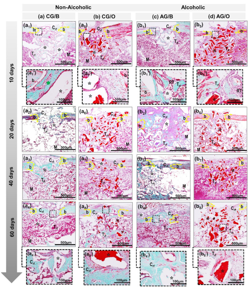
In this experimental protocol, the objective was to evaluate the biological behavior of two xenogenic scaffolds in alcohol-induced rats through histomorphometric and Picrosirius Red staining analysis of non-critical defects in the tibia of rats submitted or not to alcohol ingestion at 25% /. Eighty male rats were randomly divided into four groups ( = 20 each): CG/B (water diet + Bio-Oss graft, Geistlich Pharma AG, Wolhusen, Switzerland), CG/O (water diet + OrthoGen graft, Baumer, Mogi Mirim, Brazil), AG/B (25% / alcohol diet + Bio-Oss graft), and AG/O (25% / alcohol diet + OrthoGen graft). After 90 days of liquid diet, the rats were surgically obtained, with a defect in the tibia proximal epiphysis; filled in according to their respective groups; and euthanized at 10, 20, 40 and 60 days. In two initial periods (10 and 20 days), all groups presented biomaterial particles surrounded by disorganized collagen fibrils. Alcoholic animals (AG/B and AG/O) presented, in the cortical and medullary regions, a reactive tissue with inflammatory infiltrate. In 60 days, in the superficial area of the surgical cavities, particles of biomaterials were observed in all groups, with new compact bone tissue around them, without complete closure of the lesion, except in non-alcoholic animals treated with Bio-Oss xenograft (CG/B), where the new cortical interconnected the edges of the defect. Birefringence transition was observed in the histochemical analysis of collagen fibers by Picrosirius Red, in which all groups in periods of 10 and 20 days showed red-orange birefringence, and from 40 days onwards greenish-yellow birefringence, which demonstrates the characteristic transition from the formation of thin and disorganized collagen fibers initially to more organized and thicker later. In histomorphometric analysis, at 60 days, CG/B had the highest volume density of new bone (32.9 ± 1.15) and AG/O the lowest volume density of new bone (15.32 ± 1.71). It can be concluded that the bone neoformation occurred in the defects that received the two biomaterials, in all periods, but the Bio-Oss was superior in the results, with its groups CG/B and AG/B displaying greater bone formation (32.9 ± 1.15 and 22.74 ± 1.15, respectively) compared to the OrthoGen CG/O and AG/O groups (20.66 ± 2.12 and 15.32 ± 1.71, respectively), and that the alcoholic diet interfered negatively in the repair process and in the percentage of new bone formed.
在本实验方案中,目的是通过对摄入或未摄入25%酒精的大鼠胫骨非关键缺损进行组织形态计量学和天狼星红染色分析,评估两种异种支架在酒精诱导的大鼠中的生物学行为。80只雄性大鼠被随机分为四组(每组n = 20):CG/B组(水饮食 + Bio - Oss移植物,Geistlich Pharma AG,瑞士沃尔胡森)、CG/O组(水饮食 + OrthoGen移植物,巴西莫吉米林的鲍默公司)、AG/B组(25%酒精饮食 + Bio - Oss移植物)和AG/O组(25%酒精饮食 + OrthoGen移植物)。经过90天的流质饮食后,对大鼠进行手术,在胫骨近端骨骺处制造缺损;根据各自分组进行填充;并在第10、20、40和60天实施安乐死。在最初的两个时期(10天和20天),所有组均呈现出被杂乱的胶原纤维包围的生物材料颗粒。酒精喂养的动物(AG/B组和AG/O组)在皮质和髓质区域呈现出有炎症浸润的反应性组织。在60天时,在手术腔的表层区域,所有组均观察到生物材料颗粒,其周围有新的致密骨组织,但病变未完全闭合,除了接受Bio - Oss异种移植物治疗的非酒精喂养动物(CG/B组),其新皮质连接了缺损边缘。通过天狼星红对胶原纤维进行组织化学分析时观察到双折射转变,其中所有组在10天和20天显示橙红色双折射,从40天起显示黄绿色双折射,这表明最初形成的细而杂乱的胶原纤维逐渐转变为更有组织且更粗的胶原纤维。在组织形态计量学分析中,60天时,CG/B组新骨体积密度最高(32.9 ± 1.15),AG/O组新骨体积密度最低(15.32 ± 1.71)。可以得出结论,在所有时期,接受两种生物材料的缺损处均发生了骨新生,但Bio - Oss在结果方面更优,其CG/B组和AG/B组的骨形成量(分别为32.9 ± 1.15和22.74 ± 1.15)高于OrthoGen的CG/O组和AG/O组(分别为20.66 ± 2.12和15.32 ± 1.71),并且酒精饮食对修复过程和新形成骨的百分比产生了负面影响。