• 文献检索
  • 文档翻译
  • 深度研究
  • 学术资讯
  • Suppr Zotero 插件Zotero 插件
  • 邀请有礼
  • 套餐&价格
  • 历史记录
应用&插件
Suppr Zotero 插件Zotero 插件浏览器插件Mac 客户端Windows 客户端微信小程序
定价
高级版会员购买积分包购买API积分包
服务
文献检索文档翻译深度研究API 文档MCP 服务
关于我们
关于 Suppr公司介绍联系我们用户协议隐私条款
关注我们

Suppr 超能文献

核心技术专利:CN118964589B侵权必究
粤ICP备2023148730 号-1Suppr @ 2026

文献检索

告别复杂PubMed语法,用中文像聊天一样搜索,搜遍4000万医学文献。AI智能推荐,让科研检索更轻松。

立即免费搜索

文件翻译

保留排版,准确专业,支持PDF/Word/PPT等文件格式,支持 12+语言互译。

免费翻译文档

深度研究

AI帮你快速写综述,25分钟生成高质量综述,智能提取关键信息,辅助科研写作。

立即免费体验

基于配准的径向和线性 B 扫描复合实现高分辨率容积光学相干断层扫描的工具。

A Tool for High-Resolution Volumetric Optical Coherence Tomography by Compounding Radial-and Linear Acquired B-Scans Using Registration.

机构信息

Computer Aided Medical Procedures and Augmented Reality, Technical University Munich, 85748 Munich, Germany.

Augenklinik und Poliklinik, Klinikum Rechts der Lsar der Technische Universität München, 81675 München, Germany.

出版信息

Sensors (Basel). 2022 Feb 2;22(3):1135. doi: 10.3390/s22031135.

DOI:10.3390/s22031135
PMID:35161880
原文链接:https://pmc.ncbi.nlm.nih.gov/articles/PMC8839892/
Abstract

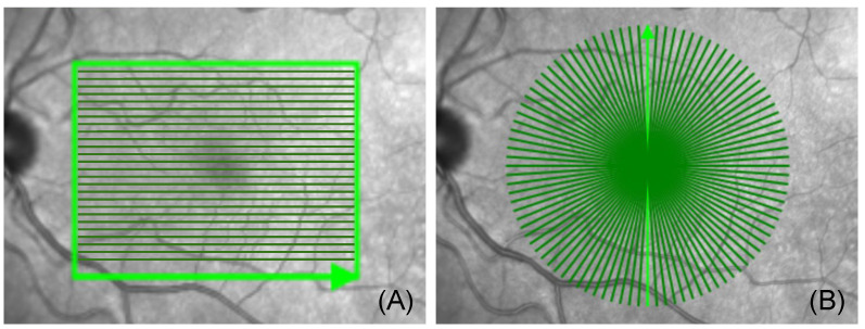
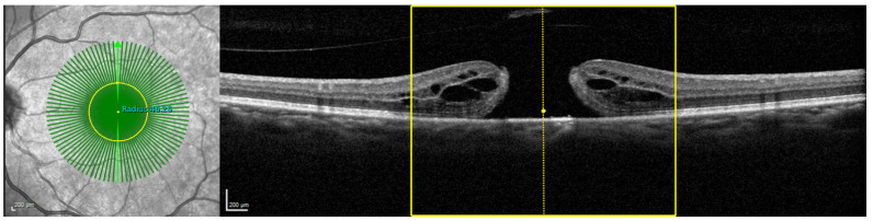
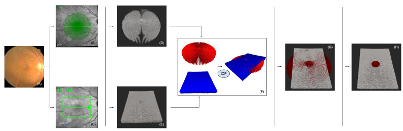
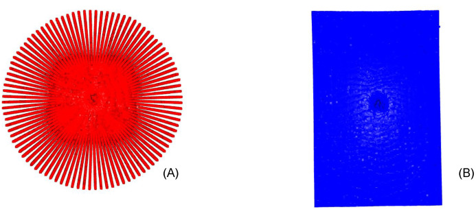
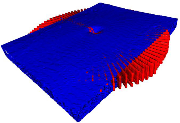
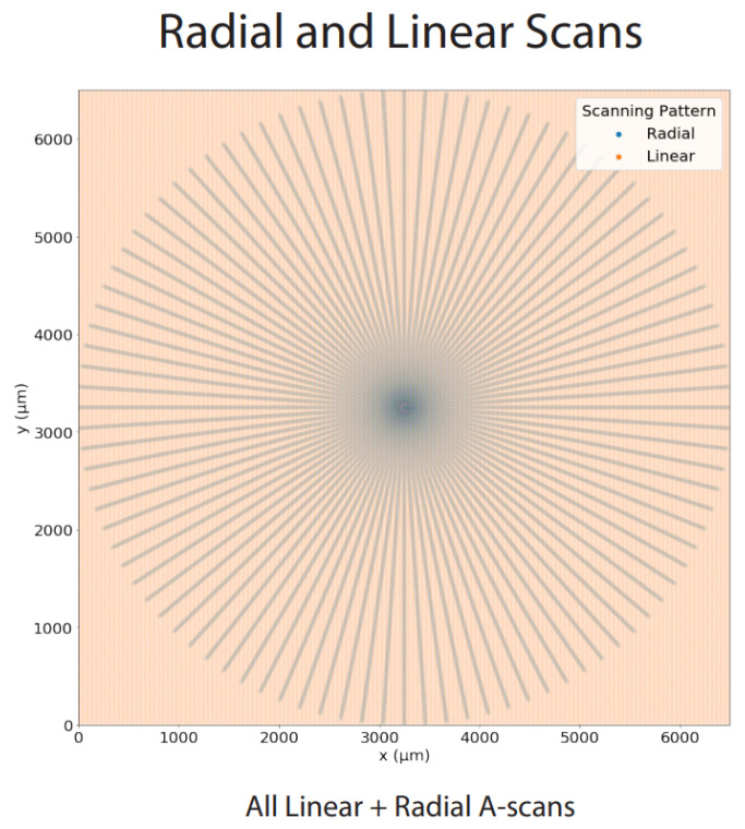
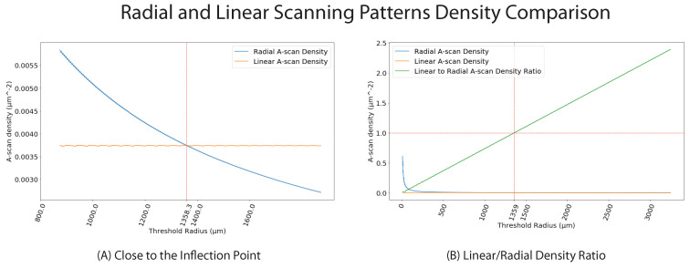
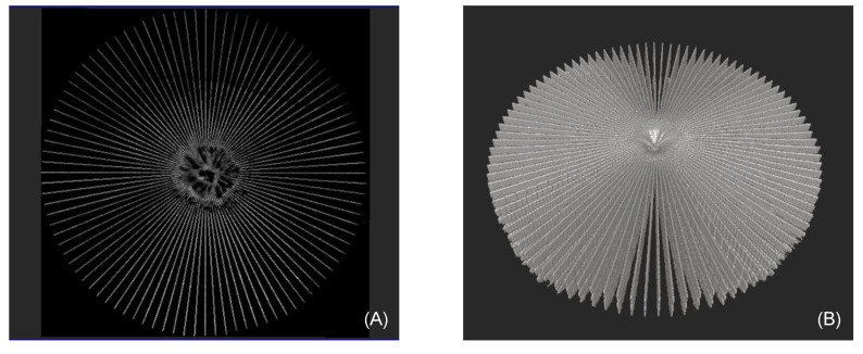
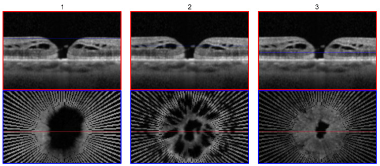
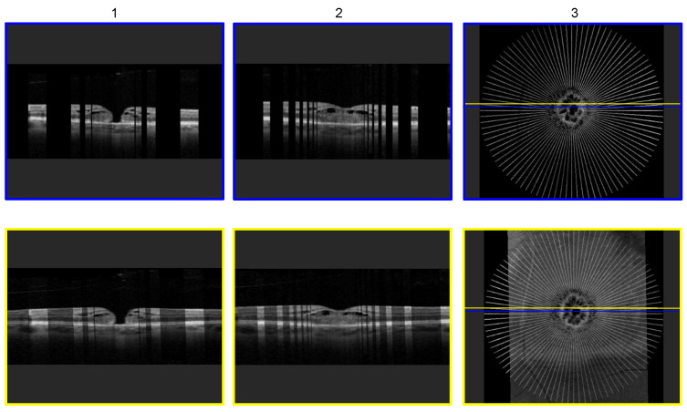
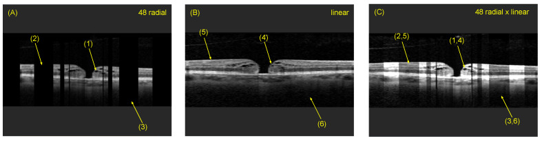
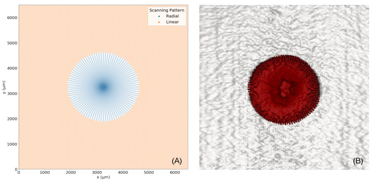
Optical coherence tomography (OCT) is a medical imaging modality that is commonly used to diagnose retinal diseases. In recent years, linear and radial scanning patterns have been proposed to acquire three-dimensional OCT data. These patterns show differences in A-scan acquisition density across the generated volumes, and thus differ in their suitability for the diagnosis of retinal diseases. While radial OCT volumes exhibit a higher A-scan sampling rate around the scan center, linear scans contain more information in the peripheral scan areas. In this paper, we propose a method to combine a linearly and radially acquired OCT volume to generate a single compound volume, which merges the advantages of both scanning patterns to increase the information that can be gained from the three-dimensional OCT data. We initially generate 3D point clouds of the linearly and radially acquired OCT volumes and use an Iterative Closest Point (ICP) variant to register both volumes. After registration, the compound volume is created by selectively exploiting linear and radial scanning data, depending on the A-scan density of the individual scans. Fusing regions from both volumes with respect to their local A-scan sampling density, we achieve improved overall anatomical OCT information in a high-resolution compound volume. We demonstrate our method on linear and radial OCT volumes for the visualization and analysis of macular holes and the surrounding anatomical structures.

摘要

光学相干断层扫描(OCT)是一种常用于诊断视网膜疾病的医学成像方式。近年来,线性和径向扫描模式已被提出用于获取三维 OCT 数据。这些模式在生成的体积中表现出 A 扫描采集密度的差异,因此在诊断视网膜疾病方面的适用性也不同。虽然径向 OCT 体积在扫描中心周围表现出更高的 A 扫描采样率,但线性扫描在周边扫描区域包含更多信息。在本文中,我们提出了一种方法,可将线性和径向采集的 OCT 体积组合生成单个复合体积,从而结合两种扫描模式的优势,增加可从三维 OCT 数据中获得的信息量。我们最初生成线性和径向采集的 OCT 体积的 3D 点云,并使用迭代最近点(ICP)变体对两个体积进行配准。配准后,根据各个扫描的 A 扫描密度,选择性地利用线性和径向扫描数据来创建复合体积。通过根据局部 A 扫描采样密度融合两个体积的区域,我们在高分辨率复合体积中实现了改进的整体解剖 OCT 信息。我们在黄斑裂孔及其周围解剖结构的线性和径向 OCT 体积上展示了我们的方法,用于可视化和分析。

https://cdn.ncbi.nlm.nih.gov/pmc/blobs/e4fb/8839892/f36c6f156ef0/sensors-22-01135-g012.jpg
https://cdn.ncbi.nlm.nih.gov/pmc/blobs/e4fb/8839892/716aa785be0b/sensors-22-01135-g001.jpg
https://cdn.ncbi.nlm.nih.gov/pmc/blobs/e4fb/8839892/af8e46939e67/sensors-22-01135-g002.jpg
https://cdn.ncbi.nlm.nih.gov/pmc/blobs/e4fb/8839892/84e45fb7e7d5/sensors-22-01135-g003.jpg
https://cdn.ncbi.nlm.nih.gov/pmc/blobs/e4fb/8839892/21776a9fd201/sensors-22-01135-g004.jpg
https://cdn.ncbi.nlm.nih.gov/pmc/blobs/e4fb/8839892/f37a023fd41c/sensors-22-01135-g005.jpg
https://cdn.ncbi.nlm.nih.gov/pmc/blobs/e4fb/8839892/7ded24bdfdbf/sensors-22-01135-g006.jpg
https://cdn.ncbi.nlm.nih.gov/pmc/blobs/e4fb/8839892/33765a75cff0/sensors-22-01135-g007.jpg
https://cdn.ncbi.nlm.nih.gov/pmc/blobs/e4fb/8839892/461780ba4e7a/sensors-22-01135-g008.jpg
https://cdn.ncbi.nlm.nih.gov/pmc/blobs/e4fb/8839892/2ee245ccbe9d/sensors-22-01135-g009.jpg
https://cdn.ncbi.nlm.nih.gov/pmc/blobs/e4fb/8839892/cd0164818447/sensors-22-01135-g010.jpg
https://cdn.ncbi.nlm.nih.gov/pmc/blobs/e4fb/8839892/3d621fac7723/sensors-22-01135-g011.jpg
https://cdn.ncbi.nlm.nih.gov/pmc/blobs/e4fb/8839892/f36c6f156ef0/sensors-22-01135-g012.jpg

相似文献

1
A Tool for High-Resolution Volumetric Optical Coherence Tomography by Compounding Radial-and Linear Acquired B-Scans Using Registration.基于配准的径向和线性 B 扫描复合实现高分辨率容积光学相干断层扫描的工具。
Sensors (Basel). 2022 Feb 2;22(3):1135. doi: 10.3390/s22031135.
2
High-definition and 3-dimensional imaging of macular pathologies with high-speed ultrahigh-resolution optical coherence tomography.利用高速超高分辨率光学相干断层扫描技术对黄斑病变进行高清三维成像。
Ophthalmology. 2006 Nov;113(11):2054.e1-14. doi: 10.1016/j.ophtha.2006.05.046.
3
Ultrahigh speed spectral / Fourier domain OCT ophthalmic imaging at 70,000 to 312,500 axial scans per second.每秒70,000至312,500次轴向扫描的超高速光谱/傅里叶域光学相干断层扫描眼科成像。
Opt Express. 2008 Sep 15;16(19):15149-69. doi: 10.1364/oe.16.015149.
4
Effect of optical coherence tomography scan pattern and density on the detection of full-thickness macular holes.光学相干断层扫描模式和密度对全层黄斑裂孔检测的影响。
Am J Ophthalmol. 2014 May;157(5):978-84. doi: 10.1016/j.ajo.2014.01.021. Epub 2014 Jan 31.
5
Three-dimensional imaging of macular holes with high-speed optical coherence tomography.利用高速光学相干断层扫描技术对黄斑裂孔进行三维成像
Ophthalmology. 2007 Apr;114(4):763-73. doi: 10.1016/j.ophtha.2006.07.055. Epub 2006 Dec 20.
6
Comparison of ultrahigh- and standard-resolution optical coherence tomography for imaging macular hole pathology and repair.超高分辨率与标准分辨率光学相干断层扫描在黄斑裂孔病变及修复成像中的比较。
Ophthalmology. 2004 Nov;111(11):2033-43. doi: 10.1016/j.ophtha.2004.05.021.
7
Comparison of retinal thickness measurements between three-dimensional and radial scans on spectral-domain optical coherence tomography.光谱域光学相干断层扫描中三维扫描与径向扫描之间视网膜厚度测量的比较。
Am J Ophthalmol. 2009 Sep;148(3):431-8. doi: 10.1016/j.ajo.2009.04.008. Epub 2009 Jun 3.
8
VISUALIZATION OF THE MACULA IN GAS-FILLED EYES: Spectral Domain Optical Coherence Tomography Versus Swept-Source Optical Coherence Tomography.充气体眼中黄斑的可视化:谱域光学相干断层扫描与扫频源光学相干断层扫描比较。
Retina. 2018 Mar;38(3):480-489. doi: 10.1097/IAE.0000000000001560.
9
Clinical application of rapid serial fourier-domain optical coherence tomography for macular imaging.快速连续傅里叶域光学相干断层扫描在黄斑成像中的临床应用。
Ophthalmology. 2006 Aug;113(8):1425-31. doi: 10.1016/j.ophtha.2006.03.020. Epub 2006 Jun 12.
10
Three-dimensional retinal imaging with high-speed ultrahigh-resolution optical coherence tomography.采用高速超高分辨率光学相干断层扫描技术的三维视网膜成像
Ophthalmology. 2005 Oct;112(10):1734-46. doi: 10.1016/j.ophtha.2005.05.023.

引用本文的文献

1
Biometric Technologies Based on Optical Coherence Tomography.基于光相干断层扫描的生物识别技术。
Sensors (Basel). 2023 Apr 5;23(7):3753. doi: 10.3390/s23073753.
2
A Series-Based Deep Learning Approach to Lung Nodule Image Classification.一种基于序列的深度学习方法用于肺结节图像分类。
Cancers (Basel). 2023 Jan 30;15(3):843. doi: 10.3390/cancers15030843.
3
Phase Unwrapping and Frequency Points Subdivision of the Frequency Sweeping Interferometry Based Absolute Ranging System.扫频干涉绝对测距系统的相位展开与频点细分

本文引用的文献

1
Repeatability and Reproducibility of Macular Hole Size Measurements Using Optical Coherence Tomography.使用光学相干断层扫描测量黄斑裂孔大小的可重复性和再现性
J Clin Med. 2021 Jun 29;10(13):2899. doi: 10.3390/jcm10132899.
2
Predictive Association of Pre-Operative Defect Areas in the Outer Retinal Layers With Visual Acuity in Macular Hole Surgery.术前外层视网膜缺损区与黄斑裂孔手术视力的预测相关性。
Transl Vis Sci Technol. 2021 Apr 1;10(4):7. doi: 10.1167/tvst.10.4.7.
3
OCT Signal Enhancement with Deep Learning.基于深度学习的光学相干断层扫描信号增强
Sensors (Basel). 2022 Apr 10;22(8):2904. doi: 10.3390/s22082904.
Ophthalmol Glaucoma. 2021 May-Jun;4(3):295-304. doi: 10.1016/j.ogla.2020.10.008. Epub 2020 Oct 15.
4
Macular hole morphology and measurement using an automated three-dimensional image segmentation algorithm.使用自动三维图像分割算法进行黄斑裂孔形态学及测量
BMJ Open Ophthalmol. 2020 Aug 16;5(1):e000404. doi: 10.1136/bmjophth-2019-000404. eCollection 2020.
5
Improving lateral resolution and image quality of optical coherence tomography by the multi-frame superresolution technique for 3D tissue imaging.通过多帧超分辨率技术提高光学相干断层扫描的横向分辨率和图像质量以用于三维组织成像
Biomed Opt Express. 2017 Oct 6;8(11):4887-4918. doi: 10.1364/BOE.8.004887. eCollection 2017 Nov 1.
6
Optical Coherence Tomography and the Development of Antiangiogenic Therapies in Neovascular Age-Related Macular Degeneration.光学相干断层扫描与新生血管性年龄相关性黄斑变性抗血管生成疗法的发展
Invest Ophthalmol Vis Sci. 2016 Jul 1;57(9):OCT14-26. doi: 10.1167/iovs.16-19969.
7
Clinical applications of spectral domain optical coherence tomography in retinal diseases.频域光学相干断层扫描在视网膜疾病中的临床应用
Biomed J. 2016 Apr;39(2):107-20. doi: 10.1016/j.bj.2016.04.003. Epub 2016 Jun 20.
8
Rapid radial optical coherence tomography image acquisition.快速径向光学相干断层扫描图像采集。
J Biomed Opt. 2013 Mar;18(3):036004. doi: 10.1117/1.JBO.18.3.036004.
9
Optical coherence tomography: future trends for imaging in glaucoma.光学相干断层扫描:青光眼成像的未来趋势
Optom Vis Sci. 2012 May;89(5):E554-62. doi: 10.1097/OPX.0b013e31824eeb43.
10
In vivo retinal imaging by optical coherence tomography.通过光学相干断层扫描进行视网膜活体成像。
Opt Lett. 1993 Nov 1;18(21):1864-6. doi: 10.1364/ol.18.001864.