Ko Tony H, Fujimoto James G, Duker Jay S, Paunescu Lelia A, Drexler Wolfgang, Baumal Caroline R, Puliafito Carmen A, Reichel Elias, Rogers Adam H, Schuman Joel S
Department of Electrical Engineering and Computer Science and Research Laboratory of Electronics, Massachusetts Institute of Technology, Cambridge, Massachusetts 02139, USA.
Ophthalmology. 2004 Nov;111(11):2033-43. doi: 10.1016/j.ophtha.2004.05.021.
To compare ultrahigh-resolution optical coherence tomography (UHR-OCT) technology to a standard-resolution OCT instrument for the imaging of macular hole pathology and repair; to identify situations where UHR-OCT provides additional information on disease morphology, pathogenesis, and management; and to use UHR-OCT as a baseline for improving the interpretation of the standard-resolution images.
Observational and interventional case series.
Twenty-nine eyes of 24 patients clinically diagnosed with macular hole in at least one eye.
A UHR-OCT system has been developed and employed in a tertiary-care ophthalmology clinic. Using a femtosecond laser as the low-coherence light source, this new UHR-OCT system can achieve an unprecedented 3-mum axial resolution for retinal OCT imaging. Comparative imaging was performed with UHR-OCT and standard 10-mum resolution OCT in 29 eyes of 24 patients with various stages of macular holes. Imaging was also performed on a subset of the population before and after macular hole surgery.
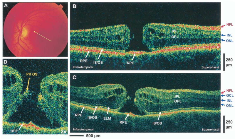
Ultrahigh- and standard-resolution cross-sectional OCT images of macular hole pathologies.
Both UHR-OCT and standard-resolution OCT exhibited comparable performance in differentiating various stages of macular holes. The UHR-OCT provided improved imaging of finer intraretinal structures, such as the external limiting membrane and photoreceptor inner segment (IS) and outer segment (OS), and identification of the anatomy of successful surgical repair. The improved resolution of UHR-OCT enabled imaging of previously unidentified changes in photoreceptor morphology associated with macular hole pathology and postoperative repair. Visualization of the junction between the photoreceptor IS and OS was found to be an important indicator of photoreceptor integrity for both standard-resolution and UHR-OCT images.
Ultrahigh-resolution optical coherence tomography improves the visualization of the macular hole architectural morphology. The increased resolution of UHR-OCT enables the visualization of photoreceptor morphology associated with macular holes. This promises to lead to a better understanding of the pathogenesis of macular holes, the causes of visual loss secondary to macular holes, the timing of surgical repair, and the evaluation of postsurgical outcome. Ultrahigh-resolution optical coherence tomography imaging of macular holes that correspond to known alterations in retinal morphology can be used to interpret retinal morphology in UHR-OCT images. Comparisons of UHR-OCT images with standard-resolution OCT images can establish a baseline for the better interpretation of clinical standard-resolution OCT images. The ability to visualize photoreceptors and their integrity or impairment is an indicator of macular hole progression and surgical outcome.

比较超高分辨率光学相干断层扫描(UHR - OCT)技术与标准分辨率光学相干断层扫描(OCT)仪器在黄斑裂孔病变及修复成像方面的差异;确定UHR - OCT在提供疾病形态、发病机制及治疗相关额外信息的情况;并将UHR - OCT作为改进标准分辨率图像解读的基线。
观察性和干预性病例系列。
24例患者的29只眼,这些患者至少一只眼临床诊断为黄斑裂孔。
已开发出一种UHR - OCT系统并应用于一家三级眼科诊所。该新型UHR - OCT系统使用飞秒激光作为低相干光源,可实现视网膜OCT成像前所未有的3微米轴向分辨率。对24例不同阶段黄斑裂孔患者的29只眼进行了UHR - OCT和标准10微米分辨率OCT的对比成像。还对部分患者在黄斑裂孔手术前后进行了成像。
黄斑裂孔病变的超高分辨率和标准分辨率横断面OCT图像。
UHR - OCT和标准分辨率OCT在区分黄斑裂孔的不同阶段表现相当。UHR - OCT能更好地显示视网膜内更细微的结构,如外界膜、光感受器内节(IS)和外节(OS),并能识别成功手术修复的解剖结构。UHR - OCT提高的分辨率能够显示与黄斑裂孔病变及术后修复相关的、以前未被识别的光感受器形态变化。对于标准分辨率和UHR - OCT图像,光感受器IS和OS之间连接的可视化被发现是光感受器完整性的重要指标。
超高分辨率光学相干断层扫描改善了黄斑裂孔结构形态的可视化。UHR - OCT提高的分辨率能够显示与黄斑裂孔相关的光感受器形态。这有望更好地理解黄斑裂孔的发病机制、黄斑裂孔导致视力丧失的原因、手术修复时机以及术后结果评估。对应视网膜形态已知改变的黄斑裂孔的超高分辨率光学相干断层扫描成像可用于解读UHR - OCT图像中的视网膜形态。将UHR - OCT图像与标准分辨率OCT图像进行比较可为更好地解读临床标准分辨率OCT图像建立基线。可视化光感受器及其完整性或损伤的能力是黄斑裂孔进展和手术结果的一个指标。