Marino Natascia, German Rana, Podicheti Ram, Rockey Pam, Sandusky George E, Temm Constance J, Nakshatri Harikrishna, Addison Rebekah J, Selman Bryce, Althouse Sandra K, Storniolo Anna Maria V
Susan G. Komen Tissue Bank at the IU Simon Comprehensive Cancer Center, Indianapolis, IN, 46202, USA.
Department of Medicine, Indiana University School of Medicine, Indianapolis, IN, 46202, USA.
Biomark Res. 2022 Feb 19;10(1):8. doi: 10.1186/s40364-022-00353-9.
Family with sequence similarity 83 member A (FAM83A) presents oncogenic properties in several cancers including breast cancer. Recently, we reported FAM83A overexpression in normal breast tissues from women at high risk of breast cancer. We now hypothesize that FAM83A is a key factor in breast cancer initiation.
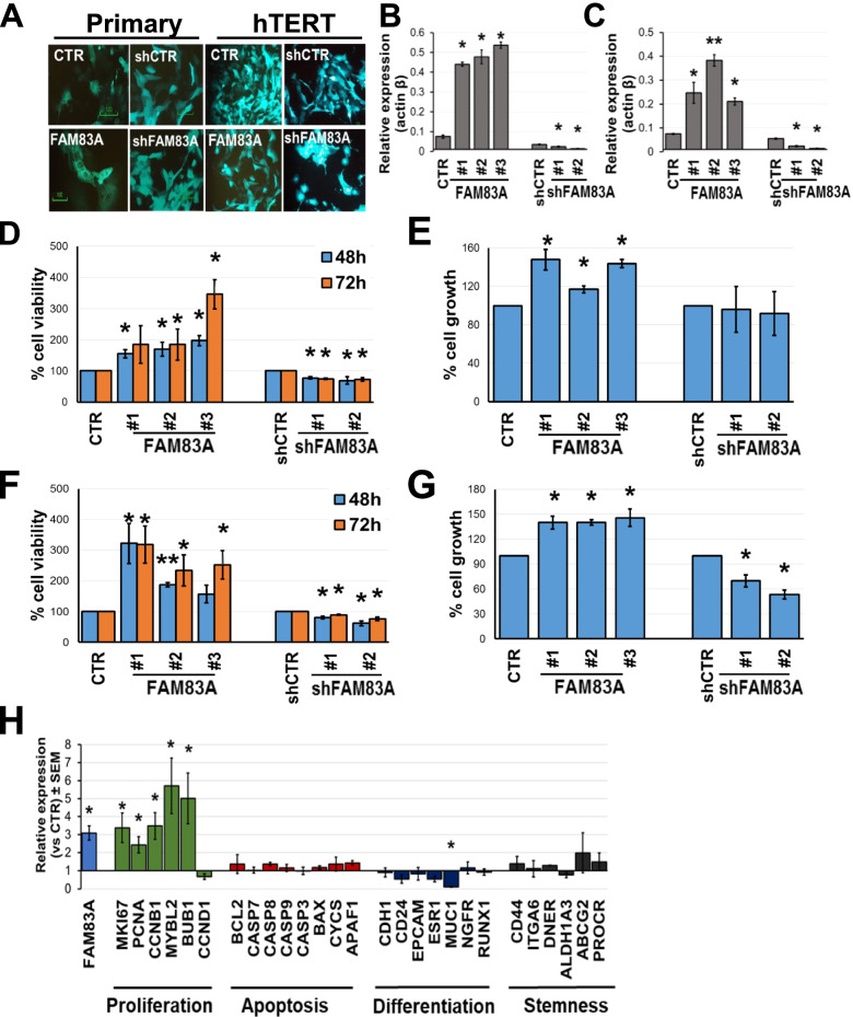
Immunohistochemical staining was used to evaluate FAM83A protein levels in both a normal breast tissue microarray (TMA, N = 411) and a breast tumor TMA (N = 349). EGFR staining and its correlation with FAM83A expression were also assessed. Lentivirus-mediated manipulation of FAM83A expression in primary and hTERT-immortalized breast epithelial cells was employed. Biological and molecular alterations upon FAM83A overexpression/downregulation and FAM83A's interaction partners were investigated.
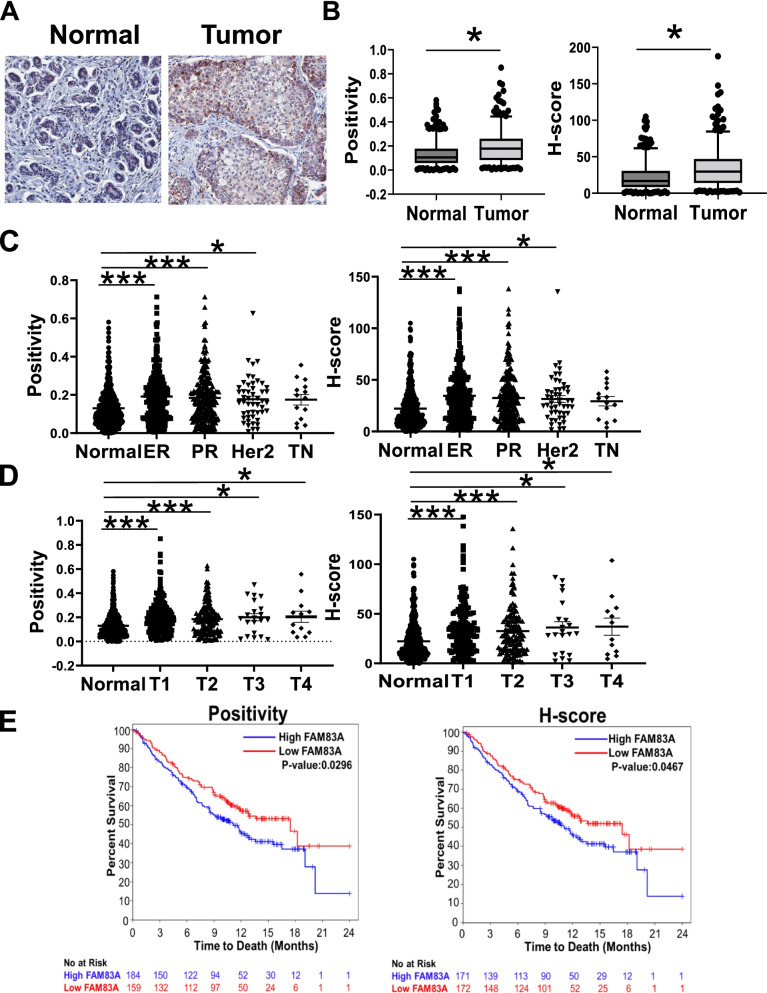
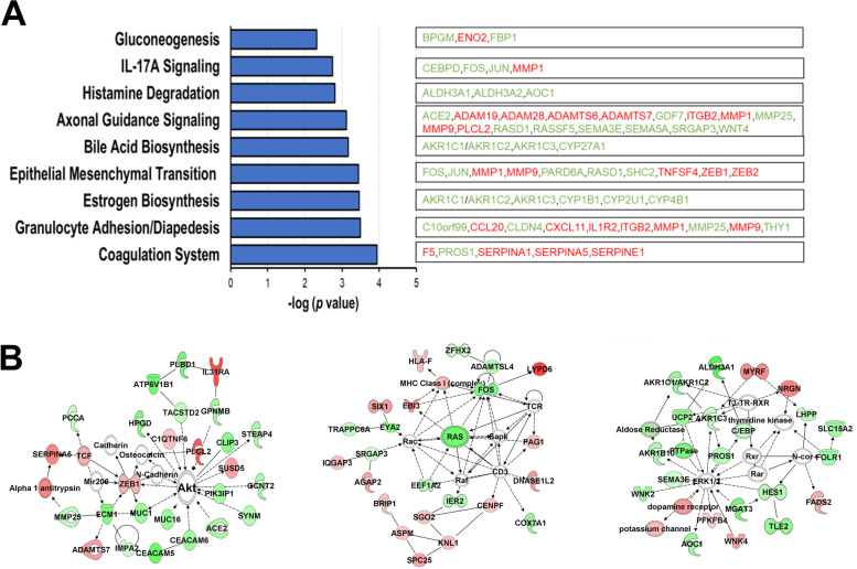
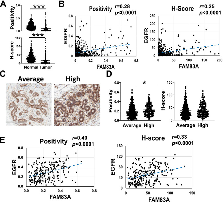
TMA analysis revealed a 1.5-fold increase in FAM83A expression level in breast cancer cases as compared with normal breast tissues (p < 0.0001). FAM83A protein expression was directly correlated with EGFR level in both normal and breast cancer tissues. In in vitro assays, exogenous expression of FAM83A in either primary or immortalized breast epithelial cells promoted cell viability and proliferation. Additionally, Ingenuity Pathway Analysis (IPA) revealed that FAM83A overexpression in primary cells affected the expression of genes involved in cellular morphology and metabolism. Mass spectrometry analysis identified DDX3X and LAMB3 as potential FAM83A interaction partners in primary cells, while we detected FAM83A interaction with cytoskeleton reorganization factors, including LIMA1, MYH10, PLEC, MYL6 in the immortalized cells.
This study shows that FAM83A promotes metabolic activation in primary breast epithelial cells and cell proliferation in both primary and immortalized cells. These findings support its role in early breast oncogenesis.
序列相似性家族83成员A(FAM83A)在包括乳腺癌在内的多种癌症中具有致癌特性。最近,我们报道了FAM83A在乳腺癌高危女性的正常乳腺组织中过表达。我们现在假设FAM83A是乳腺癌发生的关键因素。
采用免疫组织化学染色评估正常乳腺组织芯片(TMA,N = 411)和乳腺肿瘤TMA(N = 349)中FAM83A蛋白水平。还评估了表皮生长因子受体(EGFR)染色及其与FAM83A表达的相关性。采用慢病毒介导的方法在原代和hTERT永生化乳腺上皮细胞中调控FAM83A表达。研究了FAM83A过表达/下调后的生物学和分子改变以及FAM83A的相互作用伙伴。
TMA分析显示,与正常乳腺组织相比,乳腺癌病例中FAM83A表达水平增加了1.5倍(p < 0.0001)。在正常和乳腺癌组织中,FAM83A蛋白表达均与EGFR水平直接相关。在体外试验中,原代或永生化乳腺上皮细胞中外源表达FAM83A可促进细胞活力和增殖。此外, Ingenuity通路分析(IPA)显示,原代细胞中FAM83A过表达影响了参与细胞形态和代谢的基因表达。质谱分析确定DDX3X和LAMB3是原代细胞中潜在的FAM83A相互作用伙伴,而我们在永生化细胞中检测到FAM83A与细胞骨架重组因子相互作用,包括LIMA1、MYH10、PLEC、MYL6。
本研究表明,FAM83A促进原代乳腺上皮细胞的代谢激活以及原代和永生化细胞的增殖。这些发现支持了其在早期乳腺癌发生中的作用。