Abjigitova Djamila, Sadeghi Amir H, Peek Jette J, Bekkers Jos A, Bogers Ad J J C, Mahtab Edris A F
Department of Cardiothoracic Surgery, Erasmus University Medical Center, Room Rg-619, P.O. Box 2040, 3000 CA Rotterdam, The Netherlands.
J Cardiovasc Dev Dis. 2022 Jan 18;9(2):31. doi: 10.3390/jcdd9020031.
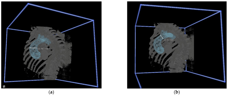
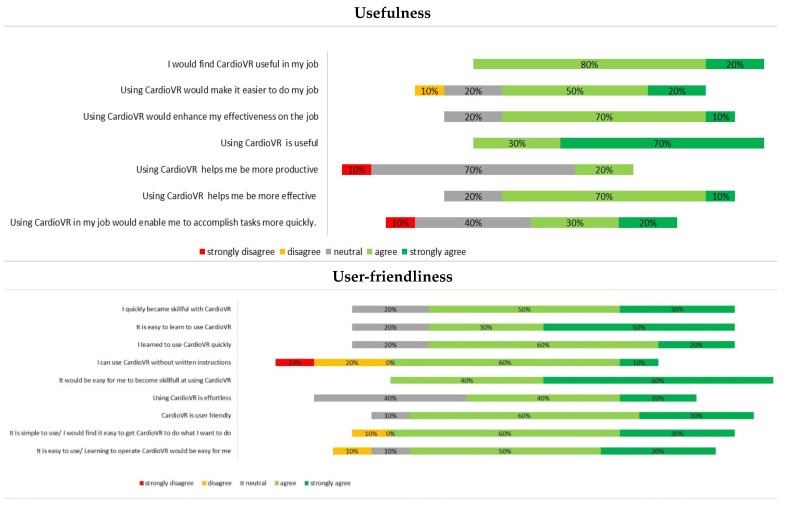
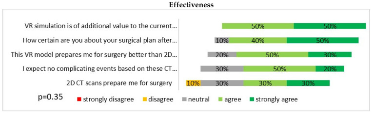
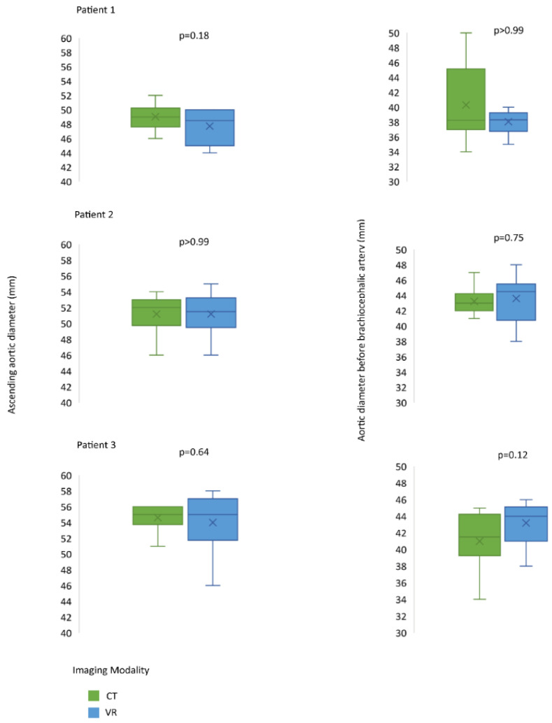
Complex aortic anatomy needs careful preoperative planning in which a patient-tailored approach with novel immersive techniques could serve as a valuable addition to current preoperative imaging. This pilot study aimed to investigate the technical feasibility of virtual reality (VR) as an additional imaging tool for preoperative planning in ascending aortic surgery. Ten cardiothoracic surgeons were presented with six patients who had each undergone a recent repair of the ascending aorta. Two-dimensional computed tomography images of each patient were assessed prior to the VR session. After three-dimensional (3D) VR rendering and 3D segmentation of the ascending aorta and aortic arch, the reconstructions were analyzed by each surgeon in VR via a head-mounted display. Each cardiothoracic surgeon completed a questionnaire after each planning procedure. The results of their assessments were compared to the performed operations. The primary endpoint of the present study was a change of surgical approach from open to clamped distal anastomosis, and vice versa. : Compared with conventional imaging, 80% of surgeons found that VR prepared them better for surgery. In 33% of cases (two out of six), the preoperative decision was adjusted due to the 3D VR-based evaluation of the anatomy. Surgeons rated CardioVR usefulness, user-friendliness, and satisfaction with median scores of 3.8 (IQR: 3.5-4.1), 4.2 (IQR: 3.8-4.6,) and 4.1 (IQR: 3.8-4.7) on a five-point Likert scale, respectively. Three-dimensional VR imaging was associated with improved anatomical understanding among surgeons and could be helpful in the future preoperative planning of ascending aortic surgery.
复杂的主动脉解剖结构需要仔细的术前规划,其中采用新颖的沉浸式技术的个性化方法可以成为当前术前成像的宝贵补充。这项前瞻性研究旨在探讨虚拟现实(VR)作为升主动脉手术术前规划的辅助成像工具的技术可行性。向十位心胸外科医生展示了六名近期均接受过升主动脉修复手术的患者。在进行VR session之前,先评估了每位患者的二维计算机断层扫描图像。在对升主动脉和主动脉弓进行三维(3D)VR渲染和3D分割后,每位外科医生通过头戴式显示器在VR中对重建图像进行分析。每位心胸外科医生在每次规划程序后都完成了一份问卷。将他们的评估结果与所进行的手术进行比较。本研究的主要终点是手术方式从开放手术改为钳夹远端吻合术,反之亦然。与传统成像相比,80%的外科医生发现VR使他们为手术做好了更好的准备。在33%的病例中(六例中的两例),由于基于3D VR的解剖结构评估,术前决策得到了调整。外科医生对CardioVR的有用性、用户友好性和满意度在五点李克特量表上的中位数评分分别为3.8(四分位间距:3.5 - 4.1)、4.2(四分位间距:3.8 - 4.6)和4.1(四分位间距:3.8 - 4.7)。三维VR成像与外科医生对解剖结构的理解改善相关,并且可能有助于未来升主动脉手术的术前规划。 (注:原文中“CardioVR”未在前面提及,可能是特定的VR相关产品或技术名称,这里直接保留英文未翻译。)