• 文献检索
  • 文档翻译
  • 深度研究
  • 学术资讯
  • Suppr Zotero 插件Zotero 插件
  • 邀请有礼
  • 套餐&价格
  • 历史记录
应用&插件
Suppr Zotero 插件Zotero 插件浏览器插件Mac 客户端Windows 客户端微信小程序
定价
高级版会员购买积分包购买API积分包
服务
文献检索文档翻译深度研究API 文档MCP 服务
关于我们
关于 Suppr公司介绍联系我们用户协议隐私条款
关注我们

Suppr 超能文献

核心技术专利:CN118964589B侵权必究
粤ICP备2023148730 号-1Suppr @ 2026

文献检索

告别复杂PubMed语法,用中文像聊天一样搜索,搜遍4000万医学文献。AI智能推荐,让科研检索更轻松。

立即免费搜索

文件翻译

保留排版,准确专业,支持PDF/Word/PPT等文件格式,支持 12+语言互译。

免费翻译文档

深度研究

AI帮你快速写综述,25分钟生成高质量综述,智能提取关键信息,辅助科研写作。

立即免费体验

基于深度学习和混合技术的血液显微镜样本的多方法诊断,用于急性淋巴细胞白血病的早期检测。

Multi-Method Diagnosis of Blood Microscopic Sample for Early Detection of Acute Lymphoblastic Leukemia Based on Deep Learning and Hybrid Techniques.

机构信息

Department of Information Systems, College of Computer and Information Sciences, Prince Sultan University, Riyadh 11586, Saudi Arabia.

Department of Computer Science & Information Technology, Dr. Babasaheb Ambedkar Marathwada University, Aurangabad 431004, India.

出版信息

Sensors (Basel). 2022 Feb 19;22(4):1629. doi: 10.3390/s22041629.

DOI:10.3390/s22041629
PMID:35214531
原文链接:https://pmc.ncbi.nlm.nih.gov/articles/PMC8876170/
Abstract

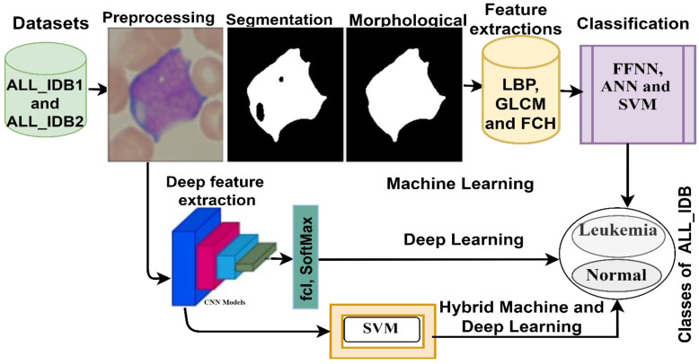
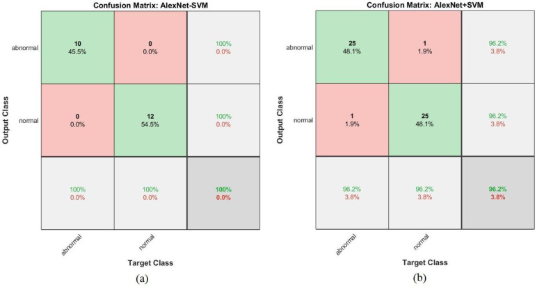
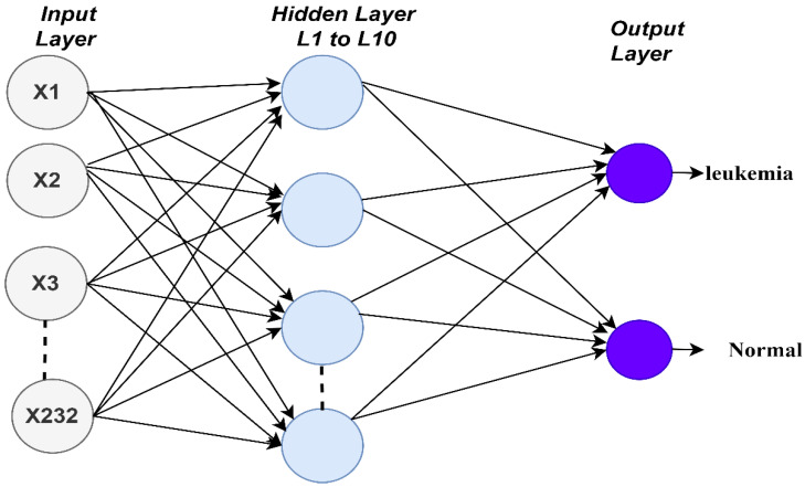
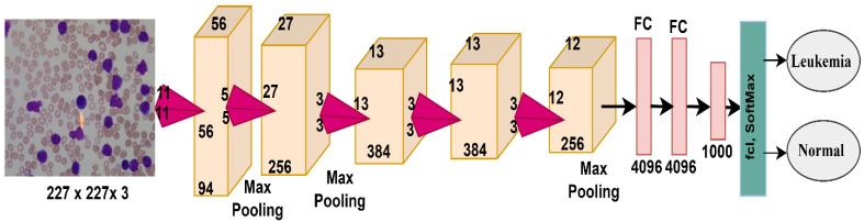
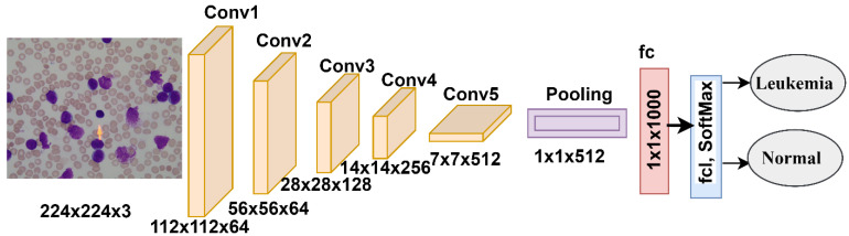
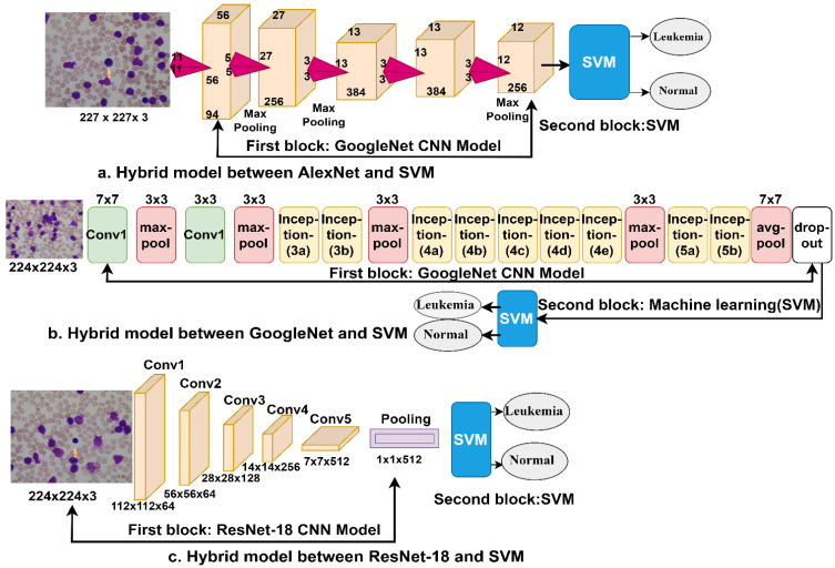
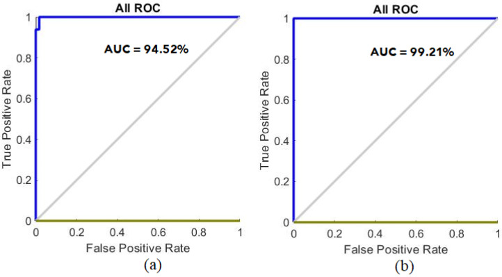
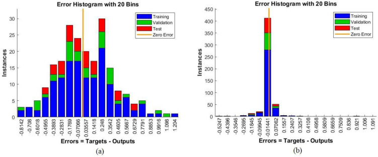
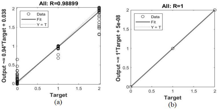
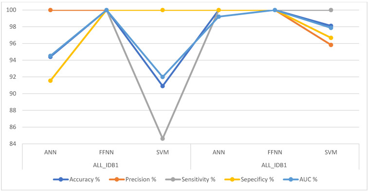
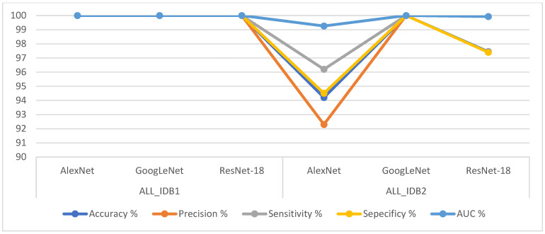
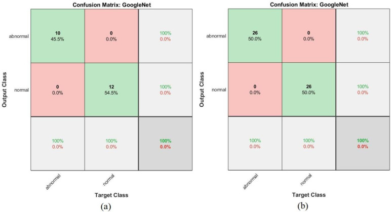
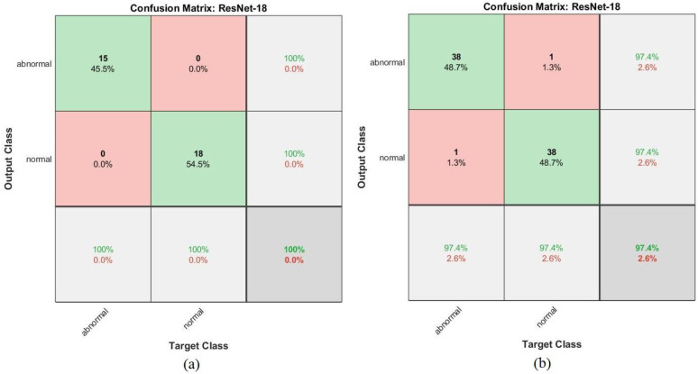
Leukemia is one of the most dangerous types of malignancies affecting the bone marrow or blood in all age groups, both in children and adults. The most dangerous and deadly type of leukemia is acute lymphoblastic leukemia (ALL). It is diagnosed by hematologists and experts in blood and bone marrow samples using a high-quality microscope with a magnifying lens. Manual diagnosis, however, is considered slow and is limited by the differing opinions of experts and other factors. Thus, this work aimed to develop diagnostic systems for two Acute Lymphoblastic Leukemia Image Databases (ALL_IDB1 and ALL_IDB2) for the early detection of leukemia. All images were optimized before being introduced to the systems by two overlapping filters: the average and Laplacian filters. This study consists of three proposed systems as follows: the first consists of the artificial neural network (ANN), feed forward neural network (FFNN), and support vector machine (SVM), all of which are based on hybrid features extracted using Local Binary Pattern (LBP), Gray Level Co-occurrence Matrix (GLCM) and Fuzzy Color Histogram (FCH) methods. Both ANN and FFNN reached an accuracy of 100%, while SVM reached an accuracy of 98.11%. The second proposed system consists of the convolutional neural network (CNN) models: AlexNet, GoogleNet, and ResNet-18, based on the transfer learning method, in which deep feature maps were extracted and classified with high accuracy. All the models obtained promising results for the early detection of leukemia in both datasets, with an accuracy of 100% for the AlexNet, GoogleNet, and ResNet-18 models. The third proposed system consists of hybrid CNN-SVM technologies, consisting of two blocks: CNN models for extracting feature maps and the SVM algorithm for classifying feature maps. All the hybrid systems achieved promising results, with AlexNet + SVM achieving 100% accuracy, Goog-LeNet + SVM achieving 98.1% accuracy, and ResNet-18 + SVM achieving 100% accuracy.

摘要

白血病是一种最危险的恶性肿瘤类型,影响所有年龄段的骨髓或血液,包括儿童和成人。最危险和致命的白血病类型是急性淋巴细胞白血病(ALL)。血液学家和血液及骨髓样本专家使用高质量显微镜和放大镜头进行诊断。然而,手动诊断被认为速度较慢,并受到专家意见和其他因素的差异限制。因此,这项工作旨在为两个急性淋巴细胞白血病图像数据库(ALL_IDB1 和 ALL_IDB2)开发诊断系统,以早期发现白血病。所有图像在引入系统之前都经过了两种重叠滤波器的优化:平均滤波器和拉普拉斯滤波器。本研究包括以下三个拟议系统:第一个系统由人工神经网络(ANN)、前馈神经网络(FFNN)和支持向量机(SVM)组成,它们都是基于使用局部二值模式(LBP)、灰度共生矩阵(GLCM)和模糊颜色直方图(FCH)方法提取的混合特征。ANN 和 FFNN 的准确率均达到 100%,而 SVM 的准确率达到 98.11%。第二个拟议系统由卷积神经网络(CNN)模型组成:AlexNet、GoogleNet 和 ResNet-18,基于迁移学习方法,提取和分类深度特征图,准确率高。所有模型在两个数据集的白血病早期检测中均取得了有前景的结果,AlexNet、GoogleNet 和 ResNet-18 模型的准确率为 100%。第三个拟议系统由混合 CNN-SVM 技术组成,包括两个模块:用于提取特征图的 CNN 模型和用于分类特征图的 SVM 算法。所有混合系统都取得了有前景的结果,其中 AlexNet+SVM 达到 100%的准确率,Goog-LeNet+SVM 达到 98.1%的准确率,ResNet-18+SVM 达到 100%的准确率。

https://cdn.ncbi.nlm.nih.gov/pmc/blobs/173f/8876170/b73981ce7cb4/sensors-22-01629-g026.jpg
https://cdn.ncbi.nlm.nih.gov/pmc/blobs/173f/8876170/daf8c72b4dd1/sensors-22-01629-g001.jpg
https://cdn.ncbi.nlm.nih.gov/pmc/blobs/173f/8876170/623b4804bc71/sensors-22-01629-g002.jpg
https://cdn.ncbi.nlm.nih.gov/pmc/blobs/173f/8876170/4f9184d173c9/sensors-22-01629-g003.jpg
https://cdn.ncbi.nlm.nih.gov/pmc/blobs/173f/8876170/2323d7d1aa8f/sensors-22-01629-g004.jpg
https://cdn.ncbi.nlm.nih.gov/pmc/blobs/173f/8876170/d5afe4d0c769/sensors-22-01629-g005.jpg
https://cdn.ncbi.nlm.nih.gov/pmc/blobs/173f/8876170/e61cc0fbed1d/sensors-22-01629-g006.jpg
https://cdn.ncbi.nlm.nih.gov/pmc/blobs/173f/8876170/9b426bd79dd4/sensors-22-01629-g007.jpg
https://cdn.ncbi.nlm.nih.gov/pmc/blobs/173f/8876170/e81819b4a400/sensors-22-01629-g008.jpg
https://cdn.ncbi.nlm.nih.gov/pmc/blobs/173f/8876170/4bd62f9dd80c/sensors-22-01629-g009.jpg
https://cdn.ncbi.nlm.nih.gov/pmc/blobs/173f/8876170/91ec947ccb0e/sensors-22-01629-g010.jpg
https://cdn.ncbi.nlm.nih.gov/pmc/blobs/173f/8876170/dfe340b491fd/sensors-22-01629-g011.jpg
https://cdn.ncbi.nlm.nih.gov/pmc/blobs/173f/8876170/7240c8b6c817/sensors-22-01629-g012.jpg
https://cdn.ncbi.nlm.nih.gov/pmc/blobs/173f/8876170/ff678ea025a8/sensors-22-01629-g013.jpg
https://cdn.ncbi.nlm.nih.gov/pmc/blobs/173f/8876170/0209f1d174f0/sensors-22-01629-g014.jpg
https://cdn.ncbi.nlm.nih.gov/pmc/blobs/173f/8876170/c76c59e2fe3f/sensors-22-01629-g015.jpg
https://cdn.ncbi.nlm.nih.gov/pmc/blobs/173f/8876170/4481fe052e49/sensors-22-01629-g016.jpg
https://cdn.ncbi.nlm.nih.gov/pmc/blobs/173f/8876170/147608f24ac6/sensors-22-01629-g017.jpg
https://cdn.ncbi.nlm.nih.gov/pmc/blobs/173f/8876170/13dd83db0018/sensors-22-01629-g018.jpg
https://cdn.ncbi.nlm.nih.gov/pmc/blobs/173f/8876170/349588670463/sensors-22-01629-g019.jpg
https://cdn.ncbi.nlm.nih.gov/pmc/blobs/173f/8876170/280e4186e2f3/sensors-22-01629-g020.jpg
https://cdn.ncbi.nlm.nih.gov/pmc/blobs/173f/8876170/0e70c28d3709/sensors-22-01629-g021.jpg
https://cdn.ncbi.nlm.nih.gov/pmc/blobs/173f/8876170/be58f50bac7c/sensors-22-01629-g022.jpg
https://cdn.ncbi.nlm.nih.gov/pmc/blobs/173f/8876170/e4f0a6c50050/sensors-22-01629-g023.jpg
https://cdn.ncbi.nlm.nih.gov/pmc/blobs/173f/8876170/844213074b0a/sensors-22-01629-g024.jpg
https://cdn.ncbi.nlm.nih.gov/pmc/blobs/173f/8876170/3f1f6d31ed4c/sensors-22-01629-g025.jpg
https://cdn.ncbi.nlm.nih.gov/pmc/blobs/173f/8876170/b73981ce7cb4/sensors-22-01629-g026.jpg

相似文献

1
Multi-Method Diagnosis of Blood Microscopic Sample for Early Detection of Acute Lymphoblastic Leukemia Based on Deep Learning and Hybrid Techniques.基于深度学习和混合技术的血液显微镜样本的多方法诊断,用于急性淋巴细胞白血病的早期检测。
Sensors (Basel). 2022 Feb 19;22(4):1629. doi: 10.3390/s22041629.
2
Hybrid and Deep Learning Approach for Early Diagnosis of Lower Gastrointestinal Diseases.混合与深度学习方法在胃肠道疾病早期诊断中的应用
Sensors (Basel). 2022 May 27;22(11):4079. doi: 10.3390/s22114079.
3
Early Diagnosis of Oral Squamous Cell Carcinoma Based on Histopathological Images Using Deep and Hybrid Learning Approaches.基于组织病理学图像,运用深度学习和混合学习方法对口腔鳞状细胞癌进行早期诊断。
Diagnostics (Basel). 2022 Aug 5;12(8):1899. doi: 10.3390/diagnostics12081899.
4
Hybrid Methods for Fundus Image Analysis for Diagnosis of Diabetic Retinopathy Development Stages Based on Fusion Features.基于融合特征的糖尿病视网膜病变发展阶段诊断的眼底图像分析混合方法
Diagnostics (Basel). 2023 Aug 28;13(17):2783. doi: 10.3390/diagnostics13172783.
5
Automated detection of leukemia by pretrained deep neural networks and transfer learning: A comparison.基于预训练深度神经网络和迁移学习的白血病自动检测:比较研究。
Med Eng Phys. 2021 Dec;98:8-19. doi: 10.1016/j.medengphy.2021.10.006. Epub 2021 Oct 13.
6
A hybrid detection model for acute lymphocytic leukemia using support vector machine and particle swarm optimization (SVM-PSO).基于支持向量机和粒子群优化算法的急性淋巴细胞白血病混合检测模型(SVM-PSO)。
Sci Rep. 2024 Oct 8;14(1):23483. doi: 10.1038/s41598-024-74889-1.
7
Mutual Information based hybrid model and deep learning for Acute Lymphocytic Leukemia detection in single cell blood smear images.基于互信息的混合模型和深度学习在单细胞血涂片图像中的急性淋巴细胞白血病检测
Comput Methods Programs Biomed. 2019 Oct;179:104987. doi: 10.1016/j.cmpb.2019.104987. Epub 2019 Jul 15.
8
Developing a Recognition System for Diagnosing Melanoma Skin Lesions Using Artificial Intelligence Algorithms.开发一种使用人工智能算法诊断黑色素瘤皮肤病变的识别系统。
Comput Math Methods Med. 2021 May 15;2021:9998379. doi: 10.1155/2021/9998379. eCollection 2021.
9
Automatic classification of acute lymphoblastic leukemia cells and lymphocyte subtypes based on a novel convolutional neural network.基于新型卷积神经网络的急性淋巴细胞白血病细胞和淋巴细胞亚型的自动分类。
Microsc Res Tech. 2024 Jul;87(7):1615-1626. doi: 10.1002/jemt.24551. Epub 2024 Mar 6.
10
Classification of acute lymphoblastic leukemia using deep learning.利用深度学习对急性淋巴细胞白血病进行分类
Microsc Res Tech. 2018 Nov;81(11):1310-1317. doi: 10.1002/jemt.23139. Epub 2018 Oct 23.

引用本文的文献

1
Classifying brain metastases originating from different pathological subtypes of lung cancer via a multimodal magnetic resonance imaging-based deep learning approach.通过基于多模态磁共振成像的深度学习方法对源自肺癌不同病理亚型的脑转移瘤进行分类。
J Thorac Dis. 2025 Jul 31;17(7):5250-5259. doi: 10.21037/jtd-2025-1285. Epub 2025 Jul 29.
2
Deep learning based semantic segmentation of leukemia effected white blood cell.基于深度学习的白血病相关白细胞语义分割
PLoS One. 2025 May 8;20(5):e0320596. doi: 10.1371/journal.pone.0320596. eCollection 2025.
3
Ultra-compact quintuple-band terahertz metamaterial biosensor for enhanced blood cancer diagnostics.

本文引用的文献

1
Score and Correlation Coefficient-Based Feature Selection for Predicting Heart Failure Diagnosis by Using Machine Learning Algorithms.基于评分和相关系数的特征选择在使用机器学习算法预测心力衰竭诊断中的应用。
Comput Math Methods Med. 2021 Dec 20;2021:8500314. doi: 10.1155/2021/8500314. eCollection 2021.
2
Automated Diagnosis of Chest X-Ray for Early Detection of COVID-19 Disease.基于胸部 X 光的 COVID-19 疾病早期检测的自动化诊断。
Comput Math Methods Med. 2021 Oct 21;2021:6919483. doi: 10.1155/2021/6919483. eCollection 2021.
3
Tens of images can suffice to train neural networks for malignant leukocyte detection.
用于增强血癌诊断的超紧凑型五频段太赫兹超材料生物传感器。
PLoS One. 2025 Jan 9;20(1):e0313874. doi: 10.1371/journal.pone.0313874. eCollection 2025.
4
Optimization of diagnosis and treatment of hematological diseases via artificial intelligence.通过人工智能优化血液疾病的诊断与治疗
Front Med (Lausanne). 2024 Nov 7;11:1487234. doi: 10.3389/fmed.2024.1487234. eCollection 2024.
5
Analysis of dermoscopy images of multi-class for early detection of skin lesions by hybrid systems based on integrating features of CNN models.基于集成卷积神经网络模型特征的混合系统对多类皮肤镜图像进行早期皮肤病变检测分析。
PLoS One. 2024 Mar 21;19(3):e0298305. doi: 10.1371/journal.pone.0298305. eCollection 2024.
6
Deep learning enhances acute lymphoblastic leukemia diagnosis and classification using bone marrow images.深度学习利用骨髓图像增强急性淋巴细胞白血病的诊断和分类。
Front Oncol. 2023 Dec 6;13:1330977. doi: 10.3389/fonc.2023.1330977. eCollection 2023.
7
RDET stacking classifier: a novel machine learning based approach for stroke prediction using imbalance data.RDET堆叠分类器:一种基于机器学习的新型方法,用于利用不平衡数据进行中风预测。
PeerJ Comput Sci. 2023 Nov 21;9:e1684. doi: 10.7717/peerj-cs.1684. eCollection 2023.
8
Hybrid Methods for Fundus Image Analysis for Diagnosis of Diabetic Retinopathy Development Stages Based on Fusion Features.基于融合特征的糖尿病视网膜病变发展阶段诊断的眼底图像分析混合方法
Diagnostics (Basel). 2023 Aug 28;13(17):2783. doi: 10.3390/diagnostics13172783.
9
DSCNet: Deep Skip Connections-Based Dense Network for ALL Diagnosis Using Peripheral Blood Smear Images.DSCNet:基于深度跳跃连接的密集网络,用于使用外周血涂片图像进行全诊断
Diagnostics (Basel). 2023 Aug 24;13(17):2752. doi: 10.3390/diagnostics13172752.
10
Blood Slide Image Analysis to Classify WBC Types for Prediction Haematology Based on a Hybrid Model of CNN and Handcrafted Features.基于卷积神经网络(CNN)和手工特征混合模型的血液涂片图像分析,用于对白细胞类型进行分类以预测血液学情况。
Diagnostics (Basel). 2023 May 29;13(11):1899. doi: 10.3390/diagnostics13111899.
数十张图像就足以训练神经网络来检测恶性白细胞。
Sci Rep. 2021 Apr 12;11(1):7995. doi: 10.1038/s41598-021-86995-5.
4
Recognition of acute lymphoblastic leukemia and lymphocytes cell subtypes in microscopic images using random forest classifier.使用随机森林分类器识别显微镜图像中的急性淋巴细胞白血病和淋巴细胞细胞亚型。
Phys Eng Sci Med. 2021 Jun;44(2):433-441. doi: 10.1007/s13246-021-00993-5. Epub 2021 Mar 22.
5
Identification of significant risks in pediatric acute lymphoblastic leukemia (ALL) through machine learning (ML) approach.通过机器学习 (ML) 方法识别小儿急性淋巴细胞白血病 (ALL) 中的显著风险。
Med Biol Eng Comput. 2020 Nov;58(11):2631-2640. doi: 10.1007/s11517-020-02245-2. Epub 2020 Aug 25.
6
Principal component analysis of blood microRNA datasets facilitates diagnosis of diverse diseases.主成分分析血液 microRNA 数据集有助于诊断多种疾病。
PLoS One. 2020 Jun 5;15(6):e0234185. doi: 10.1371/journal.pone.0234185. eCollection 2020.
7
Platelet biology and functions: new concepts and clinical perspectives.血小板生物学和功能:新概念和临床视角。
Nat Rev Cardiol. 2019 Mar;16(3):166-179. doi: 10.1038/s41569-018-0110-0.
8
A Microfluidic Device for Simultaneous Extraction of Plasma, Red Blood Cells, and On-Chip White Blood Cell Trapping.一种用于同时提取血浆、红细胞和芯片内白细胞捕获的微流控装置。
Sci Rep. 2018 Oct 18;8(1):15345. doi: 10.1038/s41598-018-33738-8.
9
Acute Lymphoblastic Leukemia Detection and Classification of Its Subtypes Using Pretrained Deep Convolutional Neural Networks.使用预训练深度卷积神经网络检测急性淋巴细胞白血病及其亚型分类
Technol Cancer Res Treat. 2018 Jan 1;17:1533033818802789. doi: 10.1177/1533033818802789.
10
Erythrocytes as a biological model for screening of xenobiotics toxicity.红细胞作为筛选外源化合物毒性的生物学模型。
Chem Biol Interact. 2018 Jan 5;279:73-83. doi: 10.1016/j.cbi.2017.11.007. Epub 2017 Nov 8.