Functional Neuroimaging Laboratory, Center for Neuroscience and Cognitive systems, Istituto Italiano di Tecnologia, Rovereto, Italy.
Center for Mind and Brain Sciences, University of Trento, Rovereto, Italy.
Nat Commun. 2022 Feb 25;13(1):1056. doi: 10.1038/s41467-022-28591-3.
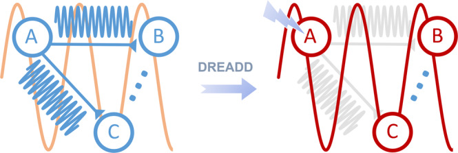
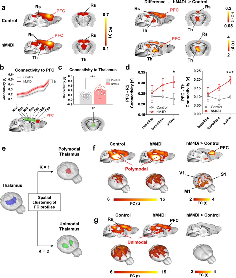
While shaped and constrained by axonal connections, fMRI-based functional connectivity reorganizes in response to varying interareal input or pathological perturbations. However, the causal contribution of regional brain activity to whole-brain fMRI network organization remains unclear. Here we combine neural manipulations, resting-state fMRI and in vivo electrophysiology to probe how inactivation of a cortical node causally affects brain-wide fMRI coupling in the mouse. We find that chronic inhibition of the medial prefrontal cortex (PFC) via overexpression of a potassium channel increases fMRI connectivity between the inhibited area and its direct thalamo-cortical targets. Acute chemogenetic inhibition of the PFC produces analogous patterns of fMRI overconnectivity. Using in vivo electrophysiology, we find that chemogenetic inhibition of the PFC enhances low frequency (0.1-4 Hz) oscillatory power via suppression of neural firing not phase-locked to slow rhythms, resulting in increased slow and δ band coherence between areas that exhibit fMRI overconnectivity. These results provide causal evidence that cortical inactivation can counterintuitively increase fMRI connectivity via enhanced, less-localized slow oscillatory processes.
虽然功能磁共振成像(fMRI)基于功能连接受到轴突连接的塑造和限制,但它会响应区域间输入或病理性干扰而重新组织。然而,区域脑活动对全脑功能磁共振成像网络组织的因果贡献仍不清楚。在这里,我们结合神经操作、静息态功能磁共振成像和在体电生理学来探究皮质节点的失活如何因果性地影响小鼠的全脑功能磁共振成像耦合。我们发现,通过过表达钾通道对内侧前额叶皮层(mPFC)进行慢性抑制会增加受抑制区域与其直接丘脑皮质靶区之间的功能磁共振成像连接。急性化学遗传抑制 mPFC 会产生类似的功能磁共振成像过度连接模式。通过在体电生理学,我们发现化学遗传抑制 mPFC 通过抑制与慢节律不同步的神经放电来增强低频(0.1-4 Hz)振荡功率,从而导致表现出功能磁共振成像过度连接的区域之间的慢波和 δ 波段相干性增加。这些结果提供了因果证据,表明皮质失活可以通过增强、非局部化的慢振荡过程反直觉地增加功能磁共振成像连接。