• 文献检索
  • 文档翻译
  • 深度研究
  • 学术资讯
  • Suppr Zotero 插件Zotero 插件
  • 邀请有礼
  • 套餐&价格
  • 历史记录
应用&插件
Suppr Zotero 插件Zotero 插件浏览器插件Mac 客户端Windows 客户端微信小程序
定价
高级版会员购买积分包购买API积分包
服务
文献检索文档翻译深度研究API 文档MCP 服务
关于我们
关于 Suppr公司介绍联系我们用户协议隐私条款
关注我们

Suppr 超能文献

核心技术专利:CN118964589B侵权必究
粤ICP备2023148730 号-1Suppr @ 2026

文献检索

告别复杂PubMed语法,用中文像聊天一样搜索,搜遍4000万医学文献。AI智能推荐,让科研检索更轻松。

立即免费搜索

文件翻译

保留排版,准确专业,支持PDF/Word/PPT等文件格式,支持 12+语言互译。

免费翻译文档

深度研究

AI帮你快速写综述,25分钟生成高质量综述,智能提取关键信息,辅助科研写作。

立即免费体验

超声组学模型与 CEUS LI-RADS v2017 对肝细胞癌与肝细胞癌-胆管细胞癌混合型的鉴别诊断效能比较。

Differentiation between combined hepatocellular carcinoma and hepatocellular carcinoma: comparison of diagnostic performance between ultrasomics-based model and CEUS LI-RADS v2017.

机构信息

Department of Medical Ultrasonics, Institute of Diagnostic and Interventional Ultrasound, The First Affiliated Hospital of Sun Yat-Sen University, 58 Zhongshan Road 2, Guangzhou, 510080, People's Republic of China.

Department of Hepatobiliary Surgery, The First Affiliated Hospital of Sun Yat-Sen University, Guangzhou, China.

出版信息

BMC Med Imaging. 2022 Mar 3;22(1):36. doi: 10.1186/s12880-022-00765-x.

DOI:10.1186/s12880-022-00765-x
PMID:35241004
原文链接:https://pmc.ncbi.nlm.nih.gov/articles/PMC8896152/
Abstract

BACKGROUND

The imaging findings of combined hepatocellular cholangiocarcinoma (CHC) may be similar to those of hepatocellular carcinoma (HCC). CEUS LI-RADS may not perform well in distinguishing CHC from HCC. Studies have shown that radiomics has an excellent imaging analysis ability. This study aimed to establish and confirm an ultrasomics model for differentiating CHC from HCC.

METHODS

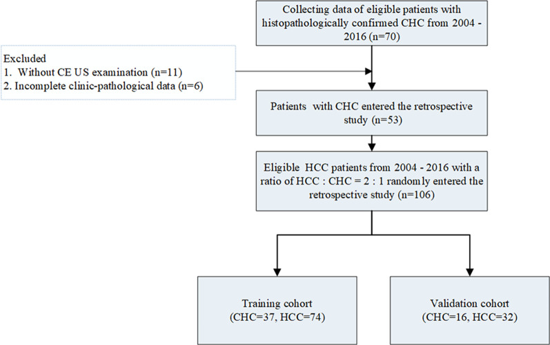
Between 2004 and 2016, we retrospectively identified 53 eligible CHC patients and randomly included 106 eligible HCC patients with a ratio of HCC:CHC = 2:1, all of whom were categorized according to Contrast-Enhanced (CE) ultrasonography (US) Liver Imaging Reporting and Data System (LI-RADS) version 2017. The model based on ultrasomics features of CE US was developed in 74 HCC and 37 CHC and confirmed in 32 HCC and 16 CHC. The diagnostic performance of the LI-RADS or ultrasomics model was assessed by the area under the curve (AUC), accuracy, sensitivity and specificity.

RESULTS

In the entire and validation cohorts, 67.0% and 81.3% of HCC cases were correctly assigned to LR-5 or LR-TIV contiguous with LR-5, and 73.6% and 87.5% of CHC cases were assigned to LR-M correctly. Up to 33.0% of HCC and 26.4% of CHC were misclassified by CE US LI-RADS. A total of 90.6% of HCC as well as 87.5% of CHC correctly diagnosed by the ultrasomics model in the validation cohort. The AUC, accuracy, sensitivity of the ultrasomics model were higher though without significant difference than those of CE US LI-RADS in the validation cohort.

CONCLUSION

The proposed ultrasomics model showed higher ability though the difference was not significantly different for differentiating CHC from HCC, which may be helpful in clinical diagnosis.

摘要

背景

混合细胞型肝癌(CHC)的影像学表现可能与肝细胞癌(HCC)相似。CEUS LI-RADS 可能无法很好地区分 CHC 与 HCC。研究表明,放射组学具有出色的影像分析能力。本研究旨在建立和验证一种超声组学模型,用于区分 CHC 与 HCC。

方法

回顾性纳入 2004 年至 2016 年间符合条件的 53 例 CHC 患者,随机纳入符合条件的 HCC 患者 106 例,HCC:CHC 比例为 2:1,并按照对比增强超声(CEUS)肝脏影像报告和数据系统(LI-RADS)版本 2017 进行分类。在 74 例 HCC 和 37 例 CHC 中建立基于 CEUS 超声组学特征的模型,并在 32 例 HCC 和 16 例 CHC 中进行验证。通过曲线下面积(AUC)、准确性、敏感性和特异性评估 LI-RADS 或超声组学模型的诊断性能。

结果

在整个队列和验证队列中,67.0%和 81.3%的 HCC 病例被正确分配到 LR-5 或与 LR-5 连续的 LR-TIV,73.6%和 87.5%的 CHC 病例被正确分配到 LR-M。CEUS LI-RADS 错误分类 HCC 为 33.0%,CHC 为 26.4%。验证队列中,超声组学模型对 HCC 的诊断准确率为 90.6%,对 CHC 的诊断准确率为 87.5%。验证队列中,超声组学模型的 AUC、准确性、敏感性均高于 CEUS LI-RADS,但差异无统计学意义。

结论

提出的超声组学模型在区分 CHC 与 HCC 方面具有更高的能力,尽管差异无统计学意义,但可能有助于临床诊断。

https://cdn.ncbi.nlm.nih.gov/pmc/blobs/50cd/8896152/72de9c88931f/12880_2022_765_Fig3_HTML.jpg
https://cdn.ncbi.nlm.nih.gov/pmc/blobs/50cd/8896152/debb9f11ec54/12880_2022_765_Fig1_HTML.jpg
https://cdn.ncbi.nlm.nih.gov/pmc/blobs/50cd/8896152/b5a7ee2c7ad2/12880_2022_765_Fig2_HTML.jpg
https://cdn.ncbi.nlm.nih.gov/pmc/blobs/50cd/8896152/72de9c88931f/12880_2022_765_Fig3_HTML.jpg
https://cdn.ncbi.nlm.nih.gov/pmc/blobs/50cd/8896152/debb9f11ec54/12880_2022_765_Fig1_HTML.jpg
https://cdn.ncbi.nlm.nih.gov/pmc/blobs/50cd/8896152/b5a7ee2c7ad2/12880_2022_765_Fig2_HTML.jpg
https://cdn.ncbi.nlm.nih.gov/pmc/blobs/50cd/8896152/72de9c88931f/12880_2022_765_Fig3_HTML.jpg

相似文献

1
Differentiation between combined hepatocellular carcinoma and hepatocellular carcinoma: comparison of diagnostic performance between ultrasomics-based model and CEUS LI-RADS v2017.超声组学模型与 CEUS LI-RADS v2017 对肝细胞癌与肝细胞癌-胆管细胞癌混合型的鉴别诊断效能比较。
BMC Med Imaging. 2022 Mar 3;22(1):36. doi: 10.1186/s12880-022-00765-x.
2
Contrast-enhanced ultrasound in association with serum biomarkers for differentiating combined hepatocellular-cholangiocarcinoma from hepatocellular carcinoma and intrahepatic cholangiocarcinoma.超声造影联合血清生物标志物鉴别肝细胞胆管癌与肝细胞癌及肝内胆管癌
World J Gastroenterol. 2020 Dec 14;26(46):7325-7337. doi: 10.3748/wjg.v26.i46.7325.
3
Combined hepatocellular-cholangiocarcinoma: can we use contrast-enhanced ultrasound Liver Imaging Reporting and Data System (LI-RADS) to predict the patient's survival?肝细胞癌-胆管细胞癌混合型:我们能否使用增强超声肝脏影像报告与数据系统(LI-RADS)来预测患者的生存情况?
Eur Radiol. 2021 Aug;31(8):6397-6405. doi: 10.1007/s00330-020-07656-1. Epub 2021 Jan 25.
4
Differentiation of hepatocellular carcinoma from intrahepatic cholangiocarcinoma and combined hepatocellular-cholangiocarcinoma in high-risk patients matched to MR field strength: diagnostic performance of LI-RADS version 2018.在与磁共振场强匹配的高危患者中,鉴别肝细胞癌、肝内胆管细胞癌和肝细胞-胆管细胞癌:LI-RADS 版本 2018 的诊断性能。
Abdom Radiol (NY). 2021 Jul;46(7):3168-3178. doi: 10.1007/s00261-021-02996-y. Epub 2021 Mar 3.
5
Contrast-enhanced ultrasound Liver Imaging Reporting and Data System: Lights and shadows in hepatocellular carcinoma and cholangiocellular carcinoma diagnosis.超声造影肝脏影像报告与数据系统:在肝细胞癌和胆管细胞癌诊断中的优缺点。
World J Gastroenterol. 2022 Jul 21;28(27):3488-3502. doi: 10.3748/wjg.v28.i27.3488.
6
LR-M Observations on Contrast-Enhanced Ultrasound: Detection of Hepatocellular Carcinoma Using Additional Features in Comparison With Current LI-RADS Criteria.LR-M 超声造影观察:与当前 LI-RADS 标准相比,利用附加特征检测肝细胞癌。
AJR Am J Roentgenol. 2022 Jul;219(1):76-85. doi: 10.2214/AJR.21.26837. Epub 2021 Dec 15.
7
LI-RADS-CEUS - Proposal for a Contrast-Enhanced Ultrasound Algorithm for the Diagnosis of Hepatocellular Carcinoma in High-Risk Populations.肝脏影像报告和数据系统-超声造影 - 关于高危人群肝细胞癌诊断的超声造影算法提案
Ultraschall Med. 2016 Dec;37(6):627-634. doi: 10.1055/s-0042-112221. Epub 2016 Aug 3.
8
Distinguishing intrahepatic cholangiocarcinoma from hepatocellular carcinoma in patients with and without risks: the evaluation of the LR-M criteria of contrast-enhanced ultrasound liver imaging reporting and data system version 2017.在有和无风险因素的患者中鉴别肝内胆管细胞癌和肝细胞癌:对比增强超声肝脏成像报告和数据系统 2017 版 LR-M 标准的评估。
Eur Radiol. 2020 Jan;30(1):461-470. doi: 10.1007/s00330-019-06317-2. Epub 2019 Jul 11.
9
Diagnostic value of CEUS LI-RADS and serum tumor markers for combined hepatocellular-cholangiocarcinoma.CEUS LI-RADS 及血清肿瘤标志物对肝细胞-胆管细胞癌的诊断价值。
Eur J Radiol. 2022 Sep;154:110415. doi: 10.1016/j.ejrad.2022.110415. Epub 2022 Jun 18.
10
Can contrast enhanced ultrasound differentiate intrahepatic cholangiocarcinoma from hepatocellular carcinoma?增强超声能区分肝内胆管细胞癌和肝细胞癌吗?
World J Gastroenterol. 2020 Jul 21;26(27):3938-3951. doi: 10.3748/wjg.v26.i27.3938.

引用本文的文献

1
Systematic Review: AI Applications in Liver Imaging with a Focus on Segmentation and Detection.系统评价:人工智能在肝脏成像中的应用,重点关注分割与检测
Life (Basel). 2025 Feb 8;15(2):258. doi: 10.3390/life15020258.
2
Bridging the Gap Between Imaging and Molecular Characterization: Current Understanding of Radiomics and Radiogenomics in Hepatocellular Carcinoma.弥合成像与分子特征之间的差距:肝细胞癌中放射组学和放射基因组学的当前认识
J Hepatocell Carcinoma. 2024 Nov 27;11:2359-2372. doi: 10.2147/JHC.S423549. eCollection 2024.
3
Comparison contrast-enhanced CT with contrast-enhanced US in diagnosing combined hepatocellular-cholangiocarcinoma: a propensity score-matched study.

本文引用的文献

1
Ultrasound-Based Radiomics Analysis for Preoperatively Predicting Different Histopathological Subtypes of Primary Liver Cancer.基于超声的影像组学分析用于术前预测原发性肝癌的不同组织病理学亚型
Front Oncol. 2020 Sep 24;10:1646. doi: 10.3389/fonc.2020.01646. eCollection 2020.
2
Ultrasound-Based Radiomic Nomogram for Predicting Lateral Cervical Lymph Node Metastasis in Papillary Thyroid Carcinoma.基于超声的放射组学列线图预测甲状腺乳头状癌侧颈淋巴结转移。
Acad Radiol. 2021 Dec;28(12):1675-1684. doi: 10.1016/j.acra.2020.07.017. Epub 2020 Aug 8.
3
Can machine learning radiomics provide pre-operative differentiation of combined hepatocellular cholangiocarcinoma from hepatocellular carcinoma and cholangiocarcinoma to inform optimal treatment planning?
对比增强CT与对比增强超声在诊断肝细胞胆管癌中的对比:一项倾向评分匹配研究。
Insights Imaging. 2024 Feb 14;15(1):44. doi: 10.1186/s13244-023-01576-6.
4
Radiomics Based on Dynamic Contrast-Enhanced Magnetic Resonance Imaging in Preoperative Differentiation of Combined Hepatocellular-Cholangiocarcinoma from Hepatocellular Carcinoma: A Multi-Center Study.基于动态对比增强磁共振成像的放射组学在术前鉴别肝细胞癌合并胆管癌与肝细胞癌中的多中心研究
J Hepatocell Carcinoma. 2023 Jun 2;10:795-806. doi: 10.2147/JHC.S406648. eCollection 2023.
5
Update on the Applications of Radiomics in Diagnosis, Staging, and Recurrence of Intrahepatic Cholangiocarcinoma.肝脏内胆管癌的影像组学在诊断、分期及复发中的应用进展
Diagnostics (Basel). 2023 Apr 20;13(8):1488. doi: 10.3390/diagnostics13081488.
6
Non-invasive imaging in the diagnosis of combined hepatocellular carcinoma and cholangiocarcinoma.联合肝细胞癌和胆管癌诊断中的非侵入性成像
Abdom Radiol (NY). 2023 Jun;48(6):2019-2037. doi: 10.1007/s00261-023-03879-0. Epub 2023 Mar 24.
7
A comparative study between deep learning and radiomics models in grading liver tumors using hepatobiliary phase contrast-enhanced MR images.深度学习与影像组学模型在肝胆期对比增强磁共振成像诊断肝脏肿瘤分级中的对比研究。
BMC Med Imaging. 2022 Dec 14;22(1):218. doi: 10.1186/s12880-022-00946-8.
8
Correction to: Differentiation between combined hepatocellular cholangiocarcinoma and hepatocellular carcinoma: comparison of diagnostic performance between ultrasomics-based model and CEUS LI-RADS v2017.对《联合肝细胞胆管癌与肝细胞癌的鉴别:基于超声组学模型与CEUS LI-RADS v2017诊断性能的比较》的更正
BMC Med Imaging. 2022 Mar 29;22(1):57. doi: 10.1186/s12880-022-00781-x.
机器学习放射组学能否提供术前鉴别肝细胞癌合并胆管细胞癌与胆管细胞癌,为最佳治疗方案提供信息?
Eur Radiol. 2021 Jan;31(1):244-255. doi: 10.1007/s00330-020-07119-7. Epub 2020 Aug 4.
4
Shear wave elastography-based ultrasomics: differentiating malignant from benign focal liver lesions.基于剪切波弹性成像的超声组学:鉴别良恶性局灶性肝病变。
Abdom Radiol (NY). 2021 Jan;46(1):237-248. doi: 10.1007/s00261-020-02614-3. Epub 2020 Jun 20.
5
Differentiation combined hepatocellular and cholangiocarcinoma from intrahepatic cholangiocarcinoma based on radiomics machine learning.基于影像组学机器学习的肝内胆管癌中肝细胞癌合并胆管癌的鉴别诊断
Ann Transl Med. 2020 Feb;8(4):119. doi: 10.21037/atm.2020.01.126.
6
Preoperative prediction of tumour deposits in rectal cancer by an artificial neural network-based US radiomics model.基于人工神经网络的超声放射组学模型预测直肠癌肿瘤沉积。
Eur Radiol. 2020 Apr;30(4):1969-1979. doi: 10.1007/s00330-019-06558-1. Epub 2019 Dec 11.
7
Ultrasound-based radiomics nomogram: A potential biomarker to predict axillary lymph node metastasis in early-stage invasive breast cancer.基于超声的放射组学列线图:预测早期浸润性乳腺癌腋窝淋巴结转移的潜在生物标志物。
Eur J Radiol. 2019 Oct;119:108658. doi: 10.1016/j.ejrad.2019.108658. Epub 2019 Sep 7.
8
CT-based peritumoral radiomics signatures to predict early recurrence in hepatocellular carcinoma after curative tumor resection or ablation.基于 CT 的肿瘤周围放射组学特征预测肝癌根治性切除或消融术后早期复发。
Cancer Imaging. 2019 Feb 27;19(1):11. doi: 10.1186/s40644-019-0197-5.
9
Hepatocellular carcinoma (HCC) versus non-HCC: accuracy and reliability of Liver Imaging Reporting and Data System v2018.肝细胞癌(HCC)与非 HCC:肝脏影像报告和数据系统 v2018 的准确性和可靠性。
Abdom Radiol (NY). 2019 Jun;44(6):2116-2132. doi: 10.1007/s00261-019-01948-x.
10
LI-RADS Classification and Prognosis of Primary Liver Cancers at Gadoxetic Acid-enhanced MRI.钆塞酸增强 MRI 对原发性肝癌的 LI-RADS 分类与预后评估。
Radiology. 2019 Feb;290(2):388-397. doi: 10.1148/radiol.2018181290. Epub 2018 Nov 13.