Rennes 1 University, SFR Biosit (UMS 3480 - US 018), Rennes, France.
Department of Radiation Oncology, Arthur G. James Hospital/Ohio State Comprehensive Cancer Center, Columbus, Ohio, United States of America.
PLoS Comput Biol. 2022 Mar 14;18(3):e1009949. doi: 10.1371/journal.pcbi.1009949. eCollection 2022 Mar.
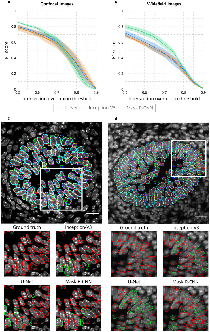
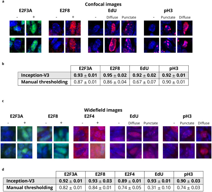
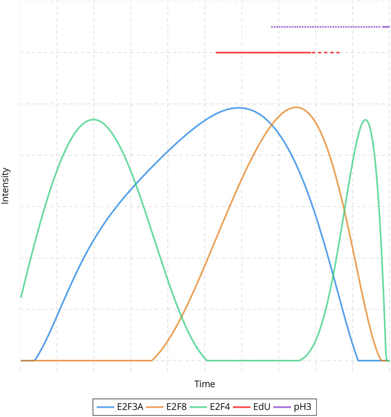
Automatic characterization of fluorescent labeling in intact mammalian tissues remains a challenge due to the lack of quantifying techniques capable of segregating densely packed nuclei and intricate tissue patterns. Here, we describe a powerful deep learning-based approach that couples remarkably precise nuclear segmentation with quantitation of fluorescent labeling intensity within segmented nuclei, and then apply it to the analysis of cell cycle dependent protein concentration in mouse tissues using 2D fluorescent still images. First, several existing deep learning-based methods were evaluated to accurately segment nuclei using different imaging modalities with a small training dataset. Next, we developed a deep learning-based approach to identify and measure fluorescent labels within segmented nuclei, and created an ImageJ plugin to allow for efficient manual correction of nuclear segmentation and label identification. Lastly, using fluorescence intensity as a readout for protein concentration, a three-step global estimation method was applied to the characterization of the cell cycle dependent expression of E2F proteins in the developing mouse intestine.
由于缺乏能够分离密集核和复杂组织模式的定量技术,完整哺乳动物组织中荧光标记的自动特征化仍然是一个挑战。在这里,我们描述了一种强大的基于深度学习的方法,该方法将核的精确分割与分割核内荧光标记强度的定量相结合,并将其应用于使用 2D 荧光静态图像分析小鼠组织中细胞周期依赖性蛋白浓度。首先,使用不同的成像模式和小的训练数据集评估了几种现有的基于深度学习的方法,以准确分割细胞核。接下来,我们开发了一种基于深度学习的方法来识别和测量分割核内的荧光标记,并创建了一个 ImageJ 插件,以允许对核分割和标记识别进行有效的手动校正。最后,使用荧光强度作为蛋白浓度的读出值,应用三步全局估计方法对发育中小鼠肠中 E2F 蛋白的细胞周期依赖性表达进行了特征描述。