• 文献检索
  • 文档翻译
  • 深度研究
  • 学术资讯
  • Suppr Zotero 插件Zotero 插件
  • 邀请有礼
  • 套餐&价格
  • 历史记录
应用&插件
Suppr Zotero 插件Zotero 插件浏览器插件Mac 客户端Windows 客户端微信小程序
定价
高级版会员购买积分包购买API积分包
服务
文献检索文档翻译深度研究API 文档MCP 服务
关于我们
关于 Suppr公司介绍联系我们用户协议隐私条款
关注我们

Suppr 超能文献

核心技术专利:CN118964589B侵权必究
粤ICP备2023148730 号-1Suppr @ 2026

文献检索

告别复杂PubMed语法,用中文像聊天一样搜索,搜遍4000万医学文献。AI智能推荐,让科研检索更轻松。

立即免费搜索

文件翻译

保留排版,准确专业,支持PDF/Word/PPT等文件格式,支持 12+语言互译。

免费翻译文档

深度研究

AI帮你快速写综述,25分钟生成高质量综述,智能提取关键信息,辅助科研写作。

立即免费体验

深度学习策略在荧光图像中细胞核分割的评估。

Evaluation of Deep Learning Strategies for Nucleus Segmentation in Fluorescence Images.

机构信息

Imaging Platform, Broad Institute of MIT and Harvard, Cambridge, Massachusetts.

Institute of Computational Biology, German Research Center for Environmental Health, Munich, Germany.

出版信息

Cytometry A. 2019 Sep;95(9):952-965. doi: 10.1002/cyto.a.23863. Epub 2019 Jul 16.

DOI:10.1002/cyto.a.23863
PMID:31313519
原文链接:https://pmc.ncbi.nlm.nih.gov/articles/PMC6771982/
Abstract

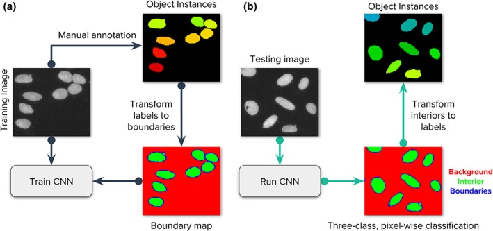
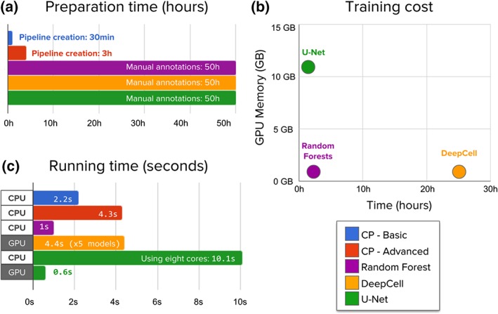
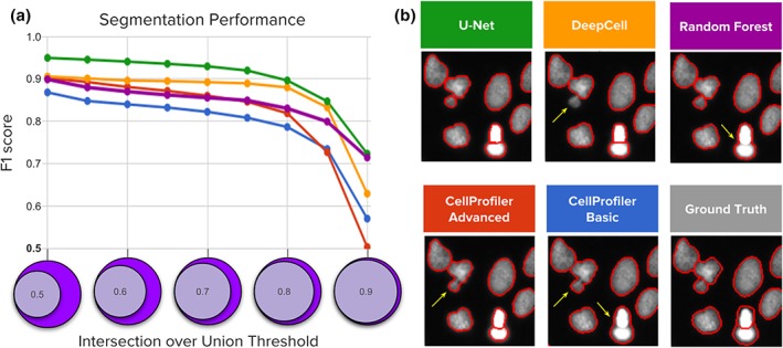
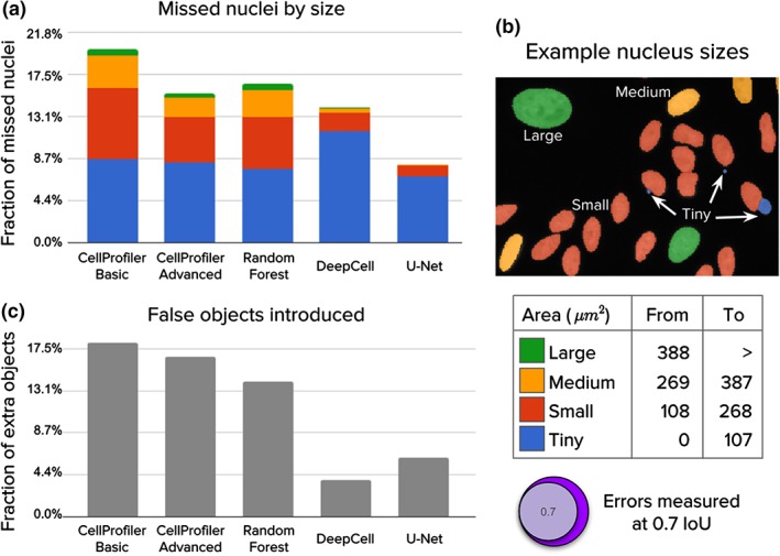
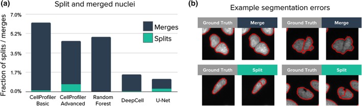
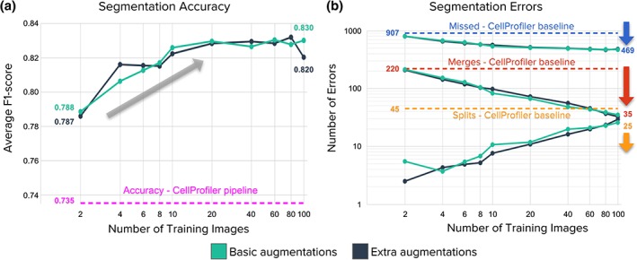
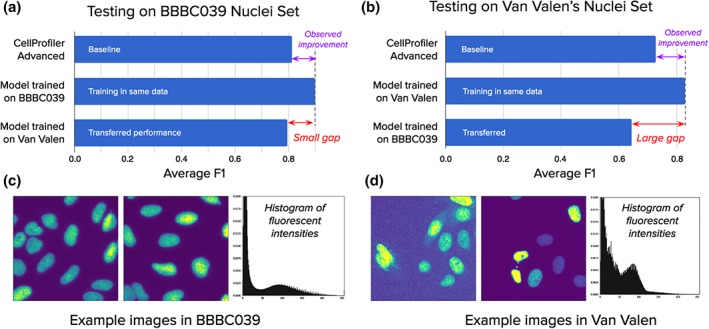
Identifying nuclei is often a critical first step in analyzing microscopy images of cells and classical image processing algorithms are most commonly used for this task. Recent developments in deep learning can yield superior accuracy, but typical evaluation metrics for nucleus segmentation do not satisfactorily capture error modes that are relevant in cellular images. We present an evaluation framework to measure accuracy, types of errors, and computational efficiency; and use it to compare deep learning strategies and classical approaches. We publicly release a set of 23,165 manually annotated nuclei and source code to reproduce experiments and run the proposed evaluation methodology. Our evaluation framework shows that deep learning improves accuracy and can reduce the number of biologically relevant errors by half. © 2019 The Authors. Cytometry Part A published by Wiley Periodicals, Inc. on behalf of International Society for Advancement of Cytometry.

摘要

识别细胞核通常是分析细胞显微镜图像的关键第一步,为此,经典的图像处理算法最为常用。深度学习的最新发展可以带来更高的准确性,但细胞核分割的典型评估指标并不能令人满意地捕捉到细胞图像中相关的错误模式。我们提出了一个评估框架来衡量准确性、错误类型和计算效率,并使用它来比较深度学习策略和经典方法。我们公开了一组 23165 个手动标注的细胞核,并提供了源代码以重现实验和运行所提出的评估方法。我们的评估框架表明,深度学习可以提高准确性,并将生物学相关错误的数量减少一半。 2019 年,作者。细胞计量学 A 版由 Wiley 期刊出版,代表国际细胞计量学促进协会。

https://cdn.ncbi.nlm.nih.gov/pmc/blobs/914b/6771982/a5ac596dd525/CYTO-95-952-g007.jpg
https://cdn.ncbi.nlm.nih.gov/pmc/blobs/914b/6771982/b6a1dbd80e19/CYTO-95-952-g001.jpg
https://cdn.ncbi.nlm.nih.gov/pmc/blobs/914b/6771982/e3b35cbee8db/CYTO-95-952-g002.jpg
https://cdn.ncbi.nlm.nih.gov/pmc/blobs/914b/6771982/def327050934/CYTO-95-952-g003.jpg
https://cdn.ncbi.nlm.nih.gov/pmc/blobs/914b/6771982/224eefc416cf/CYTO-95-952-g004.jpg
https://cdn.ncbi.nlm.nih.gov/pmc/blobs/914b/6771982/ec0bf4ee44b2/CYTO-95-952-g005.jpg
https://cdn.ncbi.nlm.nih.gov/pmc/blobs/914b/6771982/48c650a49afd/CYTO-95-952-g006.jpg
https://cdn.ncbi.nlm.nih.gov/pmc/blobs/914b/6771982/a5ac596dd525/CYTO-95-952-g007.jpg

相似文献

1
Evaluation of Deep Learning Strategies for Nucleus Segmentation in Fluorescence Images.深度学习策略在荧光图像中细胞核分割的评估。
Cytometry A. 2019 Sep;95(9):952-965. doi: 10.1002/cyto.a.23863. Epub 2019 Jul 16.
2
Deep Learning in Image Cytometry: A Review.深度学习在图像细胞检测中的应用综述。
Cytometry A. 2019 Apr;95(4):366-380. doi: 10.1002/cyto.a.23701. Epub 2018 Dec 19.
3
Improved automatic detection and segmentation of cell nuclei in histopathology images.改进组织病理学图像中细胞核的自动检测和分割。
IEEE Trans Biomed Eng. 2010 Apr;57(4):841-52. doi: 10.1109/TBME.2009.2035102. Epub 2009 Oct 30.
4
Nucleus segmentation across imaging experiments: the 2018 Data Science Bowl.跨影像实验的核分割:2018 年数据科学竞赛
Nat Methods. 2019 Dec;16(12):1247-1253. doi: 10.1038/s41592-019-0612-7. Epub 2019 Oct 21.
5
A Deep Learning Pipeline for Nucleus Segmentation.一种用于细胞核分割的深度学习管道。
Cytometry A. 2020 Dec;97(12):1248-1264. doi: 10.1002/cyto.a.24257. Epub 2020 Nov 19.
6
NuSeT: A deep learning tool for reliably separating and analyzing crowded cells.NuSeT:一种可靠分离和分析拥挤细胞的深度学习工具。
PLoS Comput Biol. 2020 Sep 14;16(9):e1008193. doi: 10.1371/journal.pcbi.1008193. eCollection 2020 Sep.
7
Attributed relational graphs for cell nucleus segmentation in fluorescence microscopy images.荧光显微镜图像中细胞核分割的归因关系图。
IEEE Trans Med Imaging. 2013 Jun;32(6):1121-31. doi: 10.1109/TMI.2013.2255309. Epub 2013 Mar 28.
8
Generic Isolated Cell Image Generator.通用细胞图像生成器。
Cytometry A. 2019 Nov;95(11):1198-1206. doi: 10.1002/cyto.a.23899. Epub 2019 Oct 8.
9
An Integrative Segmentation Framework for Cell Nucleus of Fluorescence Microscopy.荧光显微镜下细胞细胞核的综合分割框架。
Genes (Basel). 2022 Feb 26;13(3):431. doi: 10.3390/genes13030431.
10
Automatic three-dimensional segmentation of mouse embryonic stem cell nuclei by utilising multiple channels of confocal fluorescence images.利用共聚焦荧光图像的多个通道自动对小鼠胚胎干细胞核进行三维分割。
J Microsc. 2021 Jan;281(1):57-75. doi: 10.1111/jmi.12949. Epub 2020 Aug 8.

引用本文的文献

1
CellBinDB: a large-scale multimodal annotated dataset for cell segmentation with benchmarking of universal models.CellBinDB:一个用于细胞分割的大规模多模态注释数据集及通用模型基准测试
Gigascience. 2025 Jan 6;14. doi: 10.1093/gigascience/giaf069.
2
Image-based identification and isolation of micronucleated cells to dissect cellular consequences.基于图像的微核细胞识别与分离以剖析细胞后果。
Elife. 2025 Jun 2;13:RP101579. doi: 10.7554/eLife.101579.
3
Quantitative benchmarking of nuclear segmentation algorithms in multiplexed immunofluorescence imaging for translational studies.

本文引用的文献

1
U-Net: deep learning for cell counting, detection, and morphometry.U-Net:用于细胞计数、检测和形态测量学的深度学习。
Nat Methods. 2019 Jan;16(1):67-70. doi: 10.1038/s41592-018-0261-2. Epub 2018 Dec 17.
2
A Structured Tumor-Immune Microenvironment in Triple Negative Breast Cancer Revealed by Multiplexed Ion Beam Imaging.多重离子束成像揭示三阴性乳腺癌中的结构化肿瘤免疫微环境。
Cell. 2018 Sep 6;174(6):1373-1387.e19. doi: 10.1016/j.cell.2018.08.039.
3
Quanti.us: a tool for rapid, flexible, crowd-based annotation of images.Quanti.us:一种用于快速、灵活、基于众包的图像标注工具。
用于转化研究的多重免疫荧光成像中核分割算法的定量基准测试
Commun Biol. 2025 May 30;8(1):836. doi: 10.1038/s42003-025-08184-8.
4
Sensitive and Adaptable Turn-On Maturation (ATOM) Fluorescent Biosensors for Detecting Subcellular Localization of Protein Targets in Cells.用于检测细胞中蛋白质靶点亚细胞定位的灵敏且适应性开启成熟(ATOM)荧光生物传感器。
Bio Protoc. 2025 Mar 20;15(6):e5239. doi: 10.21769/BioProtoc.5239.
5
Image2Reg: Linking chromatin images to gene regulation using genetic and chemical perturbation screens.Image2Reg:利用遗传和化学扰动筛选将染色质图像与基因调控联系起来。
Cell Syst. 2025 Jun 18;16(6):101293. doi: 10.1016/j.cels.2025.101293. Epub 2025 May 12.
6
Riemannian Manifolds for Biological Imaging Applications Based on Unsupervised Learning.基于无监督学习的生物成像应用黎曼流形
J Imaging. 2025 Mar 29;11(4):103. doi: 10.3390/jimaging11040103.
7
micronuclAI enables automated quantification of micronuclei for assessment of chromosomal instability.微核人工智能可实现微核的自动定量分析,以评估染色体不稳定性。
Commun Biol. 2025 Mar 4;8(1):361. doi: 10.1038/s42003-025-07796-4.
8
State-of-the-Art Deep Learning Methods for Microscopic Image Segmentation: Applications to Cells, Nuclei, and Tissues.用于微观图像分割的前沿深度学习方法:在细胞、细胞核和组织中的应用
J Imaging. 2024 Dec 6;10(12):311. doi: 10.3390/jimaging10120311.
9
AneRBC dataset: a benchmark dataset for computer-aided anemia diagnosis using RBC images.AneRBC数据集:一个用于使用红细胞图像进行计算机辅助贫血诊断的基准数据集。
Database (Oxford). 2024 Dec 25;2024. doi: 10.1093/database/baae120.
10
Autofluorescence Virtual Staining System for H&E Histology and Multiplex Immunofluorescence Applied to Immuno-Oncology Biomarkers in Lung Cancer.用于苏木精-伊红(H&E)组织学和多重免疫荧光的自发荧光虚拟染色系统在肺癌免疫肿瘤生物标志物中的应用
Cancer Res Commun. 2025 Jan 1;5(1):54-65. doi: 10.1158/2767-9764.CRC-24-0327.
Nat Methods. 2018 Aug;15(8):587-590. doi: 10.1038/s41592-018-0069-0. Epub 2018 Jul 31.
4
CellProfiler 3.0: Next-generation image processing for biology.CellProfiler 3.0:生物学的下一代图像处理。
PLoS Biol. 2018 Jul 3;16(7):e2005970. doi: 10.1371/journal.pbio.2005970. eCollection 2018 Jul.
5
An objective comparison of cell-tracking algorithms.细胞追踪算法的客观比较。
Nat Methods. 2017 Dec;14(12):1141-1152. doi: 10.1038/nmeth.4473. Epub 2017 Oct 30.
6
The Image Data Resource: A Bioimage Data Integration and Publication Platform.图像数据资源:一个生物图像数据整合与发布平台。
Nat Methods. 2017 Aug;14(8):775-781. doi: 10.1038/nmeth.4326. Epub 2017 Jun 19.
7
Mitotic progression following DNA damage enables pattern recognition within micronuclei.DNA损伤后的有丝分裂进程能够实现微核内的模式识别。
Nature. 2017 Aug 24;548(7668):466-470. doi: 10.1038/nature23470. Epub 2017 Jul 31.
8
Fate of micronuclei and micronucleated cells.微核和有微核的细胞的命运。
Mutat Res Rev Mutat Res. 2017 Jan-Mar;771:85-98. doi: 10.1016/j.mrrev.2017.02.002. Epub 2017 Feb 13.
9
Selective Y centromere inactivation triggers chromosome shattering in micronuclei and repair by non-homologous end joining.选择性Y着丝粒失活引发微核中的染色体破碎并通过非同源末端连接进行修复。
Nat Cell Biol. 2017 Jan;19(1):68-75. doi: 10.1038/ncb3450. Epub 2016 Dec 5.
10
Deep Learning Automates the Quantitative Analysis of Individual Cells in Live-Cell Imaging Experiments.深度学习实现了活细胞成像实验中单个细胞定量分析的自动化。
PLoS Comput Biol. 2016 Nov 4;12(11):e1005177. doi: 10.1371/journal.pcbi.1005177. eCollection 2016 Nov.