Department of Thoracic Surgery, University of Pennsylvania Perelman School of Medicine, Philadelphia, PA, USA.
Department of Pathology, University of Pennsylvania Perelman School of Medicine, Philadelphia, PA, USA.
Mol Imaging Biol. 2023 Feb;25(1):156-167. doi: 10.1007/s11307-021-01699-6. Epub 2022 Mar 15.
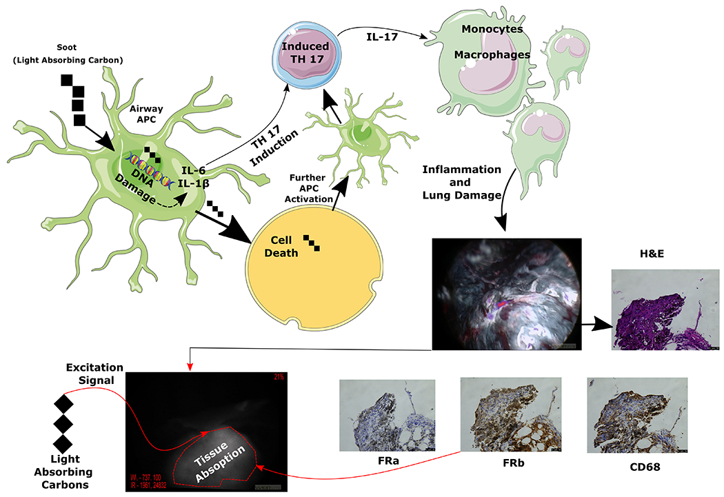
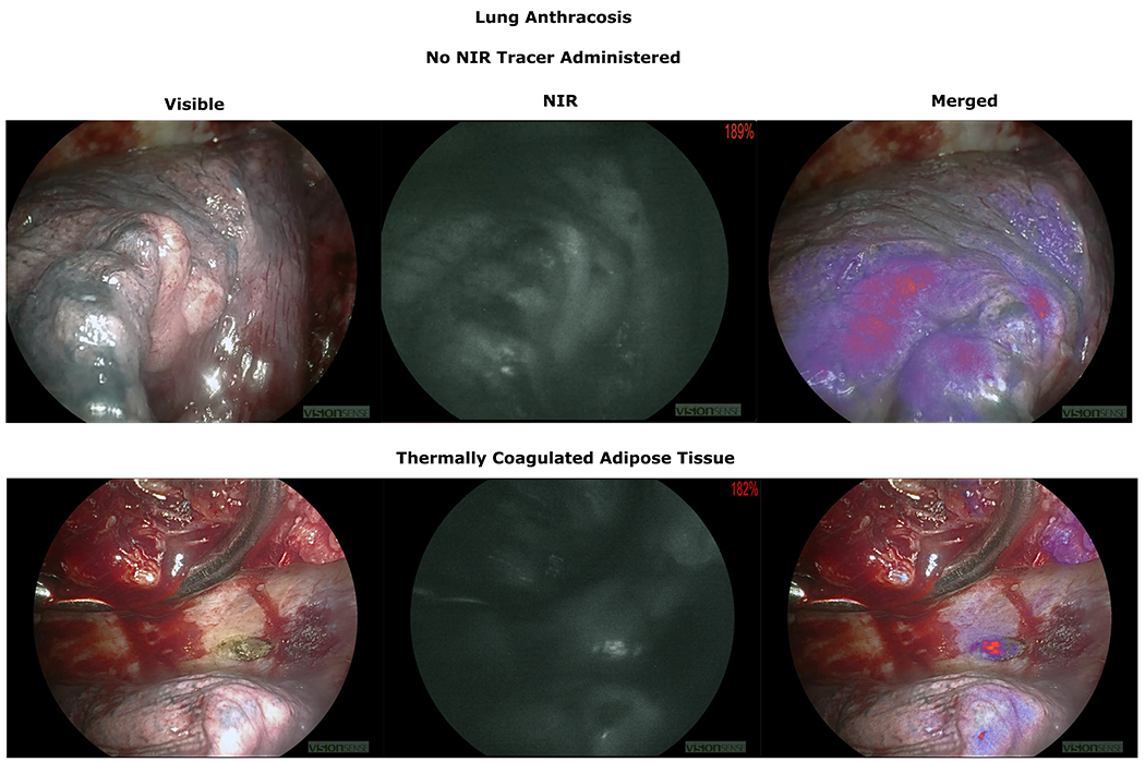
One of the novel advancements to enhance the visual aspects of lung cancer identification is intraoperative molecular imaging (IMI), which can reliably detect tumors that would otherwise be missed by standard techniques such as tactile and visual feedback, particularly for sub-centimeter or ground-glass nodules. However, there remains a subset of patients who do not benefit from IMI due to excessive background fluorescence secondary to parenchymal light-absorbing carbon deposition. Our goal was to identify the effects of these carbonaceous materials on the quality of IMI-guided lung cancer resections.
Between July 2014 and May 2021, a total of 311 patients were included in the study. Patients underwent infusion of the study drug OTL38 or ICG up to 24 h prior to VATS for lung cancer. Several factors such as age, tumor subtype, PET SUV, smoking, demographics, chronic lung conditions, patient domicile, and anthracosis were analyzed with respect to lung fluorescence during IMI. P values < 0.05 were considered statistically significant.
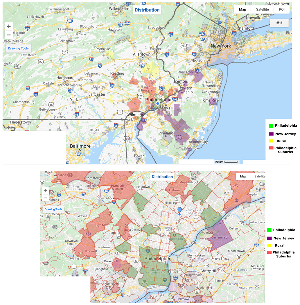
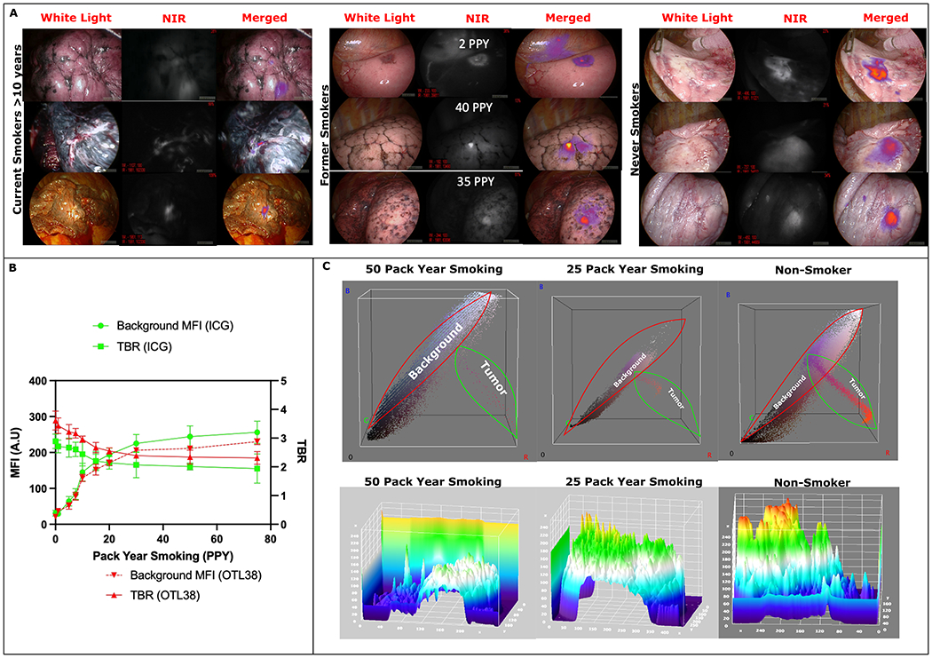
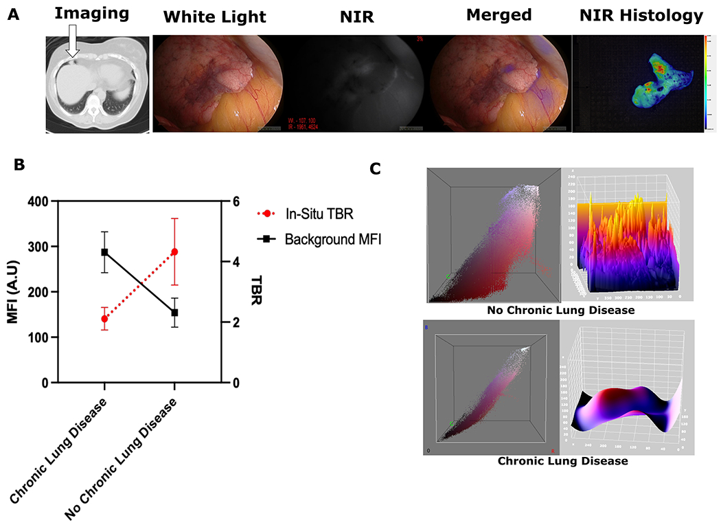
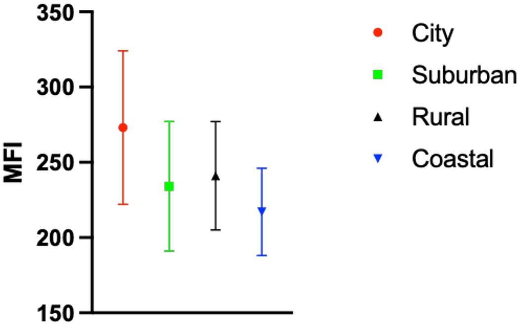
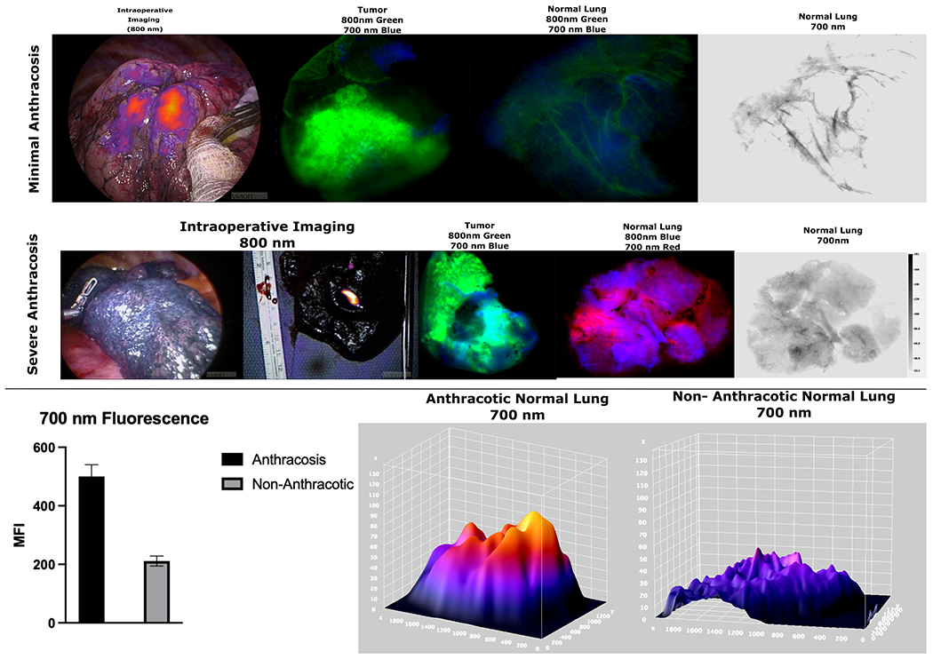
Variables such as age, sex, and race had no statistical correlation to IMI success. However, smoking status and pack year had a statistically significant correlation with background parenchymal fluorescence and lung inflammation (p < 0.05). MFI of background (lung parenchyma) correlated with smoking history (p < 0.05) which led to decreased tumor-to-background ratio (TBR) measurements for all patients with proven malignancy (p < 0.05). Patients with chronic lung disease appear to have increased background parenchymal fluorescence regardless of smoking history (287 vs. 154, p < 0.01). City dwellers compared to other groups appear to be exposed to higher pollutant load and have higher rates of anthracosis, but living location's impact on fluorescence quantification appears to be not statistically significant.
Smokers with greater than 10 PPY and those with chronic lung disease appear to have decreased lesion-to-background discrimination, significant anthracosis, and reduced IMI efficacy secondary to light-absorbing carbon deposition.
增强肺癌识别的视觉效果的新进展之一是术中分子成像(IMI),它可以可靠地检测到标准技术(如触觉和视觉反馈)可能错过的肿瘤,特别是对于亚厘米或磨玻璃结节。然而,仍有一部分患者由于实质性光吸收碳沉积引起的背景荧光过度而不能从 IMI 中受益。我们的目标是确定这些含碳物质对 IMI 引导下肺癌切除术质量的影响。
2014 年 7 月至 2021 年 5 月,共有 311 例患者纳入研究。患者在 VATS 前 24 小时内接受研究药物 OTL38 或 ICG 输注。分析了年龄、肿瘤亚型、PET SUV、吸烟、人口统计学、慢性肺部疾病、患者住所和炭沉着等因素与 IMI 期间肺部荧光之间的关系。p 值<0.05 被认为具有统计学意义。
年龄、性别和种族等变量与 IMI 成功无统计学相关性。然而,吸烟状况和吸烟年数与背景实质性荧光和肺部炎症有统计学显著相关性(p<0.05)。背景(肺实质)的 MFI 与吸烟史相关(p<0.05),这导致所有经证实患有恶性肿瘤的患者的肿瘤与背景比(TBR)测量值降低(p<0.05)。无论吸烟史如何,患有慢性肺部疾病的患者似乎都有更高的背景实质性荧光(287 比 154,p<0.01)。与其他群体相比,城市居民似乎暴露于更高的污染物负荷下,炭沉着的发生率更高,但居住地对荧光定量的影响似乎没有统计学意义。
吸烟量大于 10 PPY 的吸烟者和患有慢性肺部疾病的吸烟者似乎存在病变与背景的辨别能力下降、明显的炭沉着和由于光吸收碳沉积导致的 IMI 效果降低。