• 文献检索
  • 文档翻译
  • 深度研究
  • 学术资讯
  • Suppr Zotero 插件Zotero 插件
  • 邀请有礼
  • 套餐&价格
  • 历史记录
应用&插件
Suppr Zotero 插件Zotero 插件浏览器插件Mac 客户端Windows 客户端微信小程序
定价
高级版会员购买积分包购买API积分包
服务
文献检索文档翻译深度研究API 文档MCP 服务
关于我们
关于 Suppr公司介绍联系我们用户协议隐私条款
关注我们

Suppr 超能文献

核心技术专利:CN118964589B侵权必究
粤ICP备2023148730 号-1Suppr @ 2026

文献检索

告别复杂PubMed语法,用中文像聊天一样搜索,搜遍4000万医学文献。AI智能推荐,让科研检索更轻松。

立即免费搜索

文件翻译

保留排版,准确专业,支持PDF/Word/PPT等文件格式,支持 12+语言互译。

免费翻译文档

深度研究

AI帮你快速写综述,25分钟生成高质量综述,智能提取关键信息,辅助科研写作。

立即免费体验

周细胞的多种亚型及其在脊髓损伤修复、骨骼发育和修复中的作用。

Versatile subtypes of pericytes and their roles in spinal cord injury repair, bone development and repair.

作者信息

Zhu Sipin, Chen Min, Ying Yibo, Wu Qiuji, Huang Zhiyang, Ni Wenfei, Wang Xiangyang, Xu Huazi, Bennett Samuel, Xiao Jian, Xu Jiake

机构信息

Department of Orthopaedics, The Second Affiliated Hospital and Yuying Children's Hospital of Wenzhou Medical University, Wenzhou, Zhejiang, 325000, China.

Molecular Pharmacology Research Centre, School of Pharmaceutical Sciences, Wenzhou Medical University, Wenzhou, Zhejiang, 325035, China.

出版信息

Bone Res. 2022 Mar 16;10(1):30. doi: 10.1038/s41413-022-00203-2.

DOI:10.1038/s41413-022-00203-2
PMID:35296645
原文链接:https://pmc.ncbi.nlm.nih.gov/articles/PMC8927336/
Abstract

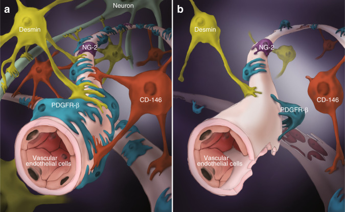
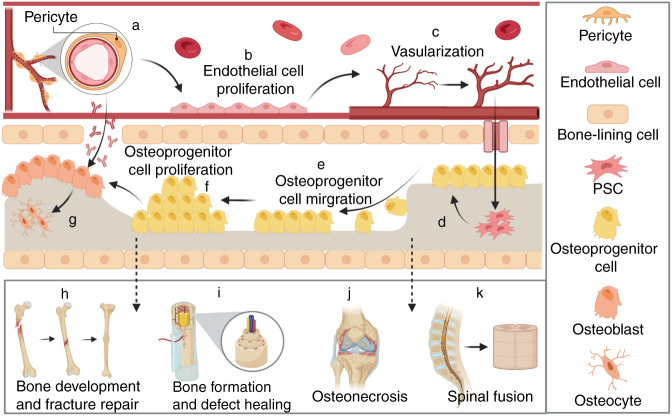
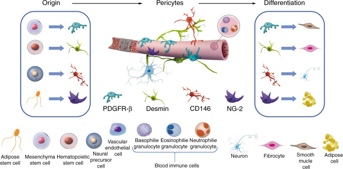
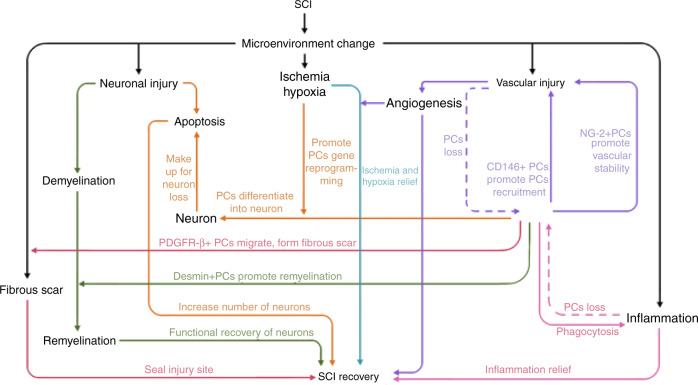
Vascular regeneration is a challenging topic in tissue repair. As one of the important components of the neurovascular unit (NVU), pericytes play an essential role in the maintenance of the vascular network of the spinal cord. To date, subtypes of pericytes have been identified by various markers, namely the PDGFR-β, Desmin, CD146, and NG2, each of which is involved with spinal cord injury (SCI) repair. In addition, pericytes may act as a stem cell source that is important for bone development and regeneration, whilst specific subtypes of pericyte could facilitate bone fracture and defect repair. One of the major challenges of pericyte biology is to determine the specific markers that would clearly distinguish the different subtypes of pericytes, and to develop efficient approaches to isolate and propagate pericytes. In this review, we discuss the biology and roles of pericytes, their markers for identification, and cell differentiation capacity with a focus on the potential application in the treatment of SCI and bone diseases in orthopedics.

摘要

血管再生是组织修复中一个具有挑战性的课题。作为神经血管单元(NVU)的重要组成部分之一,周细胞在维持脊髓血管网络方面发挥着至关重要的作用。迄今为止,已通过各种标志物鉴定出周细胞的亚型,即血小板衍生生长因子受体β(PDGFR-β)、结蛋白、CD146和NG2,它们各自都与脊髓损伤(SCI)修复有关。此外,周细胞可能作为对骨骼发育和再生很重要的干细胞来源,而特定亚型的周细胞可促进骨折和骨缺损修复。周细胞生物学的主要挑战之一是确定能够清晰区分不同亚型周细胞的特异性标志物,并开发分离和扩增周细胞的有效方法。在这篇综述中,我们讨论周细胞的生物学特性和作用、其鉴定标志物以及细胞分化能力,重点关注其在治疗脊髓损伤和骨科骨疾病中的潜在应用。

https://cdn.ncbi.nlm.nih.gov/pmc/blobs/9d43/8927336/b5ddbb2c1755/41413_2022_203_Fig4_HTML.jpg
https://cdn.ncbi.nlm.nih.gov/pmc/blobs/9d43/8927336/bc71ee6cc62d/41413_2022_203_Fig1_HTML.jpg
https://cdn.ncbi.nlm.nih.gov/pmc/blobs/9d43/8927336/8de993d20012/41413_2022_203_Fig2_HTML.jpg
https://cdn.ncbi.nlm.nih.gov/pmc/blobs/9d43/8927336/b0ee825ca12d/41413_2022_203_Fig3_HTML.jpg
https://cdn.ncbi.nlm.nih.gov/pmc/blobs/9d43/8927336/b5ddbb2c1755/41413_2022_203_Fig4_HTML.jpg

相似文献

1
Versatile subtypes of pericytes and their roles in spinal cord injury repair, bone development and repair.周细胞的多种亚型及其在脊髓损伤修复、骨骼发育和修复中的作用。
Bone Res. 2022 Mar 16;10(1):30. doi: 10.1038/s41413-022-00203-2.
2
Markers for human brain pericytes and smooth muscle cells.人脑周细胞和平滑肌细胞标志物。
J Chem Neuroanat. 2018 Oct;92:48-60. doi: 10.1016/j.jchemneu.2018.06.001. Epub 2018 Jun 7.
3
Proliferating NG2-Cell-Dependent Angiogenesis and Scar Formation Alter Axon Growth and Functional Recovery After Spinal Cord Injury in Mice.NG2 细胞依赖性血管生成和瘢痕形成促进小鼠脊髓损伤后的轴突生长和功能恢复。
J Neurosci. 2018 Feb 7;38(6):1366-1382. doi: 10.1523/JNEUROSCI.3953-16.2017. Epub 2017 Dec 26.
4
Not all MSCs can act as pericytes: functional in vitro assays to distinguish pericytes from other mesenchymal stem cells in angiogenesis.并非所有间充质干细胞都能作为周细胞发挥作用:在血管生成中体外鉴定周细胞和其他间充质干细胞功能的实验方法。
Stem Cells Dev. 2013 Sep 1;22(17):2347-55. doi: 10.1089/scd.2012.0415. Epub 2013 May 24.
5
Pericytes for the treatment of orthopedic conditions.用于治疗骨科疾病的周细胞。
Pharmacol Ther. 2017 Mar;171:93-103. doi: 10.1016/j.pharmthera.2016.08.003. Epub 2016 Aug 7.
6
Pericytes in Brain Injury and Repair After Ischemic Stroke.缺血性脑卒中后脑损伤与修复中的周细胞
Transl Stroke Res. 2017 Apr;8(2):107-121. doi: 10.1007/s12975-016-0504-4. Epub 2016 Nov 12.
7
CD133 expressing pericytes and relationship to SDF-1 and CXCR4 in spinal cord injury.CD133 阳性的周细胞与脊髓损伤中 SDF-1 和 CXCR4 的关系。
Curr Neurovasc Res. 2010 May;7(2):144-54. doi: 10.2174/156720210791184907.
8
Pericytes as a Source of Osteogenic Cells in Bone Fracture Healing.周细胞作为骨骨折愈合中成骨细胞的来源。
Int J Mol Sci. 2019 Mar 2;20(5):1079. doi: 10.3390/ijms20051079.
9
Bone Mesenchymal Stem Cell-Derived Extracellular Vesicles Promote Recovery Following Spinal Cord Injury via Improvement of the Integrity of the Blood-Spinal Cord Barrier.骨髓间充质干细胞衍生的细胞外囊泡通过改善血脊髓屏障的完整性促进脊髓损伤后的恢复。
Front Neurosci. 2019 Mar 12;13:209. doi: 10.3389/fnins.2019.00209. eCollection 2019.
10
Exogenous bone marrow derived-putative endothelial progenitor cells attenuate ischemia reperfusion-induced vascular injury and renal fibrosis in mice dependent on pericytes.外源性骨髓来源的拟血管内皮祖细胞通过周细胞依赖途径减轻小鼠缺血再灌注诱导的血管损伤和肾纤维化。
Theranostics. 2020 Oct 25;10(26):12144-12157. doi: 10.7150/thno.48562. eCollection 2020.

引用本文的文献

1
Polydopamine/Montmorillonite-Based Brushite Calcium Phosphate Cements: Synergize Mechanical and Osteogenic Capacity for Bone Regeneration.基于聚多巴胺/蒙脱石的透钙磷石磷酸钙骨水泥:增强骨再生的力学性能和成骨能力
Biomater Res. 2025 Jun 9;29:0213. doi: 10.34133/bmr.0213. eCollection 2025.
2
Generation and clearance of myelin debris after spinal cord injury.脊髓损伤后髓磷脂碎片的产生与清除
Neural Regen Res. 2026 Apr 1;21(4):1512-1527. doi: 10.4103/NRR.NRR-D-24-01405. Epub 2025 Apr 29.
3
Changes in vascular identity during vascular remodeling.

本文引用的文献

1
Extreme Diversity of the Human Vascular Mesenchymal Cell Landscape.人类血管间充质细胞景观的极端多样性。
J Am Heart Assoc. 2020 Dec;9(23):e017094. doi: 10.1161/JAHA.120.017094. Epub 2020 Nov 16.
2
Efficacy and safety of stem cell therapy for the early-stage osteonecrosis of femoral head: a systematic review and meta-analysis of randomized controlled trials.干细胞治疗早期股骨头坏死的疗效和安全性:系统评价和随机对照试验的荟萃分析。
Stem Cell Res Ther. 2020 Oct 19;11(1):445. doi: 10.1186/s13287-020-01956-5.
3
KLF4 (Kruppel-Like Factor 4)-Dependent Perivascular Plasticity Contributes to Adipose Tissue inflammation.
血管重塑过程中血管特性的变化。
JVS Vasc Sci. 2025 Feb 25;6:100282. doi: 10.1016/j.jvssci.2025.100282. eCollection 2025.
4
Single-Cell Multi-omics Assessment of Spinal Cord Injury Blocking via Cerium-doped Upconversion Antioxidant Nanoenzymes.通过铈掺杂上转换抗氧化纳米酶对脊髓损伤阻断进行单细胞多组学评估
Adv Sci (Weinh). 2025 Feb;12(8):e2412526. doi: 10.1002/advs.202412526. Epub 2025 Jan 9.
5
Physiological Insights Into the Role of Pericytes in Spinal Cord Injury.对周细胞在脊髓损伤中作用的生理学见解
J Cell Physiol. 2025 Jan;240(1):e31500. doi: 10.1002/jcp.31500.
6
Single-cell transcriptomic analysis of glioblastoma reveals pericytes contributing to the blood-brain-tumor barrier and tumor progression.胶质母细胞瘤的单细胞转录组分析揭示了周细胞对血脑肿瘤屏障和肿瘤进展的作用。
MedComm (2020). 2024 Dec 4;5(12):e70014. doi: 10.1002/mco2.70014. eCollection 2024 Dec.
7
FOXO1-mTOR pathway in vascular pericyte regulates the formation of type H vessels to control bone metabolism.血管周细胞中的FOXO1-mTOR信号通路调控H型血管的形成以控制骨代谢。
J Orthop Translat. 2024 Sep 17;49:246-263. doi: 10.1016/j.jot.2024.08.010. eCollection 2024 Nov.
8
Single-cell profiling of brain pericyte heterogeneity following ischemic stroke unveils distinct pericyte subtype-targeted neural reprogramming potential and its underlying mechanisms.脑周细胞在缺血性脑卒中后的异质性单细胞分析揭示了不同周细胞亚型的神经重编程潜力及其潜在机制。
Theranostics. 2024 Sep 23;14(16):6110-6137. doi: 10.7150/thno.97165. eCollection 2024.
9
Transcriptomic profiling of Schlemm's canal cells reveals a lymphatic-biased identity and three major cell states.对施莱姆氏管细胞的转录组谱分析揭示了其具有偏向于淋巴的特征,并存在三种主要的细胞状态。
Elife. 2024 Oct 18;13:RP96459. doi: 10.7554/eLife.96459.
10
SMAD3 mediates the specification of human induced pluripotent stem cell-derived epicardium into progenitors for the cardiac pericyte lineage.SMAD3 将人诱导多能干细胞衍生的心外膜特化为心脏周细胞谱系的祖细胞。
Stem Cell Reports. 2024 Oct 8;19(10):1399-1416. doi: 10.1016/j.stemcr.2024.08.008. Epub 2024 Sep 26.
KLF4(Kruppel 样因子 4)依赖性血管周细胞可塑性有助于脂肪组织炎症。
Arterioscler Thromb Vasc Biol. 2021 Jan;41(1):284-301. doi: 10.1161/ATVBAHA.120.314703. Epub 2020 Oct 15.
4
Comparison of skeletal and soft tissue pericytes identifies CXCR4 bone forming mural cells in human tissues.骨骼和软组织周细胞的比较确定了人类组织中形成骨的CXCR4壁细胞。
Bone Res. 2020 May 22;8(1):22. doi: 10.1038/s41413-020-0097-0. eCollection 2020.
5
Endothelial cells produce angiocrine factors to regulate bone and cartilage via versatile mechanisms.内皮细胞通过多种机制产生血管生成素以调节骨骼和软骨。
Theranostics. 2020 May 1;10(13):5957-5965. doi: 10.7150/thno.45422. eCollection 2020.
6
Autophagy modulation altered differentiation capacity of CD146 cells toward endothelial cells, pericytes, and cardiomyocytes.自噬调节改变了 CD146 细胞向血管内皮细胞、周细胞和心肌细胞分化的能力。
Stem Cell Res Ther. 2020 Mar 26;11(1):139. doi: 10.1186/s13287-020-01656-0.
7
Contribution of neural crest and GLAST Wnt1 bone marrow pericytes with liver fibrogenesis and/or regeneration.神经嵴和GLAST Wnt1骨髓周细胞对肝纤维化和/或再生的作用。
Liver Int. 2020 Apr;40(4):977-987. doi: 10.1111/liv.14401. Epub 2020 Feb 21.
8
Ninjurin 1 mediates peripheral nerve regeneration through Schwann cell maturation of NG2-positive cells.Ninjurin 1 通过 NG2 阳性细胞的施万细胞成熟来介导周围神经再生。
Biochem Biophys Res Commun. 2019 Nov 12;519(3):462-468. doi: 10.1016/j.bbrc.2019.09.007. Epub 2019 Sep 13.
9
Perivascular Mesenchymal Progenitors for Bone Regeneration.血管周间充质祖细胞在骨再生中的作用
J Orthop Res. 2019 Jun;37(6):1221-1228. doi: 10.1002/jor.24284. Epub 2019 May 17.
10
Stem cell therapy for treating osteonecrosis of the femoral head: From clinical applications to related basic research.干细胞治疗股骨头坏死:从临床应用到相关基础研究。
Stem Cell Res Ther. 2018 Oct 25;9(1):291. doi: 10.1186/s13287-018-1018-7.