• 文献检索
  • 文档翻译
  • 深度研究
  • 学术资讯
  • Suppr Zotero 插件Zotero 插件
  • 邀请有礼
  • 套餐&价格
  • 历史记录
应用&插件
Suppr Zotero 插件Zotero 插件浏览器插件Mac 客户端Windows 客户端微信小程序
定价
高级版会员购买积分包购买API积分包
服务
文献检索文档翻译深度研究API 文档MCP 服务
关于我们
关于 Suppr公司介绍联系我们用户协议隐私条款
关注我们

Suppr 超能文献

核心技术专利:CN118964589B侵权必究
粤ICP备2023148730 号-1Suppr @ 2026

文献检索

告别复杂PubMed语法,用中文像聊天一样搜索,搜遍4000万医学文献。AI智能推荐,让科研检索更轻松。

立即免费搜索

文件翻译

保留排版,准确专业,支持PDF/Word/PPT等文件格式,支持 12+语言互译。

免费翻译文档

深度研究

AI帮你快速写综述,25分钟生成高质量综述,智能提取关键信息,辅助科研写作。

立即免费体验

对施莱姆氏管细胞的转录组谱分析揭示了其具有偏向于淋巴的特征,并存在三种主要的细胞状态。

Transcriptomic profiling of Schlemm's canal cells reveals a lymphatic-biased identity and three major cell states.

机构信息

Department of Ophthalmology, Columbia University Irving Medical Center, New York, United States.

Department of Ophthalmology and Visual Sciences, The Ohio State University Medical Center, Columbus, United States.

出版信息

Elife. 2024 Oct 18;13:RP96459. doi: 10.7554/eLife.96459.

DOI:10.7554/eLife.96459
PMID:39422453
原文链接:https://pmc.ncbi.nlm.nih.gov/articles/PMC11488849/
Abstract

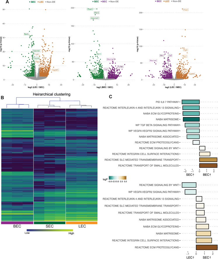
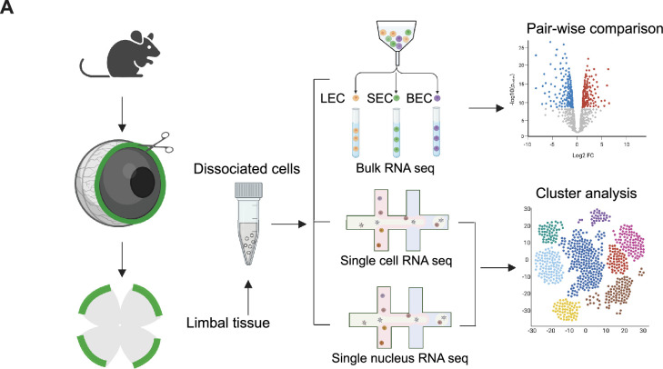
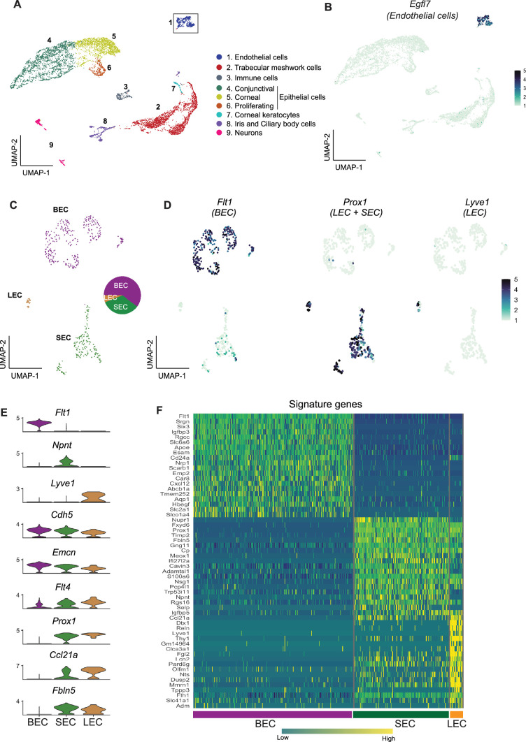
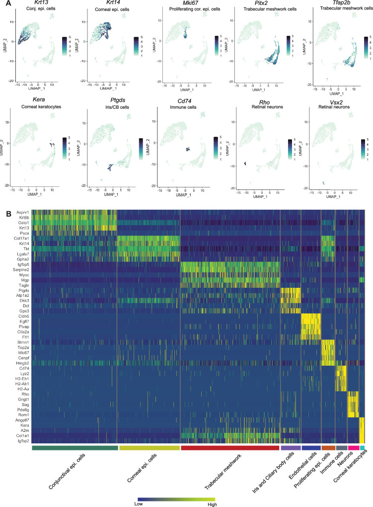
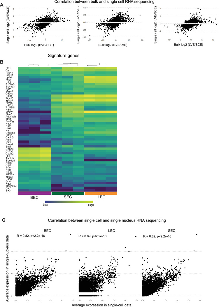
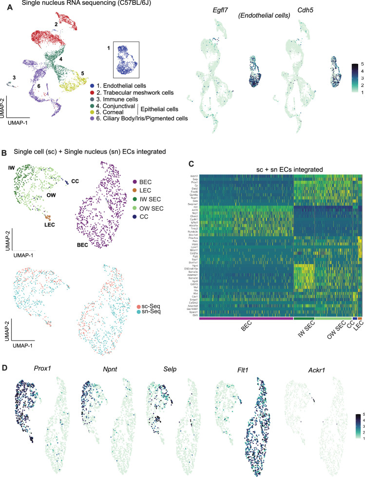
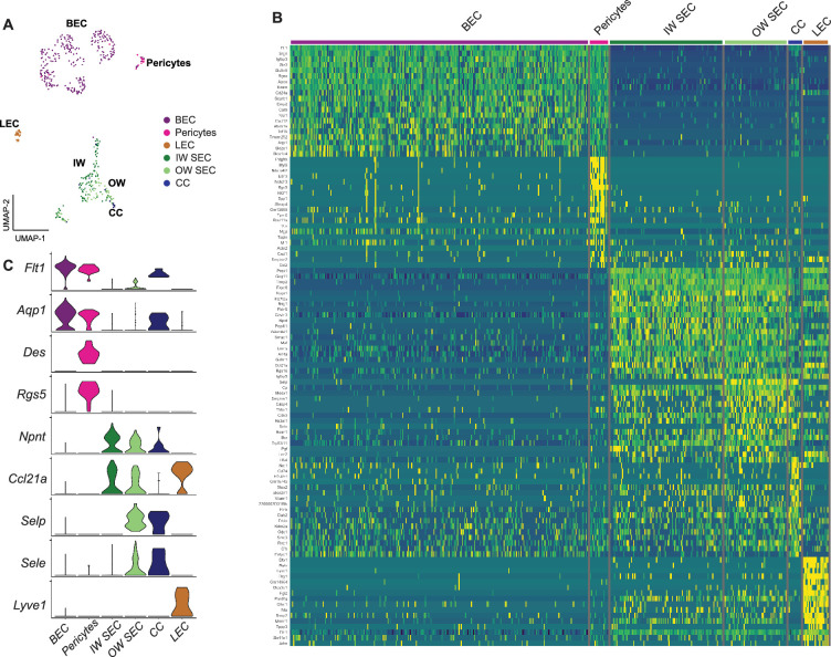
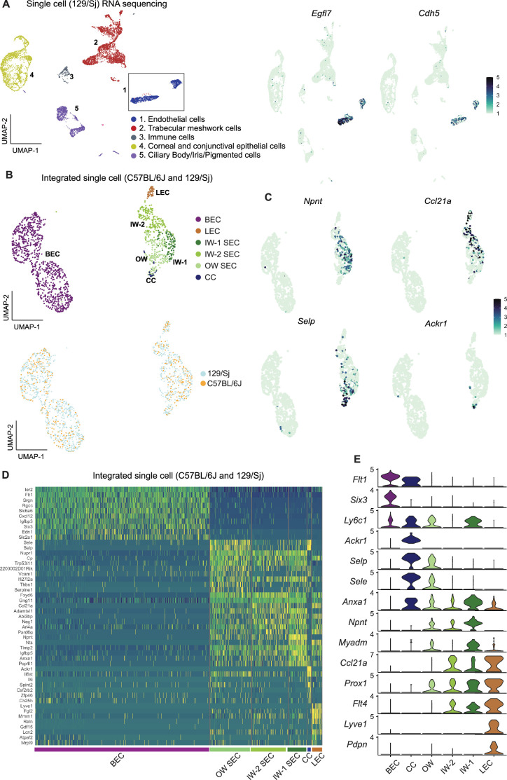
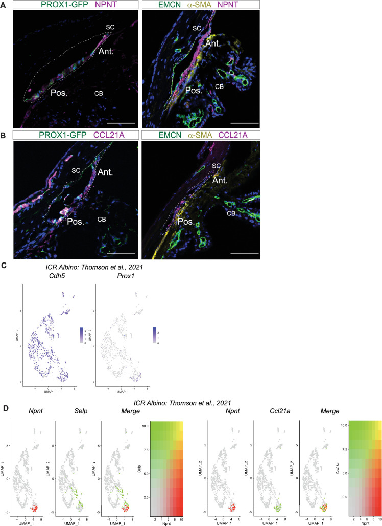
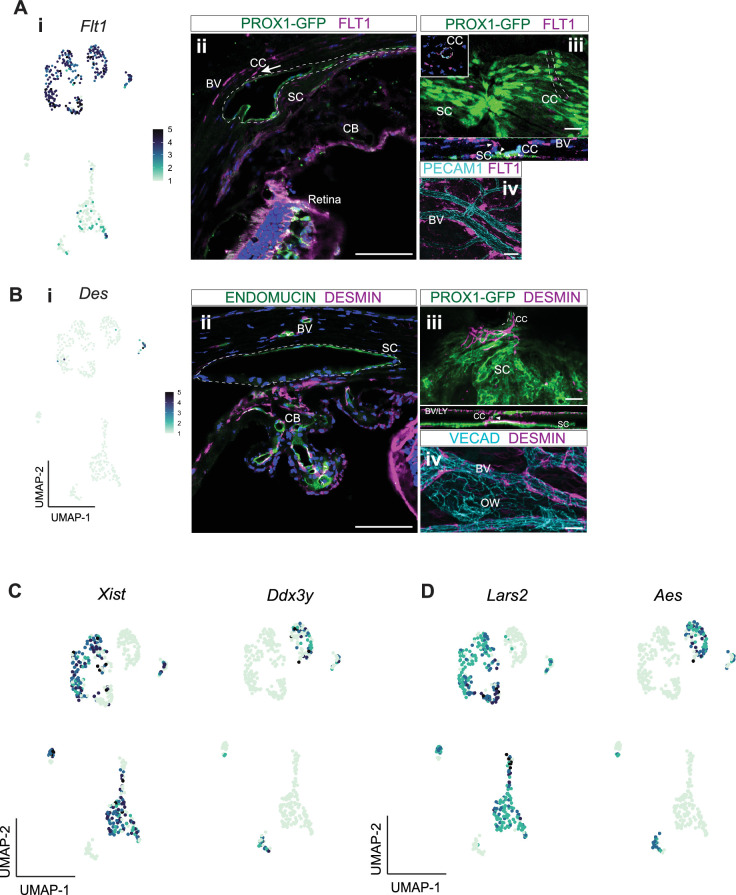
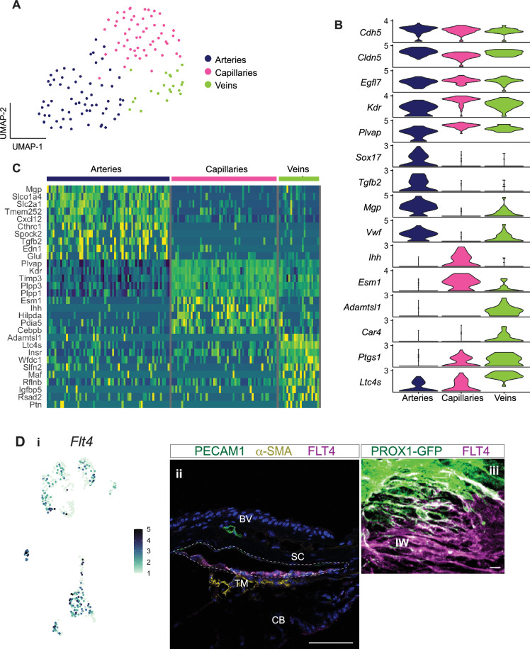
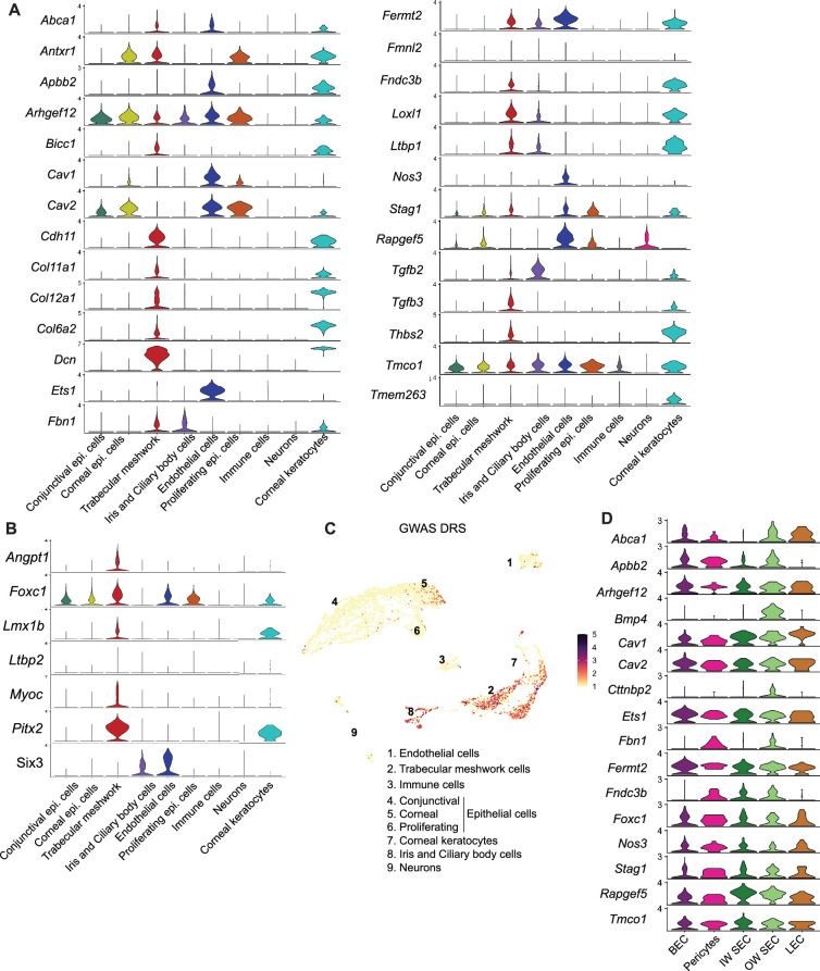
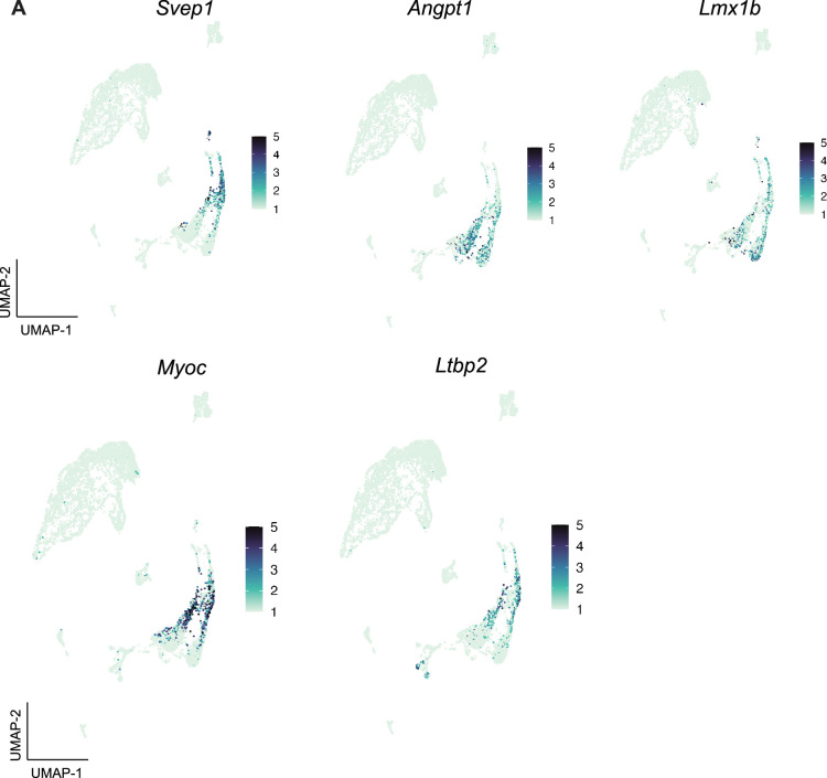
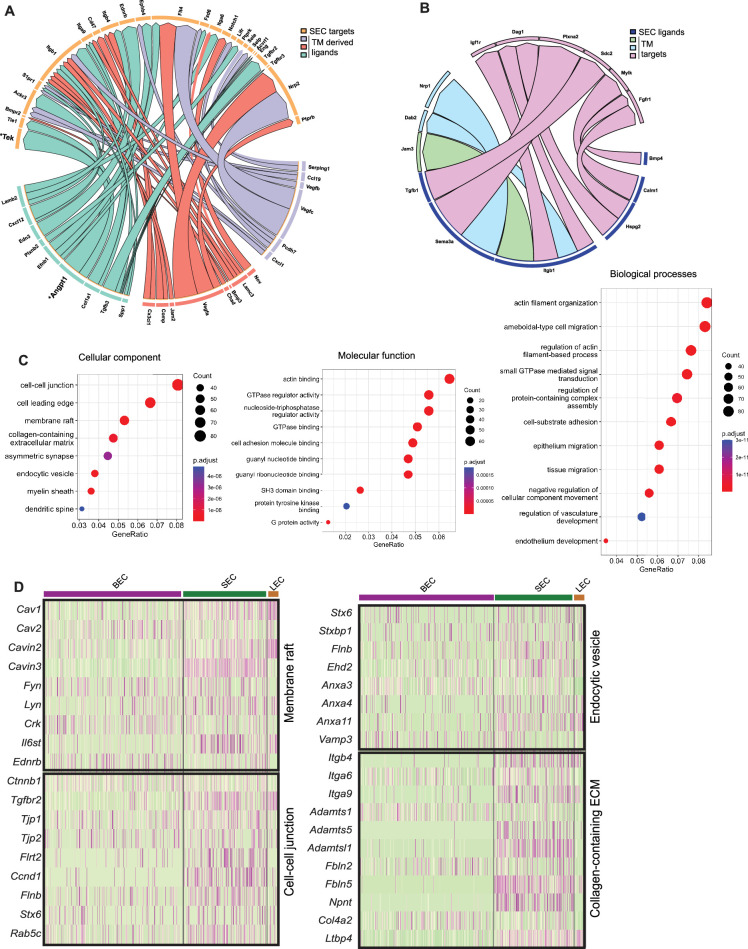
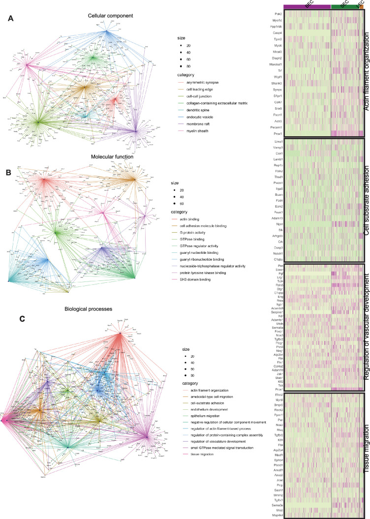
Schlemm's canal (SC) is central in intraocular pressure regulation but requires much characterization. It has distinct inner and outer walls, each composed of Schlemm's canal endothelial cells (SECs) with different morphologies and functions. Recent transcriptomic studies of the anterior segment added important knowledge, but were limited in power by SEC numbers or did not focus on SC. To gain a more comprehensive understanding of SC biology, we performed bulk RNA sequencing on C57BL/6 J SC, blood vessel, and lymphatic endothelial cells from limbal tissue (~4,500 SECs). We also analyzed mouse limbal tissues by single-cell and single-nucleus RNA sequencing (C57BL/6 J and 129/Sj strains), successfully sequencing 903 individual SECs. Together, these datasets confirm that SC has molecular characteristics of both blood and lymphatic endothelia with a lymphatic phenotype predominating. SECs are enriched in pathways that regulate cell-cell junction formation pointing to the importance of junctions in determining SC fluid permeability. Importantly, and for the first time, our analyses characterize three molecular classes of SECs, molecularly distinguishing inner wall from outer wall SECs and discovering two inner wall cell states that likely result from local environmental differences. Further, and based on ligand and receptor expression patterns, we document key interactions between SECs and cells of the adjacent trabecular meshwork (TM) drainage tissue. Also, we present cell type expression for a collection of human glaucoma genes. These data provide a new molecular foundation that will enable the functional dissection of key homeostatic processes mediated by SECs as well as the development of new glaucoma therapeutics.

摘要

施莱姆氏管 (SC) 是眼压调节的核心,但需要进一步的特征描述。它有明显的内、外壁,每一部分都由具有不同形态和功能的施莱姆氏管内皮细胞 (SECs) 组成。最近对前段的转录组学研究增加了重要的知识,但由于 SEC 的数量有限,或者没有关注 SC,因此其研究结果的说服力不足。为了更全面地了解 SC 的生物学特性,我们对 C57BL/6 J SC、血管和淋巴管内皮细胞进行了批量 RNA 测序,这些细胞来自角膜缘组织(~4500 个 SEC)。我们还通过单细胞和单核 RNA 测序(C57BL/6 J 和 129/Sj 品系)分析了小鼠角膜缘组织,成功地对 903 个单个 SEC 进行了测序。综合这些数据集,我们确认 SC 具有血液和淋巴管内皮的分子特征,以淋巴管表型为主。SECs 富含调节细胞-细胞连接形成的途径,这表明连接在决定 SC 流体渗透性方面非常重要。重要的是,也是首次,我们的分析对 3 种 SEC 分子类型进行了特征描述,从分子上区分了内、外壁 SEC,并发现了两种可能由局部环境差异引起的内壁细胞状态。此外,基于配体和受体表达模式,我们记录了 SEC 与相邻小梁网 (TM) 引流组织细胞之间的关键相互作用。同时,我们还为一系列人类青光眼基因提供了细胞类型表达数据。这些数据提供了一个新的分子基础,将使 SEC 介导的关键稳态过程的功能剖析以及新的青光眼治疗方法的开发成为可能。

https://cdn.ncbi.nlm.nih.gov/pmc/blobs/a5c0/11488849/5cc38197d6f8/elife-96459-sa3-fig1.jpg
https://cdn.ncbi.nlm.nih.gov/pmc/blobs/a5c0/11488849/216572422815/elife-96459-fig1.jpg
https://cdn.ncbi.nlm.nih.gov/pmc/blobs/a5c0/11488849/a31f4ed93b37/elife-96459-fig1-figsupp1.jpg
https://cdn.ncbi.nlm.nih.gov/pmc/blobs/a5c0/11488849/7af77500e7ec/elife-96459-fig2.jpg
https://cdn.ncbi.nlm.nih.gov/pmc/blobs/a5c0/11488849/b9746e9b216c/elife-96459-fig2-figsupp1.jpg
https://cdn.ncbi.nlm.nih.gov/pmc/blobs/a5c0/11488849/a52fc582e3af/elife-96459-fig2-figsupp2.jpg
https://cdn.ncbi.nlm.nih.gov/pmc/blobs/a5c0/11488849/29a2c7d70446/elife-96459-fig3.jpg
https://cdn.ncbi.nlm.nih.gov/pmc/blobs/a5c0/11488849/cba38f34e00f/elife-96459-fig3-figsupp1.jpg
https://cdn.ncbi.nlm.nih.gov/pmc/blobs/a5c0/11488849/79e71ba89aef/elife-96459-fig4.jpg
https://cdn.ncbi.nlm.nih.gov/pmc/blobs/a5c0/11488849/f0f723d864e5/elife-96459-fig5.jpg
https://cdn.ncbi.nlm.nih.gov/pmc/blobs/a5c0/11488849/3ceb36b8af90/elife-96459-fig5-figsupp1.jpg
https://cdn.ncbi.nlm.nih.gov/pmc/blobs/a5c0/11488849/2b805dcce608/elife-96459-fig5-figsupp2.jpg
https://cdn.ncbi.nlm.nih.gov/pmc/blobs/a5c0/11488849/52133a34d88c/elife-96459-fig5-figsupp3.jpg
https://cdn.ncbi.nlm.nih.gov/pmc/blobs/a5c0/11488849/c36aedd61fe0/elife-96459-fig6.jpg
https://cdn.ncbi.nlm.nih.gov/pmc/blobs/a5c0/11488849/3c70fe30ed05/elife-96459-fig6-figsupp1.jpg
https://cdn.ncbi.nlm.nih.gov/pmc/blobs/a5c0/11488849/ab0e30356b5b/elife-96459-fig7.jpg
https://cdn.ncbi.nlm.nih.gov/pmc/blobs/a5c0/11488849/506f02064ec3/elife-96459-fig7-figsupp1.jpg
https://cdn.ncbi.nlm.nih.gov/pmc/blobs/a5c0/11488849/b32584a43f63/elife-96459-fig7-figsupp2.jpg
https://cdn.ncbi.nlm.nih.gov/pmc/blobs/a5c0/11488849/5cc38197d6f8/elife-96459-sa3-fig1.jpg

相似文献

1
Transcriptomic profiling of Schlemm's canal cells reveals a lymphatic-biased identity and three major cell states.对施莱姆氏管细胞的转录组谱分析揭示了其具有偏向于淋巴的特征,并存在三种主要的细胞状态。
Elife. 2024 Oct 18;13:RP96459. doi: 10.7554/eLife.96459.
2
Transcriptomic profiling of Schlemm's canal cells reveals a lymphatic-biased identity and three major cell states.施莱姆管细胞的转录组分析揭示了一种偏向淋巴管的特征和三种主要细胞状态。
bioRxiv. 2024 Aug 6:2023.08.31.555823. doi: 10.1101/2023.08.31.555823.
3
Fibrotic Changes to Schlemm's Canal Endothelial Cells in Glaucoma.青光眼中海绵窦内皮细胞的纤维化改变。
Int J Mol Sci. 2021 Aug 31;22(17):9446. doi: 10.3390/ijms22179446.
4
Surface Engineering of FLT4-Targeted Nanocarriers Enhances Cell-Softening Glaucoma Therapy.靶向 FLT4 的纳米载体的表面工程增强了细胞软化青光眼治疗。
ACS Appl Mater Interfaces. 2021 Jul 21;13(28):32823-32836. doi: 10.1021/acsami.1c09294. Epub 2021 Jul 7.
5
Differential roles of FOXC2 in the trabecular meshwork and Schlemm's canal in glaucomatous pathology.叉头框蛋白 C2 在青光眼病理中的小梁网和施莱姆氏管中的差异作用。
Life Sci Alliance. 2023 Jul 6;6(9). doi: 10.26508/lsa.202201721. Print 2023 Sep.
6
Fornix-based versus limbal-based conjunctival trabeculectomy flaps for glaucoma.用于青光眼的穹窿部结膜小梁切除术瓣与角膜缘部结膜小梁切除术瓣对比
Cochrane Database Syst Rev. 2015 Nov 25;11(11):CD009380. doi: 10.1002/14651858.CD009380.pub2.
7
Digital spatial profiling of segmental outflow regions in trabecular meshwork reveals a role for ADAM15.小梁网节段性流出区域的数字空间分析揭示了 ADAM15 的作用。
PLoS One. 2024 Feb 23;19(2):e0298802. doi: 10.1371/journal.pone.0298802. eCollection 2024.
8
Schlemm's canal-selective Tie2/TEK knockdown induces sustained ocular hypertension in adult mice.Schlemm 氏管选择性 Tie2/TEK 敲低可诱导成年小鼠持续性眼压升高。
Exp Eye Res. 2024 Nov;248:110114. doi: 10.1016/j.exer.2024.110114. Epub 2024 Oct 3.
9
Fornix-based versus limbal-based conjunctival trabeculectomy flaps for glaucoma.基于穹窿部的与基于角巩膜缘的结膜小梁切除术瓣治疗青光眼。
Cochrane Database Syst Rev. 2021 Aug 26;8(8):CD009380. doi: 10.1002/14651858.CD009380.pub3.
10
Changes in Schlemm's canal morphology after trabeculectomy in open angle glaucoma assessed using anterior segment optical coherence tomography.使用眼前节光学相干断层扫描评估开角型青光眼小梁切除术后施莱姆管形态的变化。
Clin Exp Optom. 2024 Oct 14:1-6. doi: 10.1080/08164622.2024.2400319.

引用本文的文献

1
Single-Cell Characterization of Anterior Segment Development: Cell Types, Pathways, and Signals Driving Formation of the Trabecular Meshwork and Schlemm's Canal.眼前节发育的单细胞特征:驱动小梁网和施莱姆管形成的细胞类型、途径和信号
bioRxiv. 2025 Aug 21:2025.08.15.670438. doi: 10.1101/2025.08.15.670438.
2
The Advance of Single-Cell RNA Sequencing Applications in Ocular Physiology and Disease Research.单细胞RNA测序在眼生理学和疾病研究中的应用进展
Biomolecules. 2025 Aug 4;15(8):1120. doi: 10.3390/biom15081120.
3
FYN regulates aqueous humor outflow and IOP through the phosphorylation of VE-CADHERIN.

本文引用的文献

1
Integrating genetic regulation and single-cell expression with GWAS prioritizes causal genes and cell types for glaucoma.将遗传调控和单细胞表达与 GWAS 相结合,可为青光眼的因果基因和细胞类型提供优先级。
Nat Commun. 2024 Jan 9;15(1):396. doi: 10.1038/s41467-023-44380-y.
2
Transcriptomic analysis of the ocular posterior segment completes a cell atlas of the human eye.眼后部转录组分析完成了人类眼睛的细胞图谱。
Proc Natl Acad Sci U S A. 2023 Aug 22;120(34):e2306153120. doi: 10.1073/pnas.2306153120. Epub 2023 Aug 11.
3
Differential roles of FOXC2 in the trabecular meshwork and Schlemm's canal in glaucomatous pathology.
FYN通过VE-钙黏蛋白的磷酸化调节房水流出和眼压。
Nat Commun. 2025 Jan 2;16(1):51. doi: 10.1038/s41467-024-55232-8.
4
Schlemm's canal-selective Tie2/TEK knockdown induces sustained ocular hypertension in adult mice.Schlemm 氏管选择性 Tie2/TEK 敲低可诱导成年小鼠持续性眼压升高。
Exp Eye Res. 2024 Nov;248:110114. doi: 10.1016/j.exer.2024.110114. Epub 2024 Oct 3.
叉头框蛋白 C2 在青光眼病理中的小梁网和施莱姆氏管中的差异作用。
Life Sci Alliance. 2023 Jul 6;6(9). doi: 10.26508/lsa.202201721. Print 2023 Sep.
4
YAP/TAZ Mediate TGFβ2-Induced Schlemm's Canal Cell Dysfunction.YAP/TAZ 介导 TGFβ2 诱导的小梁网细胞功能障碍。
Invest Ophthalmol Vis Sci. 2022 Nov 1;63(12):15. doi: 10.1167/iovs.63.12.15.
5
Comparison of single‑nucleus and single‑cell transcriptomes in hepatocellular carcinoma tissue.肝细胞癌组织中单细胞核和单细胞转录组的比较。
Mol Med Rep. 2022 Nov;26(5). doi: 10.3892/mmr.2022.12855. Epub 2022 Sep 16.
6
Polygenic enrichment distinguishes disease associations of individual cells in single-cell RNA-seq data.多基因富集可区分单细胞 RNA-seq 数据中单个细胞的疾病关联。
Nat Genet. 2022 Oct;54(10):1572-1580. doi: 10.1038/s41588-022-01167-z. Epub 2022 Sep 1.
7
Claudin5 protects the peripheral endothelial barrier in an organ and vessel-type-specific manner.Claudin5 以器官和血管类型特异性的方式保护外周血管内皮屏障。
Elife. 2022 Jul 21;11:e78517. doi: 10.7554/eLife.78517.
8
Cell atlas of the human ocular anterior segment: Tissue-specific and shared cell types.人类眼前节细胞图谱:组织特异性和共享细胞类型。
Proc Natl Acad Sci U S A. 2022 Jul 19;119(29):e2200914119. doi: 10.1073/pnas.2200914119. Epub 2022 Jul 15.
9
A subset of macrophages and monocytes in the mouse bone marrow express atypical chemokine receptor 1.在小鼠骨髓中,一部分巨噬细胞和单核细胞表达非典型趋化因子受体 1。
Cell Stem Cell. 2022 Jul 7;29(7):1016-1017. doi: 10.1016/j.stem.2022.06.011.
10
Murine bone marrow macrophages and human monocytes do not express atypical chemokine receptor 1.小鼠骨髓巨噬细胞和人类单核细胞不表达非典型趋化因子受体1。
Cell Stem Cell. 2022 Jul 7;29(7):1013-1015. doi: 10.1016/j.stem.2021.11.010.