Department of Radiology, University of Michigan, Ann Arbor, MI.
Department of Radiology and Nuclear Medicine, Cantonal Hospital Winterthur, University of Zurich, Winterthur.
J Ultrasound Med. 2022 Nov;41(11):2867-2875. doi: 10.1002/jum.15975. Epub 2022 Mar 18.
To compare medial meniscal extrusion on weight-bearing ultrasound (US) with supine US and magnetic resonance (MR) imaging correlating with meniscal pathology and reported symptoms.
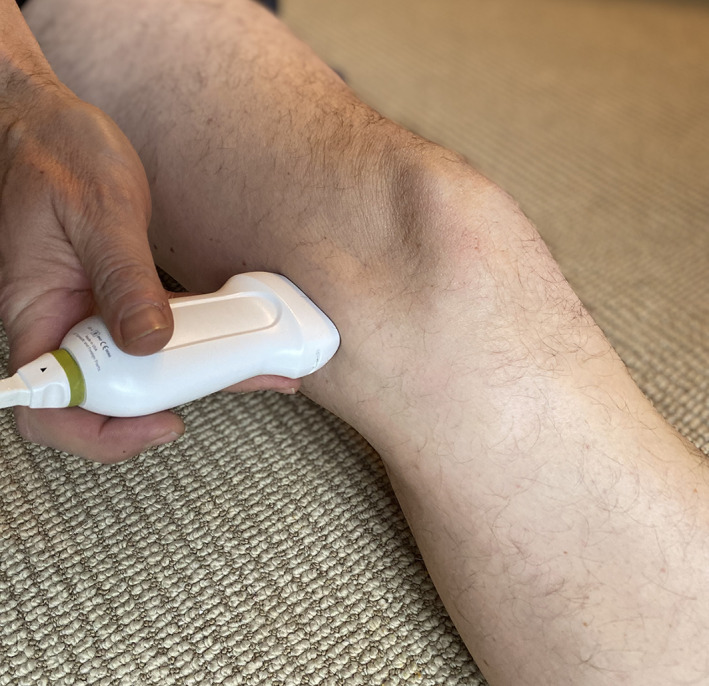
IRB approved study with informed consent. Patients obtaining routine knee MR imaging for suspected knee pathology were prospectively evaluated with supine and weight-bearing US of the medial meniscus. Meniscal extrusion was measured independently by two fellowship-trained musculoskeletal radiologists. Correlation was made to presence or absence of meniscal degeneration or tear on MR imaging, as well as reported symptoms. Statistical significance was calculated via intraclass correlation coefficient (ICC) and analysis of variance (ANOVA).
Ninety-nine knees from 95 subjects (50 males, 45 females; mean age 45 ± 15 years) were included. Mean medial meniscal extrusion measured at US for a normal meniscus (n = 36) was 0.8 mm when supine, increasing to 1.6 mm on weight-bearing. Mean meniscal extrusion in subjects with mucoid degeneration (n = 20) and those with meniscal tears (n = 43) was 1.6 mm, increasing to 2.3 mm with weight bearing. Inter-reader reliability showed ICC values of 0.853 to 0.940. There was a significant difference in medial meniscal extrusion comparing subjects with a normal medial meniscus at magnetic resonance imaging (MRI) and subjects with either meniscal degeneration or tear. There was no significant difference in degree of meniscal extrusion between subjects with meniscal degeneration or tear. There was trend of worsening symptoms and increasing functional limitations moving from normal meniscus to meniscal degeneration to meniscal tear.
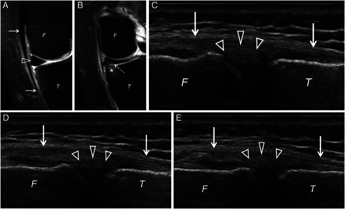
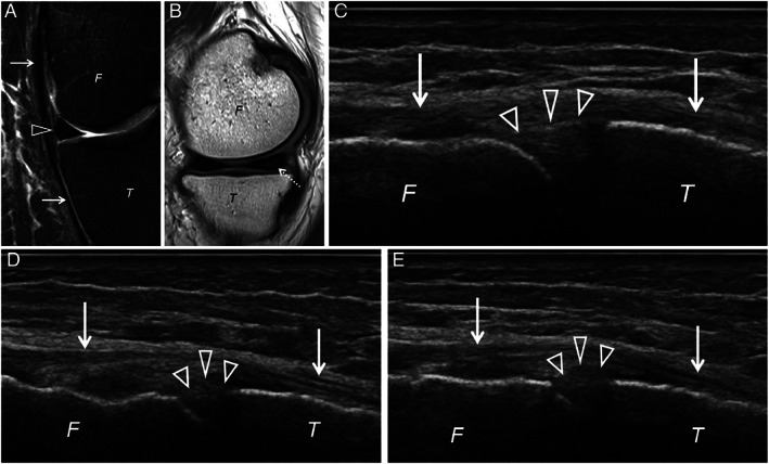
A normal meniscus shows lesser mobility between supine and upright position, than a pathologic meniscus. Both mucoid degeneration and meniscal tear demonstrate extrusion in the supine position, which increases with weight-bearing position.
比较负重超声(US)与仰卧位 US 和磁共振(MR)成像的内侧半月板挤出,与半月板病理和报告的症状相关。
经机构审查委员会批准并获得知情同意,对疑似膝关节病变患者进行常规膝关节 MR 成像,前瞻性评估内侧半月板仰卧位和负重位 US。两名接受过 fellowship培训的肌肉骨骼放射科医生独立测量半月板挤出。与 MR 成像上半月板退变或撕裂的存在或缺失以及报告的症状进行相关性分析。通过组内相关系数(ICC)和方差分析(ANOVA)进行统计学意义计算。
95 名受试者(50 名男性,45 名女性;平均年龄 45±15 岁)的 99 个膝关节纳入研究。仰卧位时正常半月板(n=36)的内侧半月板挤出平均值为 0.8mm,负重时增加到 1.6mm。黏液样退变(n=20)和半月板撕裂(n=43)患者的平均半月板挤出为 1.6mm,负重时增加到 2.3mm。两位读者之间的可靠性显示 ICC 值为 0.853 至 0.940。在 MRI 上正常内侧半月板与半月板退变或撕裂的患者相比,内侧半月板挤出有显著差异。半月板退变或撕裂患者的半月板挤出程度无显著差异。从正常半月板到半月板退变再到半月板撕裂,症状恶化和功能受限的趋势增加。
正常半月板在仰卧位和直立位之间的活动度小于病理性半月板。黏液样退变和半月板撕裂在仰卧位时均有挤出,负重位时增加。