Tortorella Fabio, Boffa Angelo, Andriolo Luca, Facchini Giancarlo, Di Carlo Maddalena, Miceli Marco, Klos Burt, Zaffagnini Stefano, Filardo Giuseppe
Applied and Translational Research (ATR) Center, IRCCS Istituto Ortopedico Rizzoli Bologna Italy.
Clinica Ortopedica e Traumatologica 2, IRCCS Istituto Ortopedico Rizzoli Bologna Italy.
J Exp Orthop. 2024 Sep 30;11(4):e70031. doi: 10.1002/jeo2.70031. eCollection 2024 Oct.
The aim of this study was to quantify meniscal extrusion through ultrasound (US) evaluation in supine and standing positions and to compare the results with those documented through magnetic resonance (MR) imaging in patients affected by knee osteoarthritis (OA).
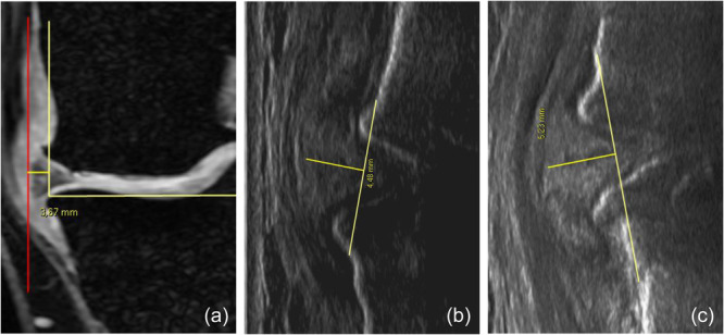
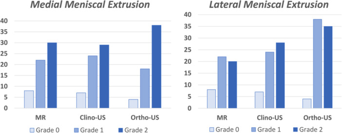
Sixty patients (38 men, 22 women, mean age 60.8 ± 9.7 years) with knee OA were enrolled and underwent a 1.5 T MR evaluation and an US examination of the symptomatic OA knee for the evaluation of the medial and lateral meniscus extrusion both in the supine clinostatic position (clino-US) with the knee fully extended and in the standing weight-bearing orthostatic position (ortho-US). For the three imaging evaluations (MR, clino-US and ortho-US), both semi-quantitative and quantitative measurements were performed.
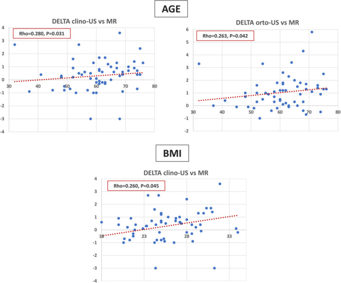
The quantitative analysis documented higher values of medial meniscal extrusion at the ortho-US evaluation (5.2 ± 2.3 mm) compared to MR (4.2 ± 2.2, < 0.0005) and clino-US (4.5 ± 2.3, < 0.0005) and of the lateral meniscus at the ortho-US evaluation (4.3 ± 1.8) compared to MR (3.3 ± 1.6, < 0.0005) and clino-US (3.8 ± 1.6, < 0.0005). The semi-quantitative analysis confirmed the same trend for both menisci. Higher extrusion values were documented in women and more advanced OA, as well as in older patients with higher body mass index, the latter being underestimated the most by the MR approach.
US outperforms MR imaging in quantifying meniscal extrusion in patients with knee OA. Moreover, the highest values of meniscal extrusion have been documented using US in standing position compared to the supine position, underlining the importance of the weight-bearing assessment of meniscal extrusion in knee OA patients.
II.
本研究旨在通过超声(US)评估在仰卧位和站立位时半月板挤出量,并将结果与膝关节骨关节炎(OA)患者通过磁共振(MR)成像记录的结果进行比较。
纳入60例膝关节OA患者(38例男性,22例女性,平均年龄60.8±9.7岁),对患侧有症状的OA膝关节进行1.5T MR评估和US检查,以评估在膝关节完全伸展的仰卧位静息状态(卧位US)和站立位负重直立状态(直立位US)下内侧和外侧半月板的挤出情况。对于三种成像评估(MR、卧位US和直立位US),均进行了半定量和定量测量。
定量分析显示,直立位US评估时内侧半月板挤出量(5.2±2.3mm)高于MR(4.2±2.2,<0.0005)和卧位US(4.5±2.3,<0.0005),直立位US评估时外侧半月板挤出量(4.3±1.8)高于MR(3.3±1.6,<0.0005)和卧位US(3.8±1.6,<0.0005)。半定量分析证实了两个半月板的相同趋势。女性、OA程度更严重者以及体重指数较高的老年患者的挤出值更高,后者在MR评估中被低估得最多。
在量化膝关节OA患者的半月板挤出量方面,US优于MR成像。此外,与仰卧位相比,站立位US记录的半月板挤出量最高,这突出了膝关节OA患者半月板挤出量负重评估的重要性。
II级