Department of Innovative Technologies in Medicine & Dentistry, "G. d'Annunzio" University, Chieti, Italy.
Department of Radiology, Stanford University School of Medicine, Stanford, CA, USA.
Abdom Radiol (NY). 2022 May;47(5):1862-1872. doi: 10.1007/s00261-022-03490-9. Epub 2022 Mar 18.
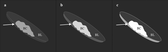
To (1) compare bladder cancer (BC) muscle invasiveness among three b-values using a contrast-free approach based on Vesical Imaging-Reporting and Data System (VI-RADS), to (2) determine if muscle-invasiveness assessment is affected by the reader experience, and to (3) compare BC conspicuity among three b-values, qualitatively and quantitatively.
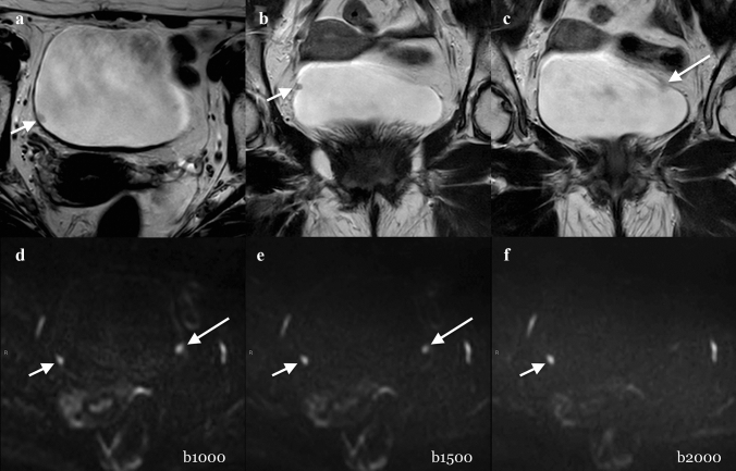
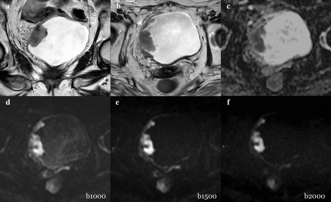
Thirty-eight patients who underwent a bladder MRI on a 3.0-T scanner were enrolled. The gold standard was histopathology report following transurethral resection of BC. Three sets of images, including T2w and different b-values for DWI, set 1 (b = 1000 s/mm), set 2 (b = 1500 s/mm), and set 3 (b = 2000 s/mm), were reviewed by three differently experienced readers. Descriptive statistics and Intraclass Correlation Coefficient (ICC) were calculated. Comparisons among readers and DWI sets were performed with the Wilcoxon test. Receiver operating characteristic (ROC) analysis was performed. Areas under the curves (AUCs) and pairwise comparison were calculated.
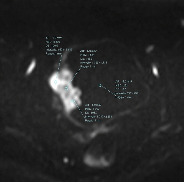
AUCs of muscle-invasiveness assessment ranged from 0.896 to 0.984 (reader 1), 0.952-0.968 (reader 2), and 0.952-0.984 (reader 3) without significant differences among different sets and readers (p > 0.05). The mean conspicuity qualitative scores were higher in Set 1 (2.21-2.33), followed by Set 2 (2-2.16) and Set 3 (1.82-2.14). The quantitative conspicuity assessment showed that mean normalized intensity of tumor was significantly higher in Set 2 (4.217-4.737) than in Set 1 (3.923-4.492) and Set 3 (3.833-3.992) (p < 0.05).
Muscle invasiveness can be assessed with high accuracy using a contrast-free protocol with T2W and DWI, regardless of reader's experience. b = 1500 s/mm showed the best tumor delineation, while b = 1000 s/mm allowed for better tumor-wall interface assessment.
(1)使用基于 Vesical Imaging-Reporting and Data System(VI-RADS)的无对比剂方法比较三种 b 值在膀胱癌(BC)肌肉侵袭性方面的差异,(2)确定肌肉侵袭性评估是否受读者经验的影响,(3)定性和定量比较三种 b 值的 BC 显著性。
共纳入 38 例在 3.0T 扫描仪上进行膀胱 MRI 的患者。金标准是经尿道切除 BC 后的组织病理学报告。三组图像,包括 T2w 和不同 b 值的 DWI,组 1(b=1000 s/mm)、组 2(b=1500 s/mm)和组 3(b=2000 s/mm),由三位不同经验的读者进行了回顾。计算了描述性统计数据和组内相关系数(ICC)。采用 Wilcoxon 检验比较读者和 DWI 组之间的差异。进行了接收器工作特征(ROC)分析。计算了曲线下面积(AUC)和两两比较。
肌肉侵袭性评估的 AUC 范围为 0.896 至 0.984(读者 1)、0.952-0.968(读者 2)和 0.952-0.984(读者 3),不同组和读者之间无显著差异(p>0.05)。组 1(2.21-2.33)的平均显著性定性评分较高,其次是组 2(2-2.16)和组 3(1.82-2.14)。定量显著性评估显示,组 2(4.217-4.737)的肿瘤归一化强度明显高于组 1(3.923-4.492)和组 3(3.833-3.992)(p<0.05)。
使用 T2W 和 DWI 的无对比剂方案可以高度准确地评估肌肉侵袭性,而与读者经验无关。b=1500 s/mm 显示出最佳的肿瘤勾画效果,而 b=1000 s/mm 则允许更好地评估肿瘤-壁界面。