文献检索文档翻译深度研究
Suppr Zotero 插件Zotero 插件
邀请有礼套餐&价格历史记录

新学期,新优惠

限时优惠:9月1日-9月22日

30天高级会员仅需29元

1天体验卡首发特惠仅需5.99元

了解详情
不再提醒
插件&应用
Suppr Zotero 插件Zotero 插件浏览器插件Mac 客户端Windows 客户端微信小程序
高级版
套餐订阅购买积分包
AI 工具
文献检索文档翻译深度研究
关于我们
关于 Suppr公司介绍联系我们用户协议隐私条款
关注我们

Suppr 超能文献

核心技术专利:CN118964589B侵权必究
粤ICP备2023148730 号-1Suppr @ 2025

Tissue Cytometry With Machine Learning in Kidney: From Small Specimens to Big Data.

作者信息

El-Achkar Tarek M, Winfree Seth, Talukder Niloy, Barwinska Daria, Ferkowicz Michael J, Al Hasan Mohammad

机构信息

Division of Nephrology, Department of Medicine, Indiana University, Indianapolis, IN, United States.

Department of Pathology and Microbiology, University of Nebraska Omaha, Omaha, NE, United States.

出版信息

Front Physiol. 2022 Mar 4;13:832457. doi: 10.3389/fphys.2022.832457. eCollection 2022.


DOI:10.3389/fphys.2022.832457
PMID:35309077
原文链接:https://pmc.ncbi.nlm.nih.gov/articles/PMC8931540/
Abstract

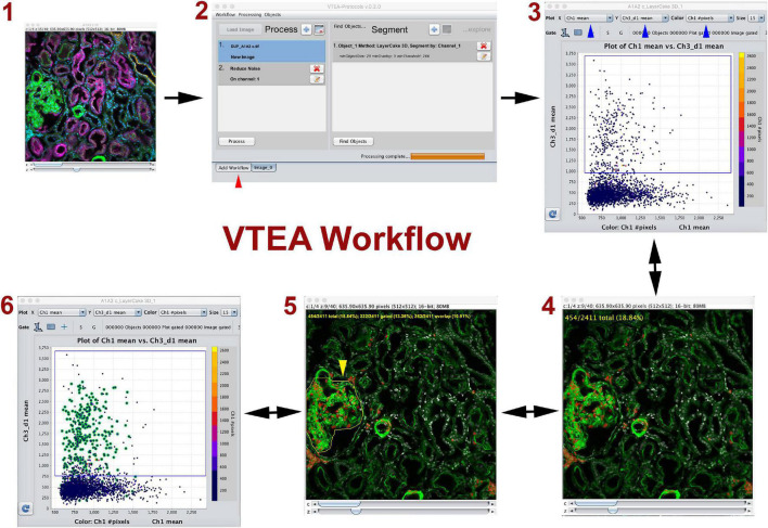
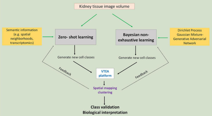
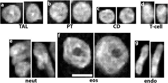
Advances in cellular and molecular interrogation of kidney tissue have ushered a new era of understanding the pathogenesis of kidney disease and potentially identifying molecular targets for therapeutic intervention. Classifying cells and identifying subtypes and states induced by injury is a foundational task in this context. High resolution Imaging-based approaches such as large-scale fluorescence 3D imaging offer significant advantages because they allow preservation of tissue architecture and provide a definition of the spatial context of each cell. We recently described the Volumetric Tissue Exploration and Analysis cytometry tool which enables an interactive analysis, quantitation and semiautomated classification of labeled cells in 3D image volumes. We also established and demonstrated an imaging-based classification using deep learning of cells in intact tissue using 3D nuclear staining with 4',6-diamidino-2-phenylindole (DAPI). In this mini-review, we will discuss recent advancements in analyzing 3D imaging of kidney tissue, and how combining machine learning with cytometry is a powerful approach to leverage the depth of content provided by high resolution imaging into a highly informative analytical output. Therefore, imaging a small tissue specimen will yield big scale data that will enable cell classification in a spatial context and provide novel insights on pathological changes induced by kidney disease.

摘要
https://cdn.ncbi.nlm.nih.gov/pmc/blobs/c79f/8931540/6f8d10e38754/fphys-13-832457-g003.jpg
https://cdn.ncbi.nlm.nih.gov/pmc/blobs/c79f/8931540/a2898c92b958/fphys-13-832457-g001.jpg
https://cdn.ncbi.nlm.nih.gov/pmc/blobs/c79f/8931540/98115d71e905/fphys-13-832457-g002.jpg
https://cdn.ncbi.nlm.nih.gov/pmc/blobs/c79f/8931540/6f8d10e38754/fphys-13-832457-g003.jpg
https://cdn.ncbi.nlm.nih.gov/pmc/blobs/c79f/8931540/a2898c92b958/fphys-13-832457-g001.jpg
https://cdn.ncbi.nlm.nih.gov/pmc/blobs/c79f/8931540/98115d71e905/fphys-13-832457-g002.jpg
https://cdn.ncbi.nlm.nih.gov/pmc/blobs/c79f/8931540/6f8d10e38754/fphys-13-832457-g003.jpg

相似文献

[1]
Tissue Cytometry With Machine Learning in Kidney: From Small Specimens to Big Data.

Front Physiol. 2022-3-4

[2]
In Situ Classification of Cell Types in Human Kidney Tissue Using 3D Nuclear Staining.

Cytometry A. 2021-7

[3]
Integrated Cytometry With Machine Learning Applied to High-Content Imaging of Human Kidney Tissue for In Situ Cell Classification and Neighborhood Analysis.

Lab Invest. 2023-6

[4]
Profiling Immune Cells in the Kidney Using Tissue Cytometry and Machine Learning.

Kidney360. 2022-5-26

[5]
Quantitative Three-Dimensional Tissue Cytometry to Study Kidney Tissue and Resident Immune Cells.

J Am Soc Nephrol. 2017-7

[6]
Quantitative Large-Scale Three-Dimensional Imaging of Human Kidney Biopsies: A Bridge to Precision Medicine in Kidney Disease.

Nephron. 2018-6-5

[7]
Probing the 3D architecture of the plant nucleus with microscopy approaches: challenges and solutions.

Nucleus. 2019-12

[8]
Automated Analysis and Classification of Histological Tissue Features by Multi-Dimensional Microscopic Molecular Profiling.

PLoS One. 2015-7-15

[9]
Deep Learning-Based Single-Cell Optical Image Studies.

Cytometry A. 2020-3

[10]
"Hi, how can i help you?": embracing artificial intelligence in kidney research.

Am J Physiol Renal Physiol. 2023-10-1

引用本文的文献

[1]
High-throughput image analysis with deep learning captures heterogeneity and spatial relationships after kidney injury.

Sci Rep. 2023-4-19

[2]
Vascular Implications of COVID-19: Role of Radiological Imaging, Artificial Intelligence, and Tissue Characterization: A Special Report.

J Cardiovasc Dev Dis. 2022-8-15

本文引用的文献

[1]
An atlas of healthy and injured cell states and niches in the human kidney.

Nature. 2023-7

[2]
Integrated Cytometry With Machine Learning Applied to High-Content Imaging of Human Kidney Tissue for In Situ Cell Classification and Neighborhood Analysis.

Lab Invest. 2023-6

[3]
Profiling Immune Cells in the Kidney Using Tissue Cytometry and Machine Learning.

Kidney360. 2022-5-26

[4]
VEGFR3 tyrosine kinase inhibition aggravates cisplatin nephrotoxicity.

Am J Physiol Renal Physiol. 2021-12-1

[5]
Highly multiplexed immunofluorescence of the human kidney using co-detection by indexing.

Kidney Int. 2022-1

[6]
Heterozygous Mutation of Vegfr3 Reduces Renal Lymphatics without Renal Dysfunction.

J Am Soc Nephrol. 2021-12-1

[7]
CellProfiler Analyst 3.0: accessible data exploration and machine learning for image analysis.

Bioinformatics. 2021-11-5

[8]
Multi-Parameter Quantitative Imaging of Tumor Microenvironments Reveals Perivascular Immune Niches Associated With Anti-Tumor Immunity.

Front Immunol. 2021

[9]
Quantitative 3-dimensional imaging and tissue cytometry reveals lymphatic expansion in acute kidney injury.

Lab Invest. 2021-9

[10]
Integration of spatial and single-cell transcriptomics localizes epithelial cell-immune cross-talk in kidney injury.

JCI Insight. 2021-6-22

文献AI研究员

20分钟写一篇综述,助力文献阅读效率提升50倍

立即体验

用中文搜PubMed

大模型驱动的PubMed中文搜索引擎

马上搜索

推荐工具

医学文档翻译智能文献检索