Department of Medicine, Division of Nephrology and Hypertension, Indiana University School of Medicine, Indianapolis, Indiana, USA.
Department of Computer Science, Indiana University Purdue University, Indianapolis, Indiana, USA.
Cytometry A. 2021 Jul;99(7):707-721. doi: 10.1002/cyto.a.24274. Epub 2020 Dec 13.
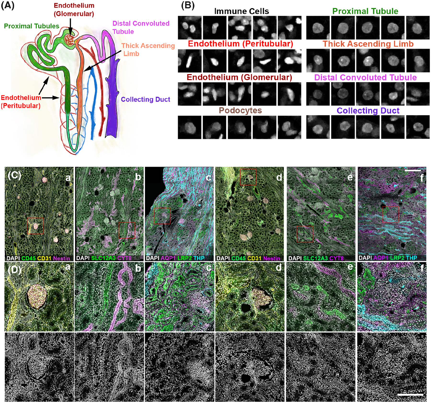
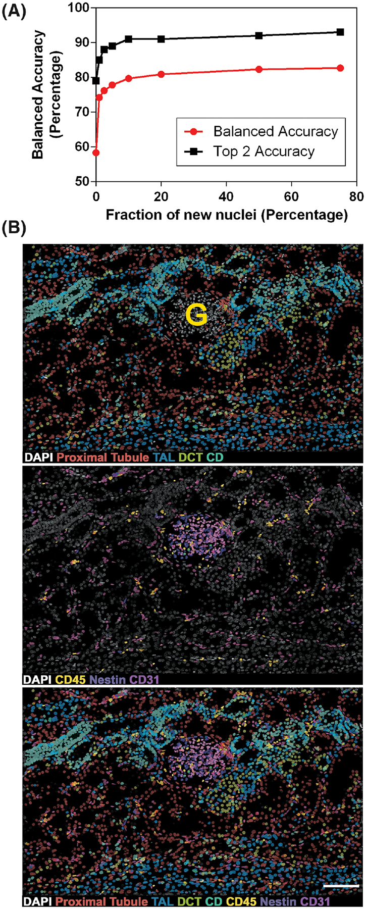
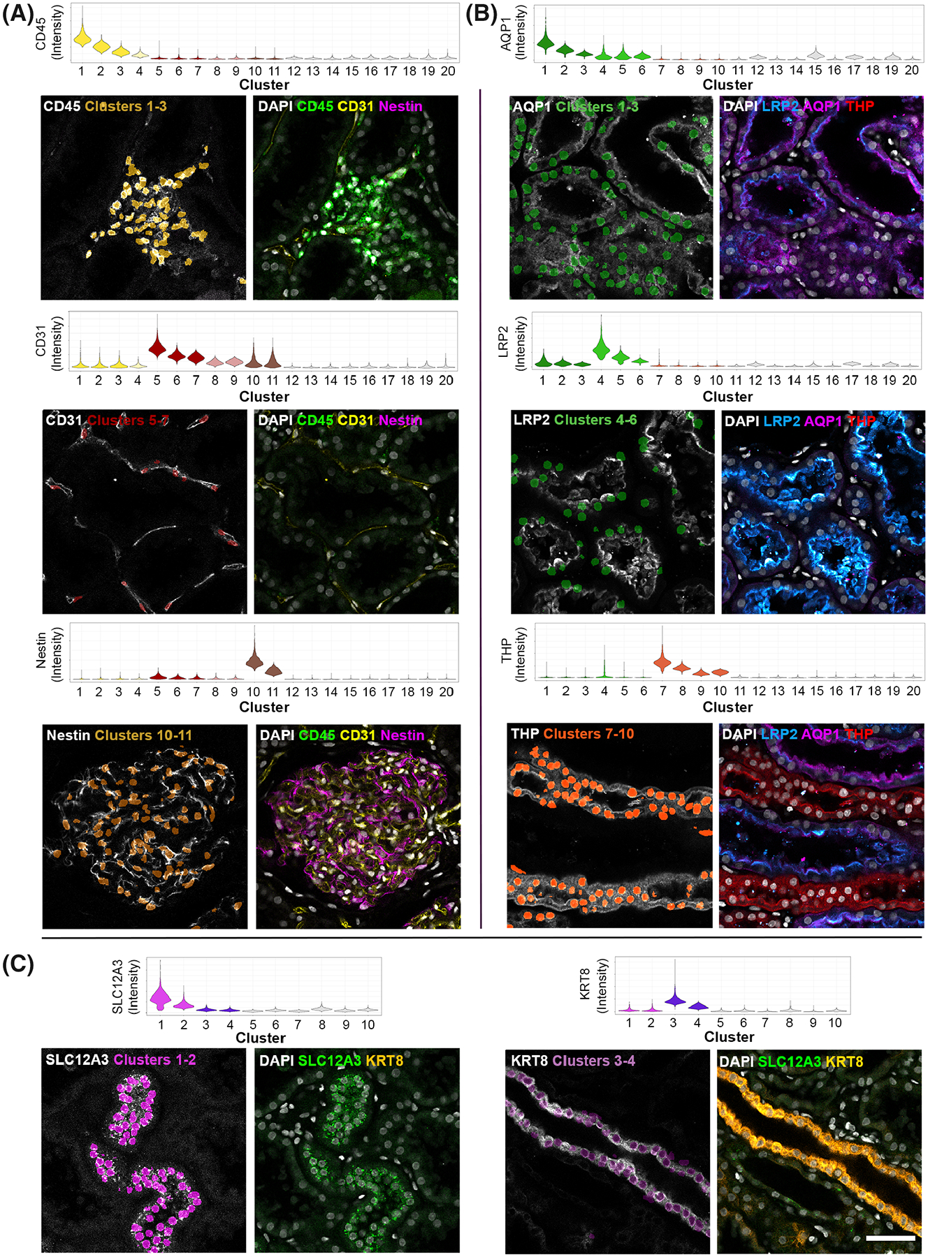
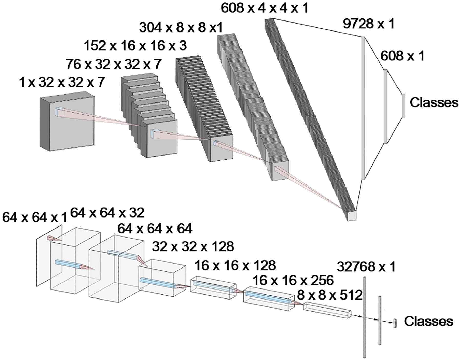
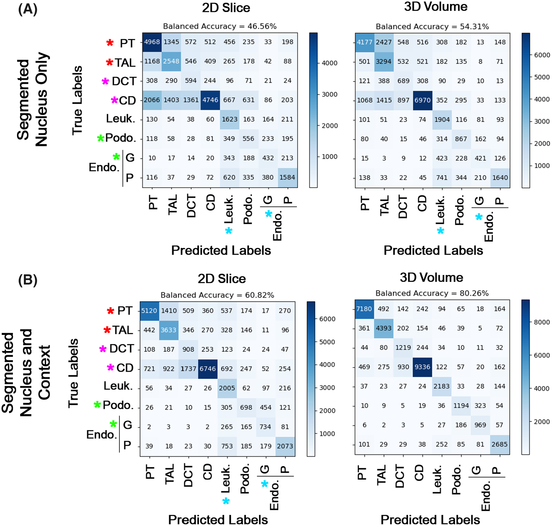
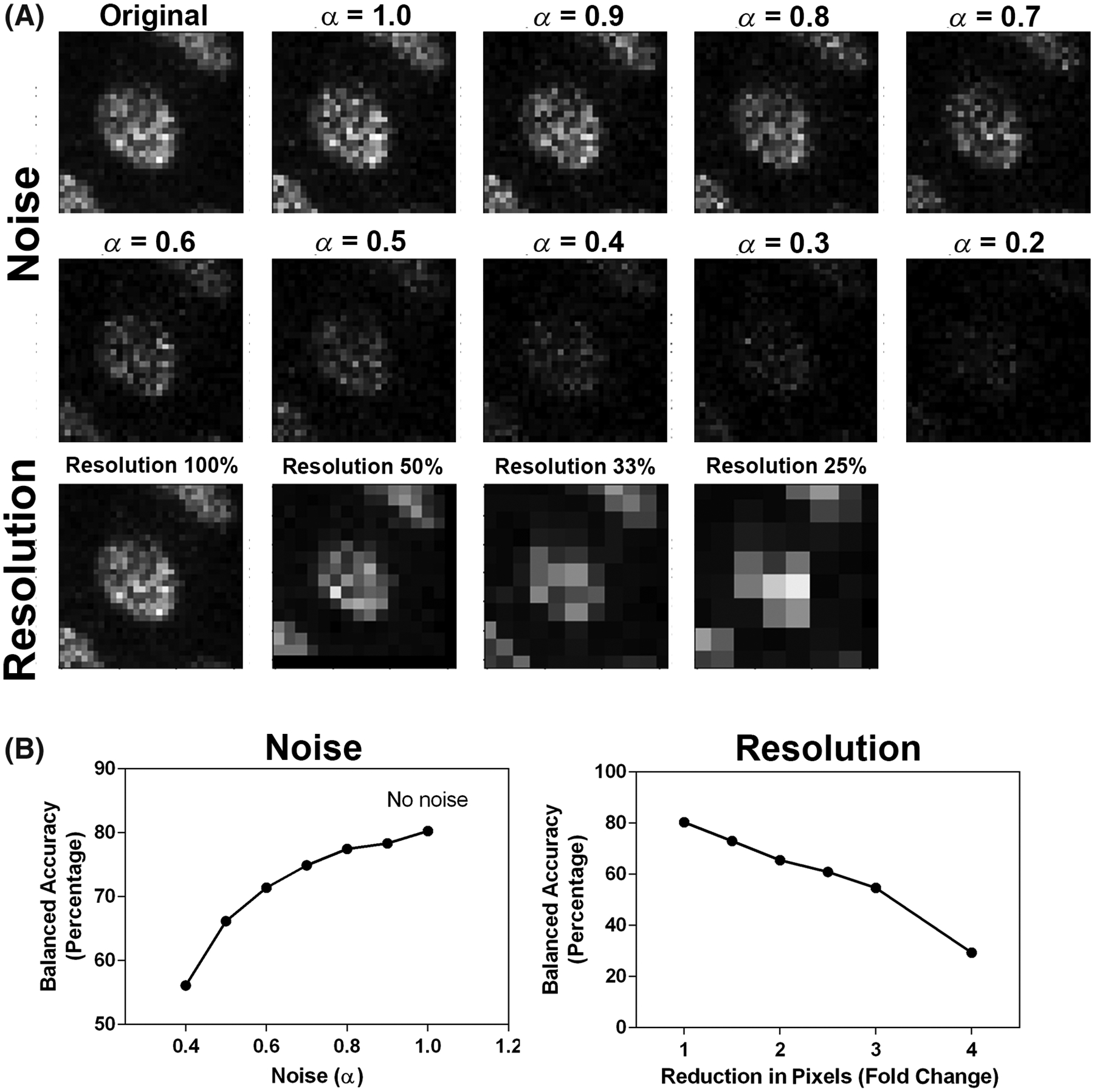
To understand the physiology and pathology of disease, capturing the heterogeneity of cell types within their tissue environment is fundamental. In such an endeavor, the human kidney presents a formidable challenge because its complex organizational structure is tightly linked to key physiological functions. Advances in imaging-based cell classification may be limited by the need to incorporate specific markers that can link classification to function. Multiplex imaging can mitigate these limitations, but requires cumulative incorporation of markers, which may lead to tissue exhaustion. Furthermore, the application of such strategies in large scale 3-dimensional (3D) imaging is challenging. Here, we propose that 3D nuclear signatures from a DNA stain, DAPI, which could be incorporated in most experimental imaging, can be used for classifying cells in intact human kidney tissue. We developed an unsupervised approach that uses 3D tissue cytometry to generate a large training dataset of nuclei images (NephNuc), where each nucleus is associated with a cell type label. We then devised various supervised machine learning approaches for kidney cell classification and demonstrated that a deep learning approach outperforms classical machine learning or shape-based classifiers. Specifically, a custom 3D convolutional neural network (NephNet3D) trained on nuclei image volumes achieved a balanced accuracy of 80.26%. Importantly, integrating NephNet3D classification with tissue cytometry allowed in situ visualization of cell type classifications in kidney tissue. In conclusion, we present a tissue cytometry and deep learning approach for in situ classification of cell types in human kidney tissue using only a DNA stain. This methodology is generalizable to other tissues and has potential advantages on tissue economy and non-exhaustive classification of different cell types.
为了理解疾病的生理学和病理学,捕捉组织环境中细胞类型的异质性是基础。在这样的努力中,人类肾脏提出了一个巨大的挑战,因为其复杂的组织结构与关键的生理功能紧密相连。基于成像的细胞分类的进展可能受到需要纳入可以将分类与功能联系起来的特定标记的限制。多重成像可以减轻这些限制,但需要累积纳入标记,这可能导致组织枯竭。此外,此类策略在大规模三维(3D)成像中的应用具有挑战性。在这里,我们提出可以从 DNA 染色剂 DAPI 获得的 3D 核特征,该特征可以整合到大多数实验成像中,用于对完整的人类肾脏组织中的细胞进行分类。我们开发了一种无监督方法,该方法使用 3D 组织细胞计量术生成大量核图像(NephNuc)的训练数据集,其中每个核都与细胞类型标签相关联。然后,我们设计了各种用于肾脏细胞分类的监督机器学习方法,并证明了深度学习方法优于经典机器学习或基于形状的分类器。具体而言,使用核图像体积训练的自定义 3D 卷积神经网络(NephNet3D)实现了 80.26%的平衡准确性。重要的是,将 NephNet3D 分类与组织细胞计量术集成允许在原位可视化肾脏组织中的细胞类型分类。总之,我们提出了一种使用仅 DNA 染色剂原位分类人类肾脏组织中细胞类型的组织细胞计量术和深度学习方法。该方法适用于其他组织,并且在组织经济性和不同细胞类型的非消耗性分类方面具有潜在优势。