Vespro Valentina, Bonanno Maria Chiara, Andrisani Maria Carmela, Ierardi Anna Maria, Phillips Alice, Tosi Davide, Mendogni Paolo, Franzi Sara, Carrafiello Gianpaolo
Department of Radiology, IRCCS Foundation Ca' Granda Ospedale Maggiore Policlinico, 20122 Milan, Italy.
Department of Health Sciences, University of Milan, 20122 Milan, Italy.
Tomography. 2022 Mar 1;8(2):617-626. doi: 10.3390/tomography8020051.
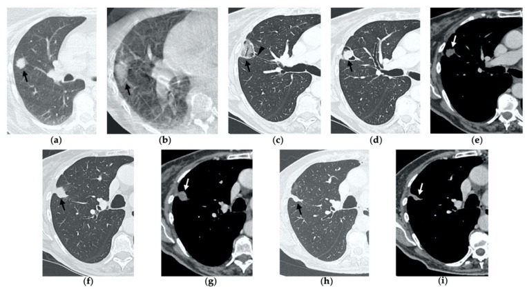
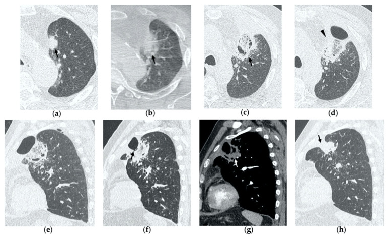
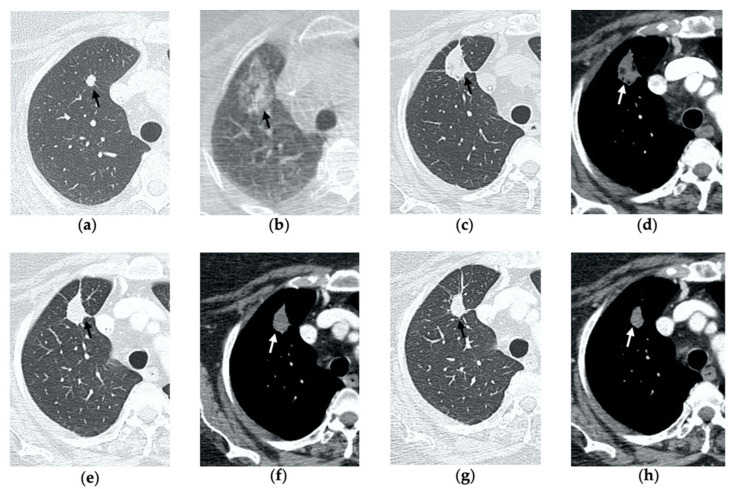
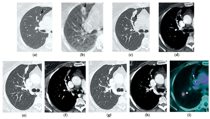
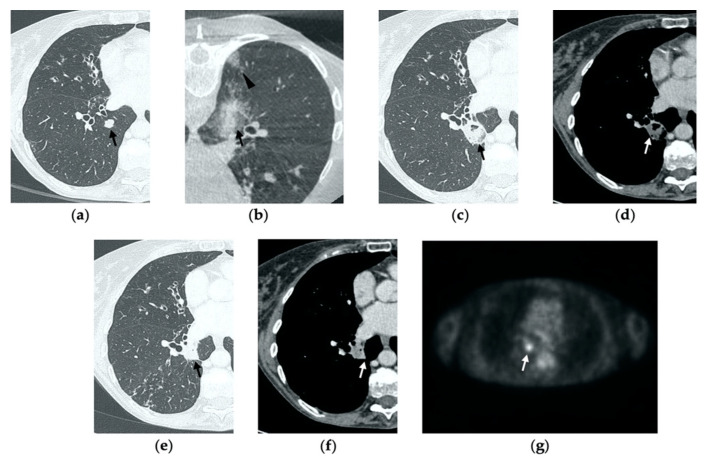
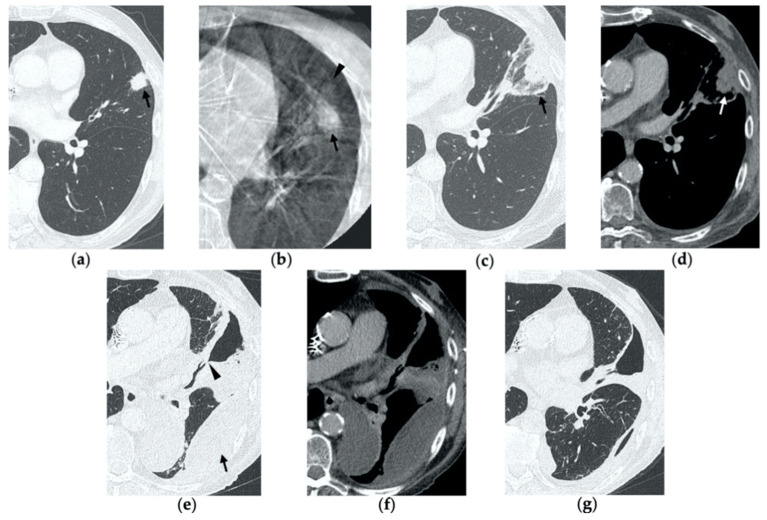
Imaging-guided percutaneous ablative treatments, such as radiofrequency ablation (RFA), cryoablation and microwave ablation (MWA), have been developed for the treatment of unresectable primary and secondary lung tumors in patients with advanced-stage disease or comorbidities contraindicating surgery. Among these therapies, MWA has recently shown promising results in the treatment of pulmonary neoplasms. The potential advantages of MWA over RFA include faster ablation times, higher intra-tumoral temperatures, larger ablation zones and lower susceptibility to the heat sink effect, resulting in greater efficacy in proximity to vascular structures. Despite encouraging results supporting its efficacy, there is a relative paucity of data in the literature regarding the role of computer tomography (CT) to monitor MWA-treated lesions, and the CT appearance of their morphologic evolution and complications. For both interventional and non-interventional radiologists, it is crucial to be familiar with the CT features of such treated lesions in order to detect incomplete therapy or recurrent disease at early stage, as well as to recognize initial signs of complications. The aim of this pictorial essay is to describe the typical CT features during follow-up of lung lesions treated with percutaneous MWA and how to interpret and differentiate them from other radiological findings, such as recurrence and complications, that are commonly encountered in this setting.
影像引导下的经皮消融治疗,如射频消融(RFA)、冷冻消融和微波消融(MWA),已被开发用于治疗晚期疾病或存在手术禁忌合并症的不可切除的原发性和继发性肺肿瘤。在这些治疗方法中,MWA最近在肺部肿瘤治疗中显示出了有前景的结果。MWA相对于RFA的潜在优势包括消融时间更快、瘤内温度更高、消融区更大以及对热沉效应的敏感性更低,从而在靠近血管结构处具有更高的疗效。尽管有支持其疗效的令人鼓舞的结果,但关于计算机断层扫描(CT)在监测MWA治疗病变中的作用以及其形态演变和并发症的CT表现,文献中的数据相对较少。对于介入和非介入放射科医生来说,熟悉此类治疗病变的CT特征至关重要,以便在早期检测到治疗不彻底或疾病复发,以及识别并发症的初始迹象。本文的目的是描述经皮MWA治疗的肺部病变随访期间的典型CT特征,以及如何将其与该情况下常见的其他影像学表现,如复发和并发症进行解释和区分。