IEEE Trans Ultrason Ferroelectr Freq Control. 2022 May;69(5):1670-1681. doi: 10.1109/TUFFC.2022.3161719. Epub 2022 Apr 27.
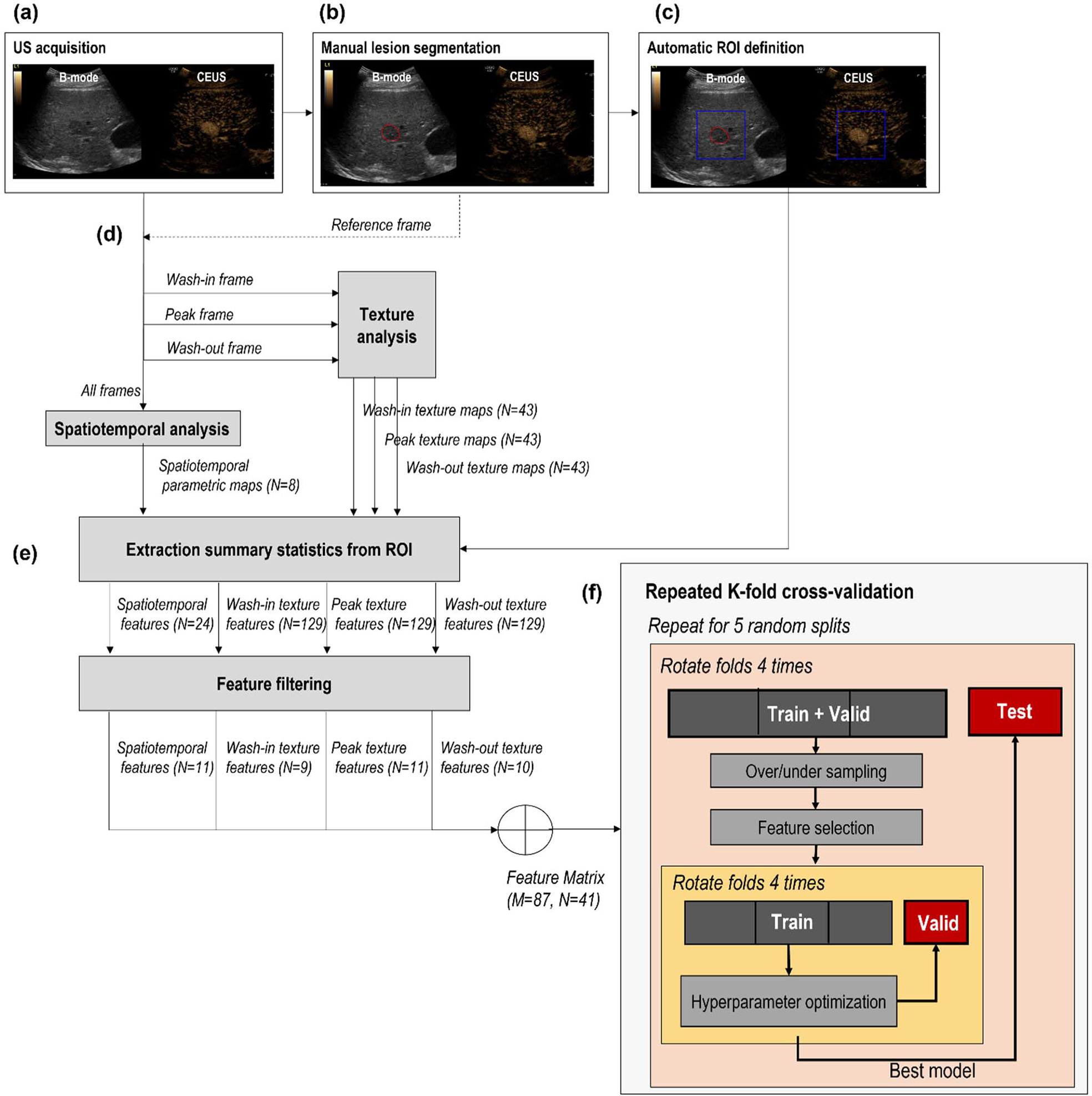
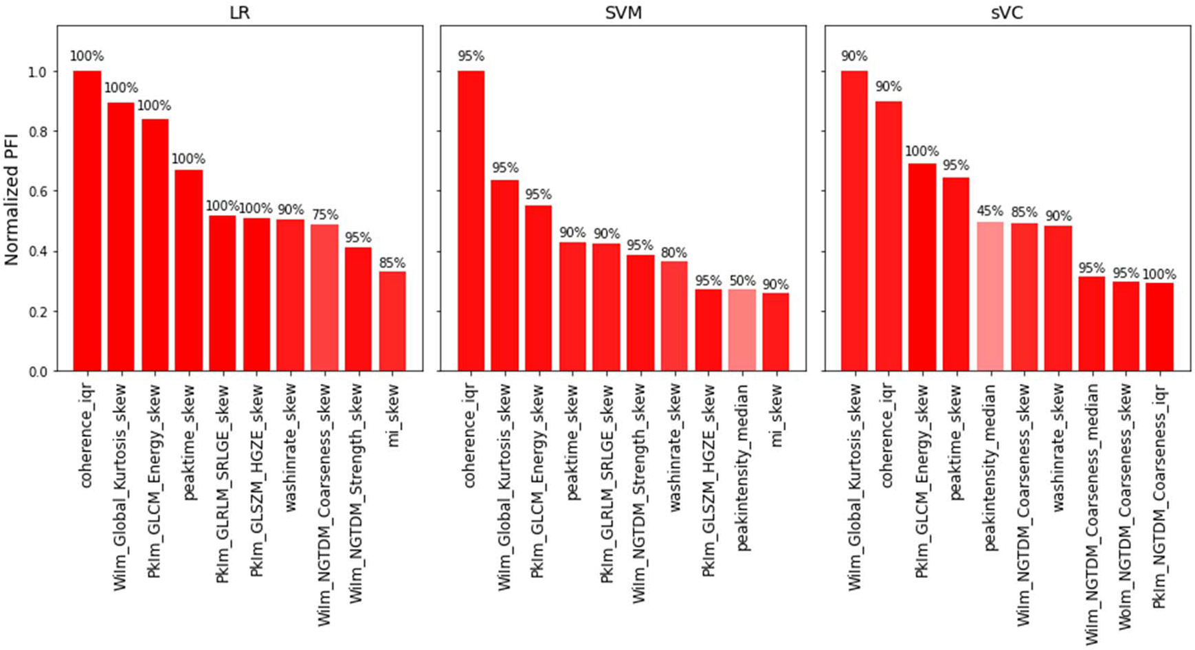
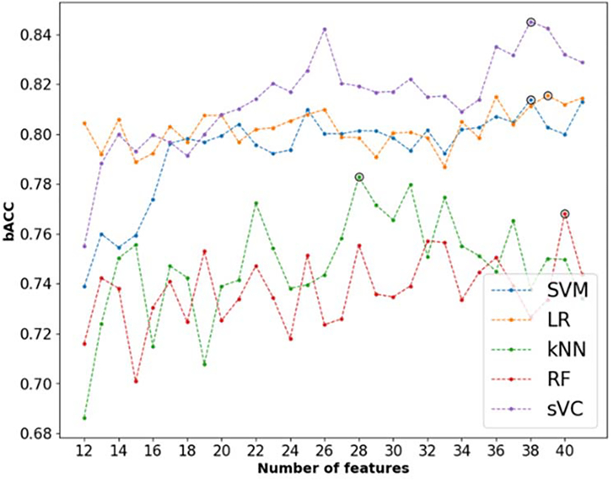
This work proposes an interpretable radiomics approach to differentiate between malignant and benign focal liver lesions (FLLs) on contrast-enhanced ultrasound (CEUS). Although CEUS has shown promise for differential FLLs diagnosis, current clinical assessment is performed only by qualitative analysis of the contrast enhancement patterns. Quantitative analysis is often hampered by the unavoidable presence of motion artifacts and by the complex, spatiotemporal nature of liver contrast enhancement, consisting of multiple, overlapping vascular phases. To fully exploit the wealth of information in CEUS, while coping with these challenges, here we propose combining features extracted by the temporal and spatiotemporal analysis in the arterial phase enhancement with spatial features extracted by texture analysis at different time points. Using the extracted features as input, several machine learning classifiers are optimized to achieve semiautomatic FLLs characterization, for which there is no need for motion compensation and the only manual input required is the location of a suspicious lesion. Clinical validation on 87 FLLs from 72 patients at risk for hepatocellular carcinoma (HCC) showed promising performance, achieving a balanced accuracy of 0.84 in the distinction between benign and malignant lesions. Analysis of feature relevance demonstrates that a combination of spatiotemporal and texture features is needed to achieve the best performance. Interpretation of the most relevant features suggests that aspects related to microvascular perfusion and the microvascular architecture, together with the spatial enhancement characteristics at wash-in and peak enhancement, are important to aid the accurate characterization of FLLs.
本研究提出了一种可解释的影像组学方法,用于区分增强超声(CEUS)上的恶性和良性局灶性肝病变(FLL)。尽管 CEUS 在鉴别 FLL 方面显示出了潜力,但目前的临床评估仅通过对比增强模式的定性分析进行。定量分析常常受到运动伪影的不可避免存在以及肝脏对比增强的复杂时空性质的限制,肝脏对比增强由多个重叠的血管期组成。为了充分利用 CEUS 中的丰富信息,同时应对这些挑战,我们在这里提出结合动脉期增强的时间和时空分析提取的特征与不同时间点的纹理分析提取的空间特征。使用提取的特征作为输入,优化了几种机器学习分类器以实现半自动 FLL 特征描述,其中不需要运动补偿,唯一需要的手动输入是可疑病变的位置。对 72 名肝癌(HCC)高危患者的 87 个 FLL 进行的临床验证显示出了有前景的性能,在良性和恶性病变之间的区分中实现了平衡准确性为 0.84。特征相关性分析表明,需要结合时空和纹理特征才能达到最佳性能。对最相关特征的分析表明,与微血管灌注和微血管结构相关的方面,以及在灌注期和峰值增强期的空间增强特征,对于准确描述 FLL 很重要。