van den Oever L B, Spoor D S, Crijns A P G, Vliegenthart R, Oudkerk M, Veldhuis R N J, de Bock G H, van Ooijen P M A
Department of Radiation Oncology, University of Groningen, University Medical Center Groningen, Hanzeplein 1, 9713GZ, Groningen, The Netherlands.
Department of Radiology and Nuclear Medicine, Radboud University Medical Center, Nijmegen, The Netherlands.
J Med Syst. 2022 Mar 25;46(5):22. doi: 10.1007/s10916-022-01810-6.
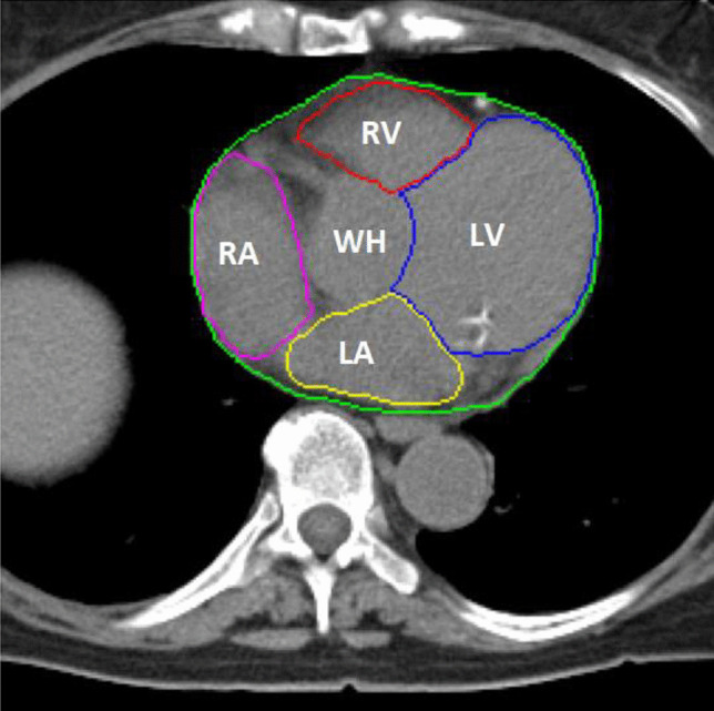
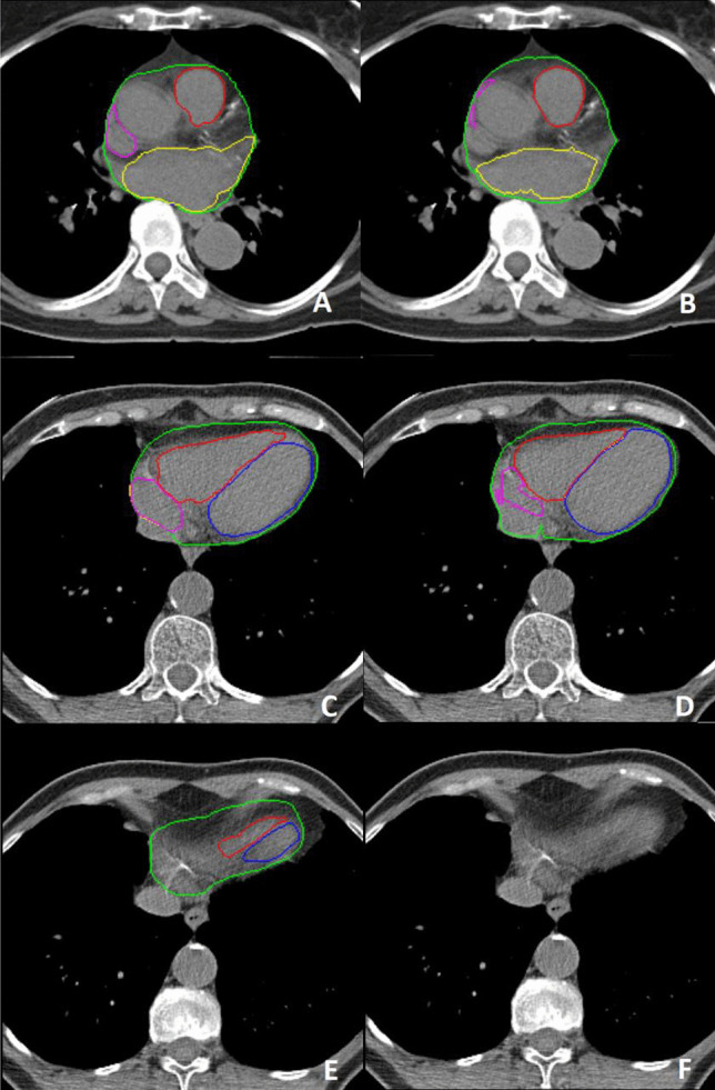
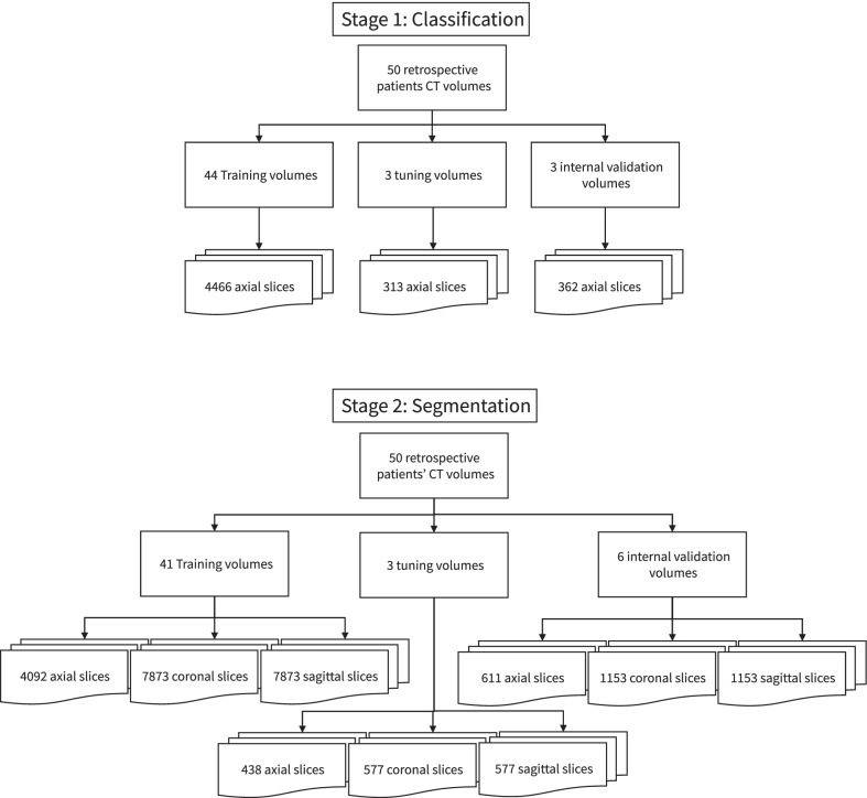
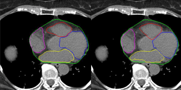
Cardiac structure contouring is a time consuming and tedious manual activity used for radiotherapeutic dose toxicity planning. We developed an automatic cardiac structure segmentation pipeline for use in low-dose non-contrast planning CT based on deep learning algorithms for small datasets. Fifty CT scans were retrospectively selected and the whole heart, ventricles and atria were contoured. A two stage deep learning pipeline was trained on 41 non contrast planning CTs, tuned with 3 CT scans and validated on 6 CT scans. In the first stage, An InceptionResNetV2 network was used to identify the slices that contained cardiac structures. The second stage consisted of three deep learning models trained on the images containing cardiac structures to segment the structures. The three deep learning models predicted the segmentations/contours on axial, coronal and sagittal images and are combined to create the final prediction. The final accuracy of the pipeline was quantified on 6 volumes by calculating the Dice similarity coefficient (DC), 95% Hausdorff distance (95% HD) and volume ratios between predicted and ground truth volumes. Median DC and 95% HD of 0.96, 0.88, 0.92, 0.80 and 0.82, and 1.86, 2.98, 2.02, 6.16 and 6.46 were achieved for the whole heart, right and left ventricle, and right and left atria respectively. The median differences in volume were -4, -1, + 5, -16 and -20% for the whole heart, right and left ventricle, and right and left atria respectively. The automatic contouring pipeline achieves good results for whole heart and ventricles. Robust automatic contouring with deep learning methods seems viable for local centers with small datasets.
心脏结构轮廓勾画是一项耗时且繁琐的手工活动,用于放射治疗剂量毒性规划。我们基于深度学习算法开发了一种自动心脏结构分割流程,用于基于小数据集的低剂量非增强规划CT。回顾性选取了50例CT扫描,对整个心脏、心室和心房进行轮廓勾画。在41例非增强规划CT上训练了一个两阶段深度学习流程,用3例CT扫描进行调整,并在6例CT扫描上进行验证。在第一阶段,使用InceptionResNetV2网络识别包含心脏结构的切片。第二阶段由三个深度学习模型组成,这些模型在包含心脏结构的图像上进行训练以分割结构。这三个深度学习模型在轴向、冠状和矢状图像上预测分割/轮廓,并将其组合以创建最终预测。通过计算Dice相似系数(DC)、95%豪斯多夫距离(95%HD)以及预测体积与真实体积之间的体积比,对6个容积的流程最终准确性进行了量化。整个心脏、右心室、左心室、右心房和左心房的DC中位数分别为0.96、0.88、0.92、0.80和0.82,95%HD中位数分别为1.86、2.98、2.02、6.16和6.46。整个心脏、右心室、左心室、右心房和左心房的体积中位数差异分别为-4%、-1%、+5%、-16%和-20%。自动轮廓勾画流程在整个心脏和心室方面取得了良好结果。对于拥有小数据集的本地中心而言,采用深度学习方法进行稳健的自动轮廓勾画似乎是可行的。