Department of General Surgery & Guangdong Provincial Key Laboratory of Precision Medicine for Gastrointestinal Tumor, Nanfang Hospital, Southern Medical University, Guangzhou, China.
J Immunol Res. 2022 Mar 16;2022:6011632. doi: 10.1155/2022/6011632. eCollection 2022.
Microbes have been shown to contribute to gastric cancer (GC), gastric bacteria and viruses are associated with gastric carcinogenesis. However, the relationship between gastric fungi and GC is still unclear. Our aim was to evaluate the gastric fungal microbiota in the GC microenvironment.
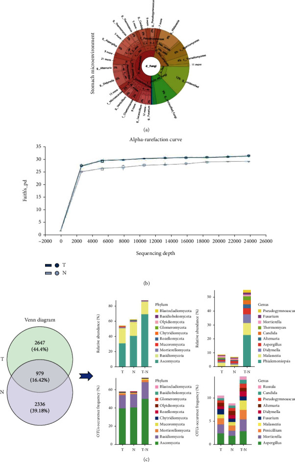
Gastric fungal microbiome profiling was performed with internal transcribed spacer (ITS) rDNA sequencing in primary tumor and corresponding paired normal mucosal tissues from 61 GC patients. Differences in microbial composition, taxa diversity, and predicted function were further analyzed.
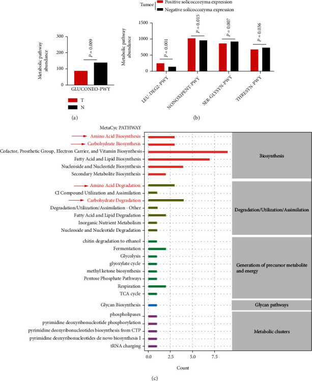
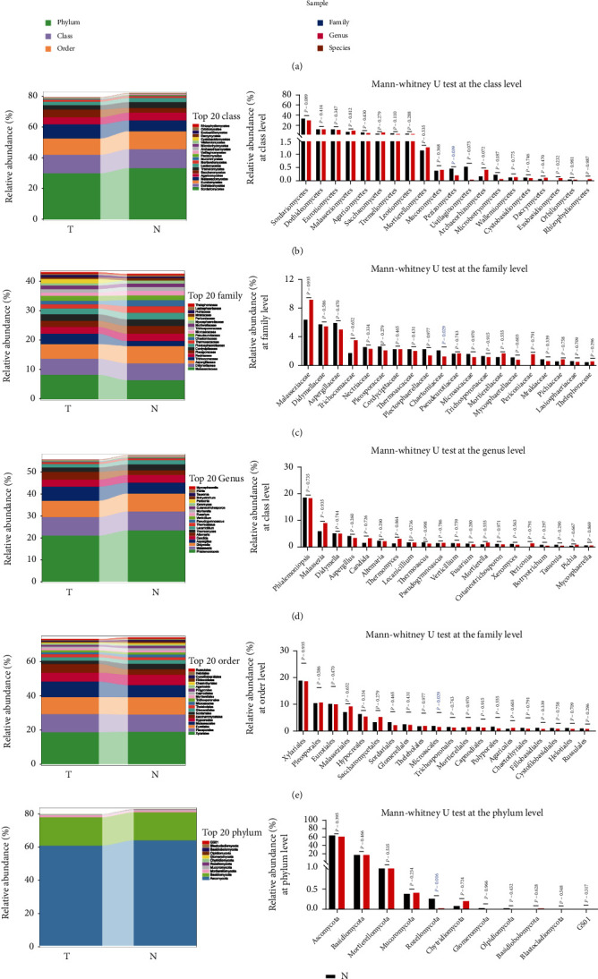
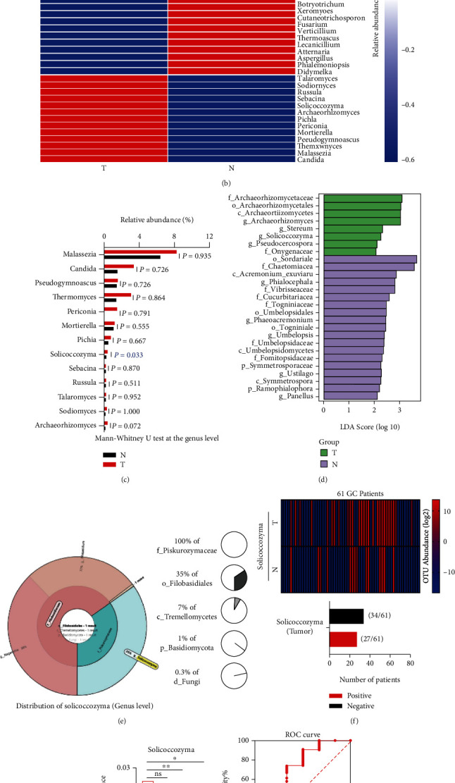
Dysbiosis of gastric mucosal fungal microbiome was observed between the tumor and normal groups in GC. The tumor group had a higher abundance of certain taxa than the normal group. In the taxa classification, the abundances of , , , and were lower in the tumor group than in the normal group. At the genus level, ( = 0.033) was found in higher abundance and was differentially enriched in the tumor group with Lefse analysis. Additionally, accounted for 0.3% of gastric fungi in the GC microenvironment. Twenty-seven of the 61 GC patients showed positive expression in tumors. -positive expression in tumors was associated with the Bormann classification and nerve invasion. was considered a gastric fungal marker to classify stage I and stage II-IV GC patients with an area under the receiver-operating curve (AUC) of 0.7061, as well as to classify the nerve invasive and nonnerve invasive tumors from GC patients with an AUC of 0.6978. Functional prediction indicated that the positive expression of in tumors was associated with the amino acid- and carbohydrate-related metabolic pathways in GC.
This study revealed a novel perspective on the role of in tumors and a theoretical basis for therapeutic targets against GC.
微生物已被证明与胃癌(GC)有关,胃细菌和病毒与胃癌的发生有关。然而,胃真菌与 GC 之间的关系尚不清楚。我们的目的是评估 GC 微环境中的胃真菌微生物组。
对 61 例 GC 患者的原发肿瘤及其相应配对的正常黏膜组织进行内部转录间隔区(ITS)rDNA 测序,进行胃真菌微生物组谱分析。进一步分析微生物组成、分类群多样性和预测功能的差异。
GC 患者肿瘤组和正常组胃黏膜真菌微生物组发生失调。与正常组相比,肿瘤组中某些分类群的丰度更高。在分类群分类中,肿瘤组中 、 、 、 的丰度低于正常组。在属水平上,发现 ( = 0.033)丰度更高,且在肿瘤组中通过 Lefse 分析发现差异富集。此外, 占 GC 微环境中胃真菌的 0.3%。61 例 GC 患者中有 27 例肿瘤中 呈阳性表达。肿瘤中 阳性表达与 Bormann 分类和神经浸润有关。被认为是一种胃真菌标志物,可将 I 期和 II-IV 期 GC 患者分为阳性和阴性,曲线下面积(AUC)为 0.7061,也可将 GC 患者的神经浸润和非神经浸润肿瘤分为阳性和阴性,AUC 为 0.6978。功能预测表明,肿瘤中 的阳性表达与 GC 中与氨基酸和碳水化合物相关的代谢途径有关。
本研究揭示了 在肿瘤中的作用的新视角,并为针对 GC 的治疗靶点提供了理论依据。