• 文献检索
  • 文档翻译
  • 深度研究
  • 学术资讯
  • Suppr Zotero 插件Zotero 插件
  • 邀请有礼
  • 套餐&价格
  • 历史记录
应用&插件
Suppr Zotero 插件Zotero 插件浏览器插件Mac 客户端Windows 客户端微信小程序
定价
高级版会员购买积分包购买API积分包
服务
文献检索文档翻译深度研究API 文档MCP 服务
关于我们
关于 Suppr公司介绍联系我们用户协议隐私条款
关注我们

Suppr 超能文献

核心技术专利:CN118964589B侵权必究
粤ICP备2023148730 号-1Suppr @ 2026

文献检索

告别复杂PubMed语法,用中文像聊天一样搜索,搜遍4000万医学文献。AI智能推荐,让科研检索更轻松。

立即免费搜索

文件翻译

保留排版,准确专业,支持PDF/Word/PPT等文件格式,支持 12+语言互译。

免费翻译文档

深度研究

AI帮你快速写综述,25分钟生成高质量综述,智能提取关键信息,辅助科研写作。

立即免费体验

用于诊断左心室肥厚患者心脏淀粉样变性的 T1 mapping 技术。

Native T1 mapping for the diagnosis of cardiac amyloidosis in patients with left ventricular hypertrophy.

机构信息

Klinik und Poliklinik für Kardiologie, Universitätsklinikum Leipzig, Liebigstrasse 20, 04103, Leipzig, Germany.

Klinik und Poliklinik für Diagnostische und Interventionelle Radiologie, Universitätsklinikum Leipzig, Liebigstrasse 20, 04103, Leipzig, Germany.

出版信息

Clin Res Cardiol. 2023 Mar;112(3):334-342. doi: 10.1007/s00392-022-02005-2. Epub 2022 Mar 31.

DOI:10.1007/s00392-022-02005-2
PMID:35355115
原文链接:https://pmc.ncbi.nlm.nih.gov/articles/PMC9998594/
Abstract

BACKGROUND

Cardiac magnetic resonance (CMR) with parametric mapping can improve the characterization of myocardial tissue. We studied the diagnostic value of native T1 mapping to detect cardiac amyloidosis in patients with left ventricular (LV) hypertrophy.

METHODS

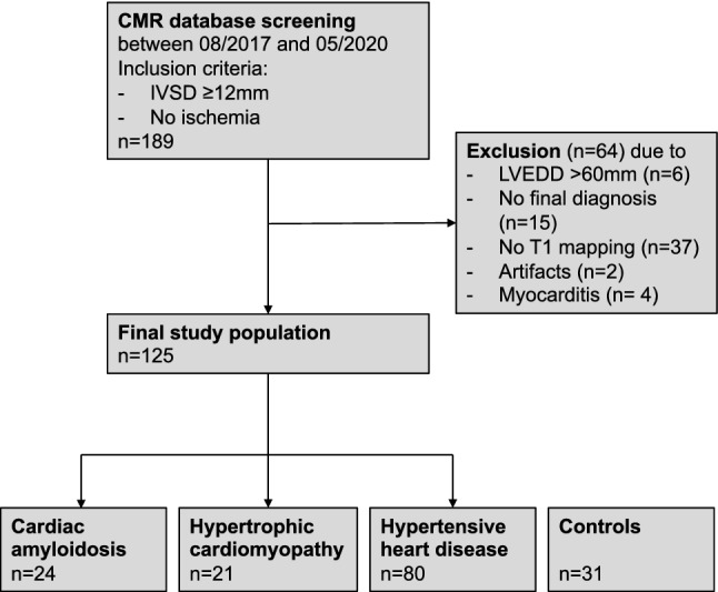
One hundred twenty-five patients with increased LV wall thickness (≥ 12 mm end-diastole) who received clinical CMR in a 3 T scanner between 2017 and 2020 were included. 31 subjects without structural heart disease served as controls. Native T1 was measured as global mean value from 3 LV short axis slices. The study was registered at German clinical trial registry (DRKS00022048).

RESULTS

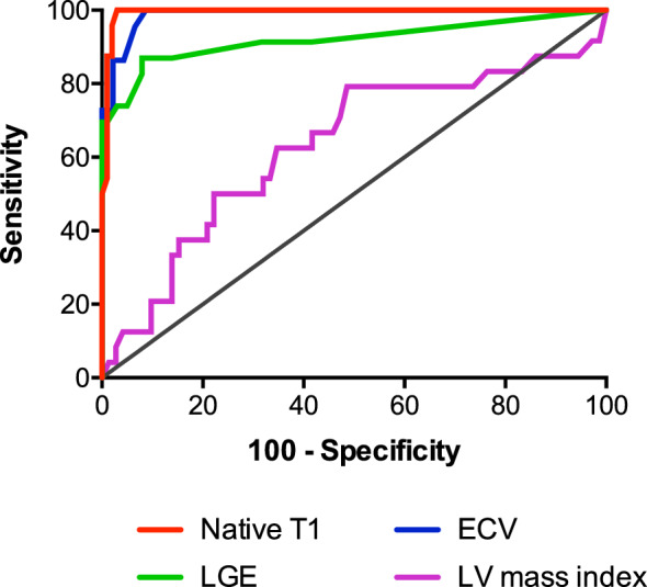
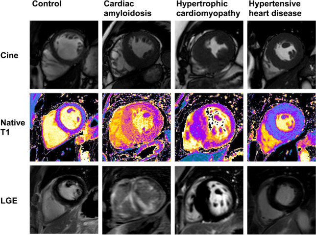
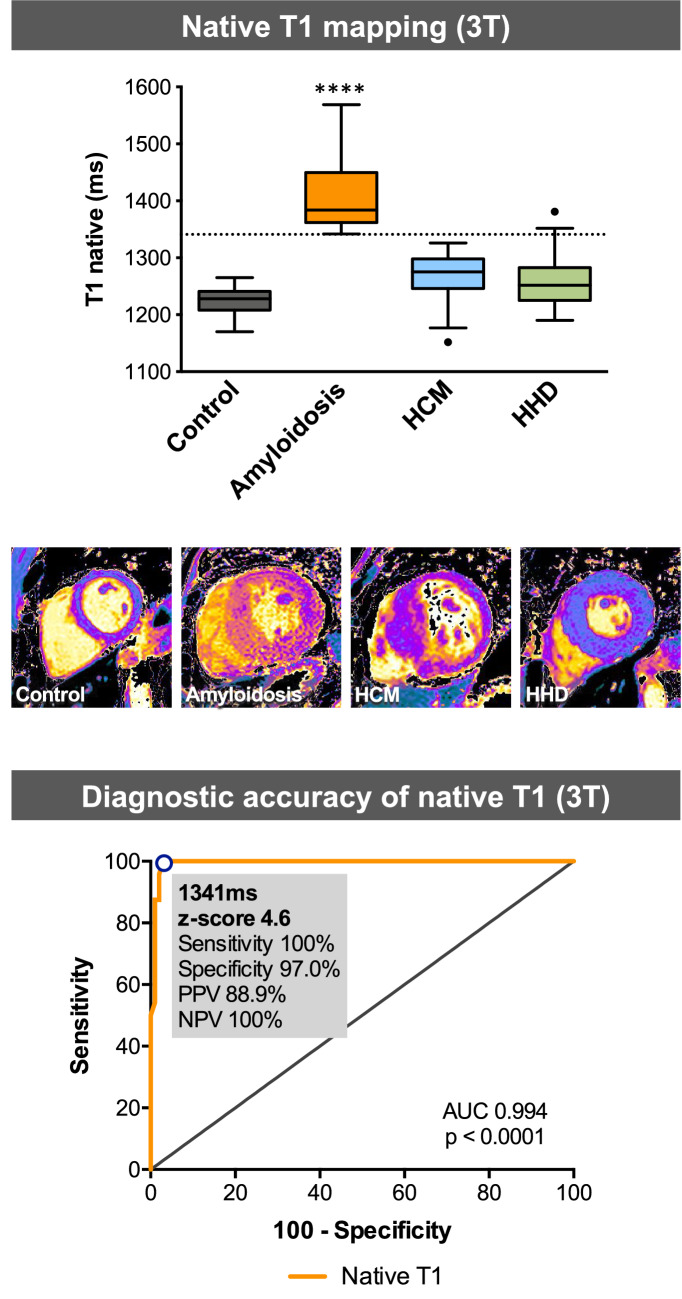
Mean age of the patients was 66 ± 14 years, 83% were males. CA was present in 24 patients, 21 patients had hypertrophic cardiomyopathy (HCM), 80 patients suffered from hypertensive heart disease (HHD). Native T1 times were higher in patients with CA (1409 ± 59 ms, p < 0.0001) compared to healthy controls (1225 ± 21 ms), HCM (1266 ± 44 ms) and HHD (1257 ± 41 ms). HCM and HHD patients did not differ in their native T1 times but were increased compared to control (p < 0.01). ROC analysis of native T1 demonstrated an area under the curve for the detection of CA vs. HCM and HHD of 0.9938 (p < 0.0001), which was higher than that of extracellular volume (0.9876) or quantitative late gadolinium enhancement (0.9406; both p < 0.0001). The optimal cut-off value of native T1 to diagnose CA was 1341 ms (sensitivity 100%, specificity 97%).

CONCLUSION

Non-contrast CMR imaging with native T1 mapping provides high diagnostic accuracy to diagnose cardiac amyloidosis in patients with left ventricular hypertrophy.

摘要

背景

心脏磁共振(CMR)结合参数图可提高心肌组织特征描述的准确性。我们研究了心脏磁共振的心肌 T1 mapping 技术在诊断左心室肥厚患者心脏淀粉样变性中的作用。

方法

2017 年至 2020 年期间,在 3T 扫描仪中进行临床 CMR 检查的 125 例左心室壁增厚(舒张末期≥12mm)的患者被纳入研究。31 例无结构性心脏病的患者作为对照组。从 3 个左心室短轴切片中测量整体平均 T1 值。该研究在德国临床试验注册中心(DRKS00022048)注册。

结果

患者的平均年龄为 66±14 岁,83%为男性。24 例患者存在心脏淀粉样变性,21 例患者患有肥厚型心肌病(HCM),80 例患者患有高血压性心脏病(HHD)。与健康对照组(1225±21ms)、HCM(1266±44ms)和 HHD(1257±41ms)相比,心脏淀粉样变性患者的 T1 值更高(1409±59ms,p<0.0001)。HCM 和 HHD 患者的 T1 值无差异,但均高于对照组(p<0.01)。T1 值的 ROC 分析显示,用于检测心脏淀粉样变性与 HCM 和 HHD 的曲线下面积为 0.9938(p<0.0001),高于细胞外容积(0.9876)或定量延迟钆增强(0.9406;均 p<0.0001)。诊断心脏淀粉样变性的 T1 值最佳截断值为 1341ms(敏感性 100%,特异性 97%)。

结论

非对比增强心脏磁共振成像 T1 mapping 可提供诊断左心室肥厚患者心脏淀粉样变性的高准确性。

https://cdn.ncbi.nlm.nih.gov/pmc/blobs/016b/9998594/39265a25c2f9/392_2022_2005_Fig4_HTML.jpg
https://cdn.ncbi.nlm.nih.gov/pmc/blobs/016b/9998594/ca3faa97cd47/392_2022_2005_Fig1_HTML.jpg
https://cdn.ncbi.nlm.nih.gov/pmc/blobs/016b/9998594/8eecf7d735c3/392_2022_2005_Fig2_HTML.jpg
https://cdn.ncbi.nlm.nih.gov/pmc/blobs/016b/9998594/e51743000893/392_2022_2005_Fig3_HTML.jpg
https://cdn.ncbi.nlm.nih.gov/pmc/blobs/016b/9998594/39265a25c2f9/392_2022_2005_Fig4_HTML.jpg

相似文献

1
Native T1 mapping for the diagnosis of cardiac amyloidosis in patients with left ventricular hypertrophy.用于诊断左心室肥厚患者心脏淀粉样变性的 T1 mapping 技术。
Clin Res Cardiol. 2023 Mar;112(3):334-342. doi: 10.1007/s00392-022-02005-2. Epub 2022 Mar 31.
2
T1 Mapping in Discrimination of Hypertrophic Phenotypes: Hypertensive Heart Disease and Hypertrophic Cardiomyopathy: Findings From the International T1 Multicenter Cardiovascular Magnetic Resonance Study.T1 Mapping在肥厚型表型鉴别中的应用:高血压性心脏病和肥厚型心肌病:国际T1多中心心血管磁共振研究结果
Circ Cardiovasc Imaging. 2015 Dec;8(12). doi: 10.1161/CIRCIMAGING.115.003285.
3
Multi-parametric assessment of left ventricular hypertrophy using late gadolinium enhancement, T1 mapping and strain-encoded cardiovascular magnetic resonance.应用钆延迟增强、T1 mapping 及应变编码心血管磁共振技术对左心室肥厚进行多参数评估。
J Cardiovasc Magn Reson. 2021 Jul 12;23(1):92. doi: 10.1186/s12968-021-00775-8.
4
Diagnostic value of the novel CMR parameter "myocardial transit-time" (MyoTT) for the assessment of microvascular changes in cardiac amyloidosis and hypertrophic cardiomyopathy.新型心脏磁共振参数“心肌通过时间”(MyoTT)评估心脏淀粉样变和肥厚型心肌病微血管改变的诊断价值。
Clin Res Cardiol. 2021 Jan;110(1):136-145. doi: 10.1007/s00392-020-01661-6. Epub 2020 May 5.
5
Hypertensive heart disease versus hypertrophic cardiomyopathy: multi-parametric cardiovascular magnetic resonance discriminators when end-diastolic wall thickness ≥ 15 mm.高血压性心脏病与肥厚型心肌病:舒张末期室壁厚度≥15mm时的多参数心血管磁共振鉴别指标
Eur Radiol. 2017 Mar;27(3):1125-1135. doi: 10.1007/s00330-016-4468-2. Epub 2016 Jul 1.
6
The diagnostic value of multiparameter cardiovascular magnetic resonance for early detection of light-chain amyloidosis from hypertrophic cardiomyopathy patients.多参数心血管磁共振对肥厚型心肌病患者早期检测轻链淀粉样变性的诊断价值。
Front Cardiovasc Med. 2022 Oct 18;9:1017097. doi: 10.3389/fcvm.2022.1017097. eCollection 2022.
7
[Late gadolinium enhancement and T1 mapping for the diagnosis of cardiac amyloidosis].[延迟钆增强及T1成像在心脏淀粉样变性诊断中的应用]
Zhonghua Wei Zhong Bing Ji Jiu Yi Xue. 2019 Dec;31(12):1538-1541. doi: 10.3760/cma.j.issn.2095-4352.2019.12.021.
8
Cardiovascular magnetic resonance feature tracking strain analysis for discrimination between hypertensive heart disease and hypertrophic cardiomyopathy.心血管磁共振特征追踪应变分析在鉴别高血压性心脏病与肥厚型心肌病中的应用。
PLoS One. 2019 Aug 21;14(8):e0221061. doi: 10.1371/journal.pone.0221061. eCollection 2019.
9
Radiomic Analysis of Myocardial Native T Imaging Discriminates Between Hypertensive Heart Disease and Hypertrophic Cardiomyopathy.心肌固有 T 成像的放射组学分析可区分高血压性心脏病与肥厚型心肌病。
JACC Cardiovasc Imaging. 2019 Oct;12(10):1946-1954. doi: 10.1016/j.jcmg.2018.11.024. Epub 2019 Jan 16.
10
MRI differentiation of cardiomyopathy showing left ventricular hypertrophy and heart failure: differentiation between cardiac amyloidosis, hypertrophic cardiomyopathy, and hypertensive heart disease.MRI 鉴别表现为左心室肥厚和心力衰竭的心肌病:心脏淀粉样变性、肥厚型心肌病和高血压性心脏病的鉴别。
Jpn J Radiol. 2013 Oct;31(10):693-700. doi: 10.1007/s11604-013-0238-0. Epub 2013 Aug 31.

引用本文的文献

1
Amide proton transfer-weighted imaging and T1 mapping as diagnostic tools to evaluate high-risk histopathologic phenotypes before surgery in rectal adenocarcinoma and their correlation with Ki-67 expression:a two-center study.酰胺质子转移加权成像和T1映射作为诊断工具在术前评估直肠腺癌高危组织病理学表型及其与Ki-67表达的相关性:一项双中心研究
Front Oncol. 2025 May 19;15:1403890. doi: 10.3389/fonc.2025.1403890. eCollection 2025.
2
Mid-ventricular hypertrophic cardiomyopathy with apical aneurysm: a multimodality imaging case report.伴有心尖部动脉瘤的心室中部肥厚型心肌病:一例多模态影像学病例报告
Arch Peru Cardiol Cir Cardiovasc. 2025 Feb 12;6(1):44-48. doi: 10.47487/apcyccv.v6i1.452. eCollection 2025 Jan-Mar.
3

本文引用的文献

1
2021 ESC Guidelines for the diagnosis and treatment of acute and chronic heart failure.2021年欧洲心脏病学会急性和慢性心力衰竭诊断与治疗指南。
Eur Heart J. 2021 Sep 21;42(36):3599-3726. doi: 10.1093/eurheartj/ehab368.
2
Diagnosis and treatment of cardiac amyloidosis: a position statement of the ESC Working Group on Myocardial and Pericardial Diseases.心脏淀粉样变性的诊断与治疗:欧洲心脏病学会心肌与心包疾病工作组的立场声明
Eur Heart J. 2021 Apr 21;42(16):1554-1568. doi: 10.1093/eurheartj/ehab072.
3
Diagnosis and treatment of cardiac amyloidosis: position statement of the German Cardiac Society (DGK).
Role of Cardiac Magnetic Resonance in Inflammatory and Infiltrative Cardiomyopathies: A Narrative Review.
心脏磁共振在炎症性和浸润性心肌病中的作用:一项叙述性综述。
J Clin Med. 2024 Aug 12;13(16):4733. doi: 10.3390/jcm13164733.
4
Cardiac magnetic resonance imaging for discrimination of hypertensive heart disease and hypertrophic cardiomyopathy: a systematic review and meta-analysis.心脏磁共振成像用于鉴别高血压性心脏病和肥厚型心肌病:一项系统评价和荟萃分析。
Front Cardiovasc Med. 2024 Aug 2;11:1421013. doi: 10.3389/fcvm.2024.1421013. eCollection 2024.
5
Multi-parametric non-contrast cardiac magnetic resonance for the differentiation between cardiac amyloidosis and hypertrophic cardiomyopathy.多参数非对比心脏磁共振成像在心脏淀粉样变性和肥厚型心肌病鉴别诊断中的应用。
Clin Res Cardiol. 2024 Mar;113(3):469-480. doi: 10.1007/s00392-023-02348-4. Epub 2023 Dec 14.
6
Certainties and Uncertainties of Cardiac Magnetic Resonance Imaging in Athletes.运动员心脏磁共振成像的确定性与不确定性
J Cardiovasc Dev Dis. 2022 Oct 20;9(10):361. doi: 10.3390/jcdd9100361.
心脏淀粉样变性的诊断和治疗:德国心脏病学会(DGK)立场声明。
Clin Res Cardiol. 2021 Apr;110(4):479-506. doi: 10.1007/s00392-020-01799-3. Epub 2021 Jan 18.
4
Diagnostic value of cardiovascular magnetic resonance in comparison to endomyocardial biopsy in cardiac amyloidosis: a multi-centre study.心血管磁共振诊断价值与心肌活检在心脏淀粉样变性中的比较:一项多中心研究。
Clin Res Cardiol. 2021 Apr;110(4):555-568. doi: 10.1007/s00392-020-01771-1. Epub 2020 Nov 10.
5
Native T1 Mapping, Extracellular Volume Mapping, and Late Gadolinium Enhancement in Cardiac Amyloidosis: A Meta-Analysis.心肌淀粉样变性中心肌 T1 mapping、细胞外容积 mapping 和钆延迟增强:荟萃分析。
JACC Cardiovasc Imaging. 2020 Jun;13(6):1299-1310. doi: 10.1016/j.jcmg.2020.03.010.
6
Cardiac Amyloidosis: Evolving Diagnosis and Management: A Scientific Statement From the American Heart Association.心脏淀粉样变性:不断发展的诊断和治疗——美国心脏协会的科学声明。
Circulation. 2020 Jul 7;142(1):e7-e22. doi: 10.1161/CIR.0000000000000792. Epub 2020 Jun 1.
7
Multimodality imaging in cardiac amyloidosis: a primer for cardiologists.心脏淀粉样变的多模态影像学:心内科医生的入门指南。
Eur Heart J Cardiovasc Imaging. 2020 Aug 1;21(8):833-844. doi: 10.1093/ehjci/jeaa063.
8
Standardized image interpretation and post-processing in cardiovascular magnetic resonance - 2020 update : Society for Cardiovascular Magnetic Resonance (SCMR): Board of Trustees Task Force on Standardized Post-Processing.心血管磁共振标准化图像解读和后处理 - 2020 年更新:心血管磁共振学会(SCMR):标准化后处理董事会信托基金工作组。
J Cardiovasc Magn Reson. 2020 Mar 12;22(1):19. doi: 10.1186/s12968-020-00610-6.
9
Z-score mapping for standardized analysis and reporting of cardiovascular magnetic resonance modified Look-Locker inversion recovery (MOLLI) T1 data: Normal behavior and validation in patients with amyloidosis.Z 分数映射用于心血管磁共振改良 Look-Locker 反转恢复(MOLLI)T1 数据的标准化分析和报告:淀粉样变性患者的正常表现和验证。
J Cardiovasc Magn Reson. 2020 Jan 20;22(1):6. doi: 10.1186/s12968-019-0595-7.
10
How to Image Cardiac Amyloidosis: A Practical Approach.如何成像心脏淀粉样变性:实用方法。
JACC Cardiovasc Imaging. 2020 Jun;13(6):1368-1383. doi: 10.1016/j.jcmg.2019.07.015. Epub 2019 Oct 11.