Department of Clinical Airway Research, Fraunhofer Institute for Toxicology and Experimental Medicine ITEM, 30625, Hannover, Germany.
German Center for Lung Research (BREATH), Hannover, Germany.
Sci Rep. 2022 Apr 4;12(1):5620. doi: 10.1038/s41598-022-09399-z.
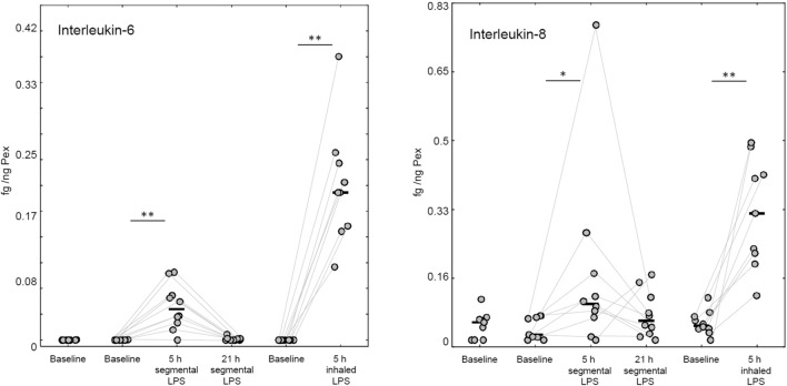
Particles in exhaled air (PEx) are generated when collapsed small airways reopen during breathing. PEx can be noninvasively collected by particle impaction, allowing the analysis of undiluted epithelial lining fluid (ELF). We used the endotoxin (LPS) challenge model to proof the concept that PEx can be used to monitor inflammatory changes in the lung. In this pilot study PEx were collected from ten healthy nonsmoking subjects using the PExA instrument twice before and twice after a segmental LPS challenge (5, 21 h). Following a 4-week washout period, PEx were collected during the week before and 5 h after a whole lung LPS inhalation challenge. PEx biomarkers were compared to blood, bronchoalveolar lavage (BAL) following segmental challenge and induced sputum (ISP) following inhalation challenge. A clear LPS-induced inflammatory response was detectable in BAL fluid, ISP and blood. Albumin and surfactant-protein D were detectable in all PEx samples. While most baseline cytokines were close to or below the detection limit, the median (IQR) IL-6 and IL-8 concentrations in PEx increased significantly after segmental (0.04 (0.03; 0.06) fg/ng PEx; 0.10 (0.08; 0.17) fg/ng PEx) and inhalation LPS challenge (0.19 (0.15; 0.23) fg/ng PEx; 0.32 (0.23; 0.42) fg/ng PEx). Using a highly sensitive analysis platform, we were able to detect a cytokine response in PEx during the early phase of LPS-induced inflammation. This will broaden the spectrum of applications for this noninvasive method to monitor inflammatory processes in the lung, including its use in clinical trials for respiratory drug development.Trial registration: The study has been registered on 07.02.2017 at Clinicaltrials.gov (NCT03044327).
呼气颗粒(PEx)是在呼吸过程中小气道塌陷再开放时产生的。PEx 可以通过颗粒撞击非侵入性地收集,从而可以对未稀释的上皮衬液(ELF)进行分析。我们使用内毒素(LPS)挑战模型证明了这样一个概念,即 PEx 可用于监测肺部炎症变化。在这项初步研究中,使用 PExA 仪器在十名健康不吸烟的受试者进行了五次分段 LPS 挑战前和两次挑战后(5、21 小时)收集 PEx。经过四周的洗脱期后,在整个肺 LPS 吸入挑战前一周和 5 小时后收集 PEx。将 PEx 生物标志物与血液、分段挑战后的支气管肺泡灌洗液(BAL)和吸入挑战后的诱导痰(ISP)进行比较。在 BAL 液、ISP 和血液中均能检测到明显的 LPS 诱导的炎症反应。白蛋白和表面活性蛋白 D 可在所有 PEx 样本中检测到。虽然大多数基线细胞因子接近或低于检测限,但在分段 LPS 挑战后(0.04(0.03;0.06)fg/ng PEx;0.10(0.08;0.17)fg/ng PEx)和吸入 LPS 挑战后(0.19(0.15;0.23)fg/ng PEx;0.32(0.23;0.42)fg/ng PEx),PEx 中 IL-6 和 IL-8 浓度中位数(IQR)显著增加。使用高灵敏度分析平台,我们能够在 LPS 诱导的炎症早期阶段检测到 PEx 中的细胞因子反应。这将拓宽这种非侵入性方法监测肺部炎症过程的应用范围,包括将其用于呼吸药物开发的临床试验。
该研究于 2017 年 2 月 7 日在 Clinicaltrials.gov 上注册(NCT03044327)。