Peel Brandon, Lee Whal, Hussein Nabil, Yoo Shi-Joon
Center for Image-Guided Innovation and Therapeutic Intervention, The Hospital for Sick Children, University of Toronto, Toronto, Ontario, Canada.
Department of Radiology, Seoul National University Hospital, Seoul, Korea.
JTCVS Tech. 2022 Jan 19;12:132-142. doi: 10.1016/j.xjtc.2021.12.009. eCollection 2022 Apr.
Three-dimensional (3D) printed models are widely accepted for use in training of various surgical procedures for congenital heart disease; however, their physical properties have been considered suboptimum for procedures. We created silicone molded models produced using a novel "parting and assembly" strategy and compared their suitability for hands-on training with that of conventional 3D printed models.
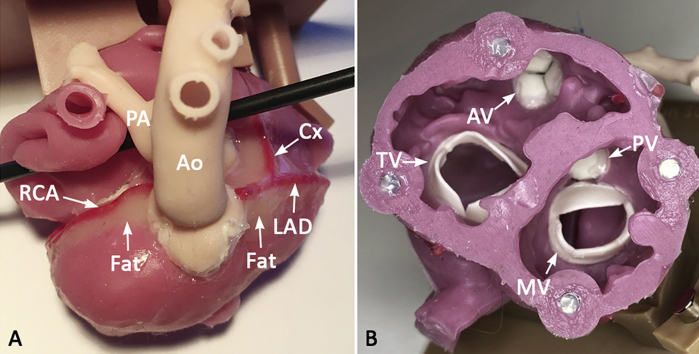
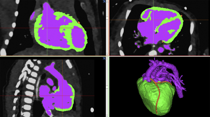
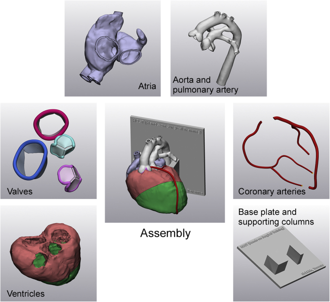
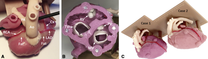
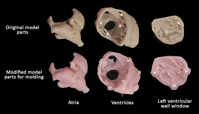
Computed tomography imaging data from 2 patients with transposition of the great arteries were used. The heart was divided into multiple parts (atria, ventricles, great arteries, coronary arteries, and valves), and molds of each part were created. The parts reproduced by silicone molding were assembled using an adhesive agent. In an online course, 2 silicone molded models and 1 3D printed model were used for training of 34 surgeons. A questionnaire was distributed to these surgeons aimed at assessing the suitability of the models for the arterial switch operation (ASO).
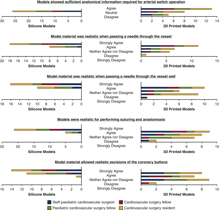
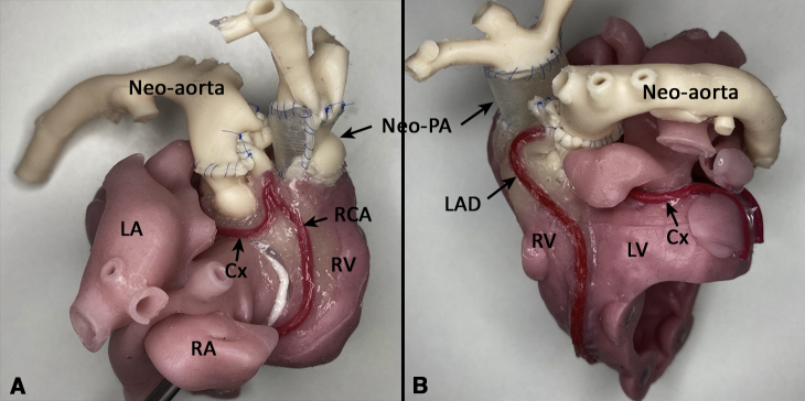
The silicone molded models showed excellent anatomic detail, high elasticity, and high resistance to tearing. The cost per model, based on the production of 50 models, was slightly higher for the silicone molded models compared with the 3D printed models. All 26 surgeons who completed the questionnaire reported that the silicone molded models provided sufficient anatomic information, but only 19% said the same for the 3D printed models. All surgeons also considered the silicone models to be realistic when passing a needle, cutting vessels, suturing, and excision of the coronary buttons, as opposed to <46% for the 3D printed models.
Silicone molding of models for the ASO is feasible by applying a "parting and assembly" strategy. Silicone molded models provide excellent physical properties that are far superior to those of 3D printed models for surgical simulation.
三维(3D)打印模型在先天性心脏病各种手术操作培训中被广泛接受;然而,其物理特性被认为在手术操作中并非最佳。我们创建了采用新型“分离与组装”策略生产的硅胶模制模型,并将其用于实践培训的适用性与传统3D打印模型进行了比较。
使用来自2例大动脉转位患者的计算机断层扫描成像数据。将心脏分为多个部分(心房、心室、大动脉、冠状动脉和瓣膜),并制作每个部分的模具。通过硅胶模制复制的部分使用粘合剂进行组装。在一个在线课程中,使用2个硅胶模制模型和1个3D打印模型对34名外科医生进行培训。向这些外科医生发放了一份问卷,旨在评估模型对动脉调转术(ASO)的适用性。
硅胶模制模型显示出出色的解剖细节、高弹性和高抗撕裂性。基于生产50个模型计算,每个硅胶模制模型的成本比3D打印模型略高。所有26名完成问卷的外科医生报告称,硅胶模制模型提供了足够的解剖信息,但只有19%的人对3D打印模型给出了相同评价。所有外科医生还认为,在穿针、切割血管、缝合以及切除冠状动脉纽扣时,硅胶模型更逼真,而认为3D打印模型逼真的比例不到46%。
通过应用“分离与组装”策略,对ASO模型进行硅胶模制是可行的。硅胶模制模型具有出色的物理特性,在手术模拟方面远优于3D打印模型。