Ilié Marius, Benzaquen Jonathan, Tourniaire Paul, Heeke Simon, Ayache Nicholas, Delingette Hervé, Long-Mira Elodie, Lassalle Sandra, Hamila Marame, Fayada Julien, Otto Josiane, Cohen Charlotte, Gomez-Caro Abel, Berthet Jean-Philippe, Marquette Charles-Hugo, Hofman Véronique, Bontoux Christophe, Hofman Paul
Laboratory of Clinical and Experimental Pathology, Centre Hospitalier Universitaire de Nice, FHU OncoAge, Université Côte d'Azur, 06000 Nice, France.
Hospital-Related Biobank (BB-0033-00025), Centre Hospitalier Universitaire de Nice, FHU OncoAge, Université Côte d'Azur, 06000 Nice, France.
Cancers (Basel). 2022 Mar 29;14(7):1740. doi: 10.3390/cancers14071740.
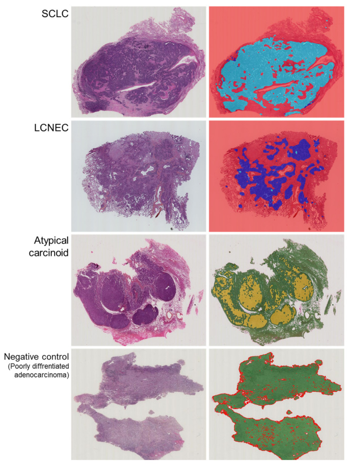
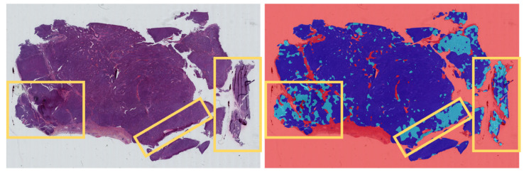
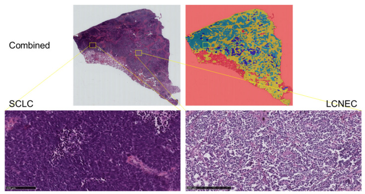
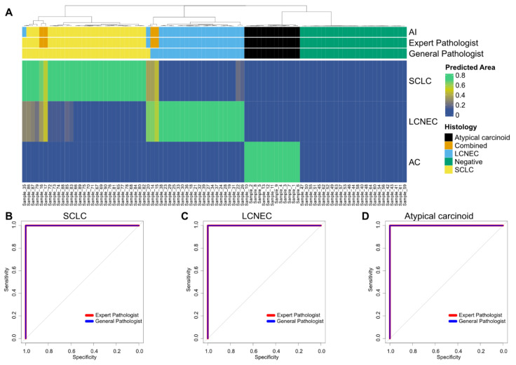
The histological distinction of lung neuroendocrine carcinoma, including small cell lung carcinoma (SCLC), large cell neuroendocrine carcinoma (LCNEC) and atypical carcinoid (AC), can be challenging in some cases, while bearing prognostic and therapeutic significance. To assist pathologists with the differentiation of histologic subtyping, we applied a deep learning classifier equipped with a convolutional neural network (CNN) to recognize lung neuroendocrine neoplasms. Slides of primary lung SCLC, LCNEC and AC were obtained from the Laboratory of Clinical and Experimental Pathology (University Hospital Nice, France). Three thoracic pathologists blindly established gold standard diagnoses. The HALO-AI module (Indica Labs, UK) trained with 18,752 image tiles extracted from 60 slides (SCLC = 20, LCNEC = 20, AC = 20 cases) was then tested on 90 slides (SCLC = 26, LCNEC = 22, AC = 13 and combined SCLC with LCNEC = 4 cases; NSCLC = 25 cases) by F1-score and accuracy. A HALO-AI correct area distribution (AD) cutoff of 50% or more was required to credit the CNN with the correct diagnosis. The tumor maps were false colored and displayed side by side to original hematoxylin and eosin slides with superimposed pathologist annotations. The trained HALO-AI yielded a mean F1-score of 0.99 (95% CI, 0.939-0.999) on the testing set. Our CNN model, providing further larger validation, has the potential to work side by side with the pathologist to accurately differentiate between the different lung neuroendocrine carcinoma in challenging cases.
肺神经内分泌癌的组织学区分,包括小细胞肺癌(SCLC)、大细胞神经内分泌癌(LCNEC)和非典型类癌(AC),在某些情况下可能具有挑战性,但具有预后和治疗意义。为了帮助病理学家进行组织学亚型的鉴别,我们应用了一个配备卷积神经网络(CNN)的深度学习分类器来识别肺神经内分泌肿瘤。原发性肺SCLC、LCNEC和AC的切片取自临床与实验病理学实验室(法国尼斯大学医院)。三位胸科病理学家在不知情的情况下确立了金标准诊断。然后,使用从60张切片(SCLC = 20例、LCNEC = 20例、AC = 20例)中提取的18,752个图像块训练的HALO-AI模块(英国Indica Labs公司),通过F1分数和准确率在90张切片(SCLC = 26例、LCNEC = 22例、AC = 13例以及SCLC与LCNEC合并 = 4例;非小细胞肺癌 = 25例)上进行测试。要认定CNN诊断正确,HALO-AI正确面积分布(AD)的截断值需达到50%或更高。肿瘤图谱进行了伪彩色处理,并与带有病理学家叠加注释的原始苏木精和伊红切片并排显示。经过训练的HALO-AI在测试集上的平均F1分数为0.99(95%可信区间,0.939 - 0.999)。我们的CNN模型经过了进一步更大规模的验证,有潜力在具有挑战性的病例中与病理学家并肩工作,以准确区分不同类型的肺神经内分泌癌。