Department of Surgery, Cedars-Sinai Medical Center, Los Angeles, California, USA.
Department of Pathology and Laboratory Medicine, Cedars-Sinai Medical Center, Los Angeles, California, USA.
Sci Rep. 2019 Feb 6;9(1):1483. doi: 10.1038/s41598-018-37638-9.
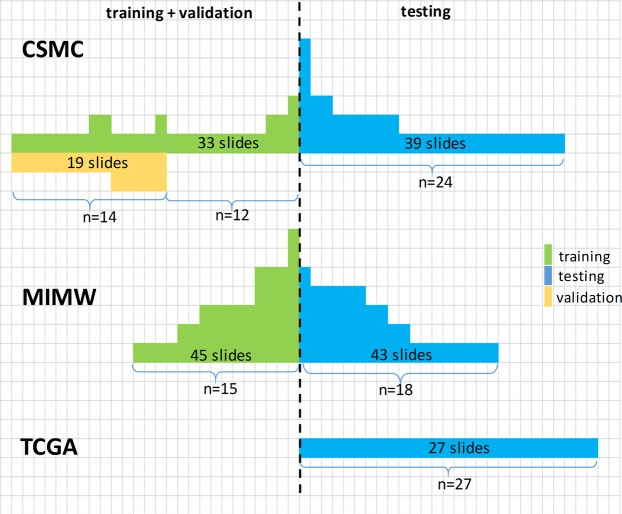
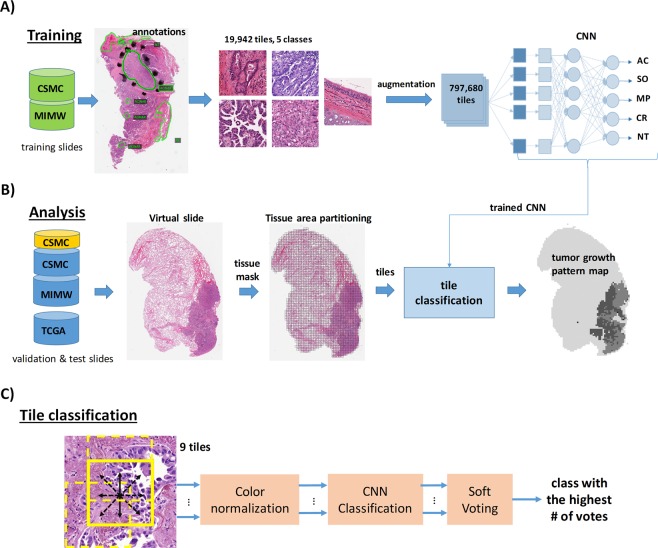
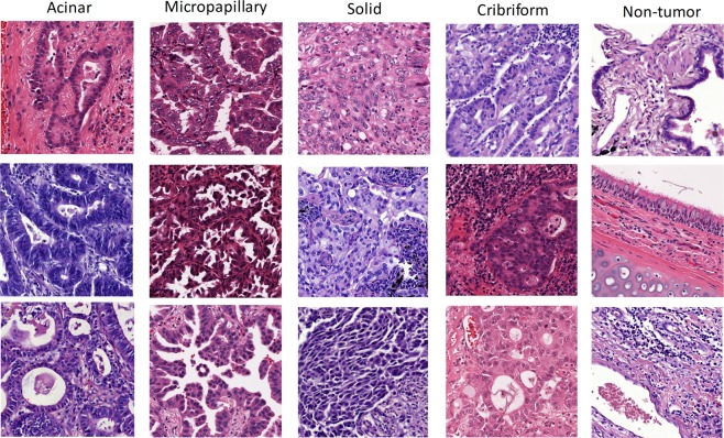
During the diagnostic workup of lung adenocarcinomas (LAC), pathologists evaluate distinct histological tumor growth patterns. The percentage of each pattern on multiple slides bears prognostic significance. To assist with the quantification of growth patterns, we constructed a pipeline equipped with a convolutional neural network (CNN) and soft-voting as the decision function to recognize solid, micropapillary, acinar, and cribriform growth patterns, and non-tumor areas. Slides of primary LAC were obtained from Cedars-Sinai Medical Center (CSMC), the Military Institute of Medicine in Warsaw and the TCGA portal. Several CNN models trained with 19,924 image tiles extracted from 78 slides (MIMW and CSMC) were evaluated on 128 test slides from the three sites by F1-score and accuracy using manual tumor annotations by pathologist. The best CNN yielded F1-scores of 0.91 (solid), 0.76 (micropapillary), 0.74 (acinar), 0.6 (cribriform), and 0.96 (non-tumor) respectively. The overall accuracy of distinguishing the five tissue classes was 89.24%. Slide-based accuracy in the CSMC set (88.5%) was significantly better (p < 2.3E-4) than the accuracy in the MIMW (84.2%) and TCGA (84%) sets due to superior slide quality. Our model can work side-by-side with a pathologist to accurately quantify the percentages of growth patterns in tumors with mixed LAC patterns.
在肺腺癌(LAC)的诊断工作中,病理学家评估不同的组织学肿瘤生长模式。多种切片上每种模式的百分比具有预后意义。为了协助量化生长模式,我们构建了一个配备卷积神经网络(CNN)和软投票作为决策函数的管道,以识别实体、微乳头状、腺泡和筛状生长模式以及非肿瘤区域。原发 LAC 的切片来自雪松西奈医疗中心(CSMC)、华沙军事医学研究所和 TCGA 门户。从 MIMW 和 CSMC 的 78 张切片中提取 19,924 个图像块,用多个 CNN 模型进行训练,然后用 F1 评分和病理学家手动肿瘤标注的准确性对来自三个地点的 128 个测试切片进行评估。最佳的 CNN 在实体(0.91)、微乳头状(0.76)、腺泡(0.74)、筛状(0.6)和非肿瘤(0.96)方面的 F1 评分分别为 0.91。区分这五个组织类别的总体准确率为 89.24%。CSMC 组的切片准确率(88.5%)明显优于 MIMW 组(84.2%)和 TCGA 组(84%),因为 CSMC 组的切片质量更高。我们的模型可以与病理学家并肩工作,准确地量化混合 LAC 模式肿瘤中生长模式的百分比。