Gottlieb Assaf, Toledano-Furman Naama, Prabhakara Karthik S, Kumar Akshita, Caplan Henry W, Bedi Supinder, Cox Charles S, Olson Scott D
Center for Precision Health, School of Biomedical Informatics, University of Texas Health Science Center, Houston, TX, 77030, USA.
Department of Pediatric Surgery, McGovern School of Medicine, University of Texas Health Science Center at Houston, 6431 Fannin St., Houston, TX, 77030, USA.
Sci Rep. 2022 Apr 15;12(1):6289. doi: 10.1038/s41598-022-10419-1.
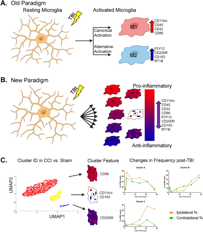
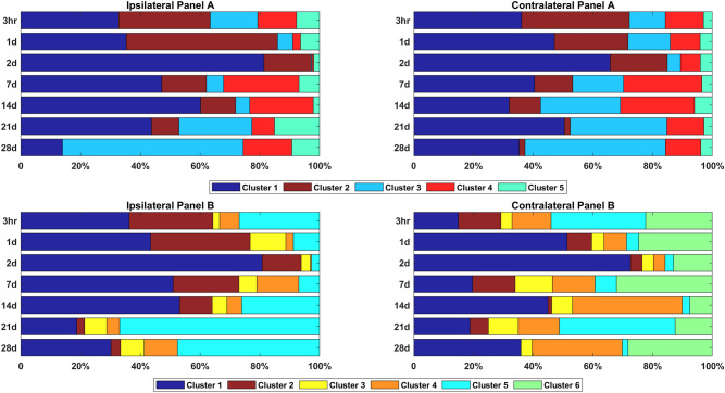
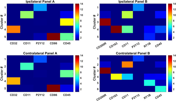
Traumatic brain injury (TBI) results in a cascade of cellular responses, which produce neuroinflammation, partly due to the activation of microglia. Accurate identification of microglial populations is key to understanding therapeutic approaches that modify microglial responses to TBI and improve long-term outcome measures. Notably, previous studies often utilized an outdated convention to describe microglial phenotypes. We conducted a temporal analysis of the response to controlled cortical impact (CCI) in rat microglia between ipsilateral and contralateral hemispheres across seven time points, identified microglia through expression of activation markers including CD45, CD11b/c, and p2y12 receptor and evaluated their activation state using additional markers of CD32, CD86, RT1B, CD200R, and CD163. We identified unique sub-populations of microglial cells that express individual or combination of activation markers across time points. We further portrayed how the size of these sub-populations changes through time, corresponding to stages in TBI response. We described longitudinal changes in microglial population after CCI in two different locations using activation markers, showing clear separation into cellular sub-populations that feature different temporal patterns of markers after injury. These changes may aid in understanding the symptomatic progression following TBI and help define microglial subpopulations beyond the outdated M1/M2 paradigm.
创伤性脑损伤(TBI)会引发一系列细胞反应,进而导致神经炎症,部分原因是小胶质细胞的激活。准确识别小胶质细胞群体是理解改变小胶质细胞对TBI反应并改善长期预后指标的治疗方法的关键。值得注意的是,以往的研究常常采用过时的惯例来描述小胶质细胞表型。我们对大鼠小胶质细胞在同侧和对侧半球对控制性皮质撞击(CCI)的反应进行了时间分析,跨越七个时间点,通过包括CD45、CD11b/c和p2y12受体在内的激活标志物的表达来识别小胶质细胞,并使用CD32、CD86、RT1B、CD200R和CD163等其他标志物评估它们的激活状态。我们识别出了在各个时间点表达单个或组合激活标志物的独特小胶质细胞亚群。我们进一步描绘了这些亚群的大小如何随时间变化,这与TBI反应的阶段相对应。我们使用激活标志物描述了CCI后两个不同位置小胶质细胞群体的纵向变化,显示出明显分离为具有损伤后不同标志物时间模式的细胞亚群。这些变化可能有助于理解TBI后的症状进展,并有助于定义超越过时的M1/M2范式的小胶质细胞亚群。