• 文献检索
  • 文档翻译
  • 深度研究
  • 学术资讯
  • Suppr Zotero 插件Zotero 插件
  • 邀请有礼
  • 套餐&价格
  • 历史记录
应用&插件
Suppr Zotero 插件Zotero 插件浏览器插件Mac 客户端Windows 客户端微信小程序
定价
高级版会员购买积分包购买API积分包
服务
文献检索文档翻译深度研究API 文档MCP 服务
关于我们
关于 Suppr公司介绍联系我们用户协议隐私条款
关注我们

Suppr 超能文献

核心技术专利:CN118964589B侵权必究
粤ICP备2023148730 号-1Suppr @ 2026

文献检索

告别复杂PubMed语法,用中文像聊天一样搜索,搜遍4000万医学文献。AI智能推荐,让科研检索更轻松。

立即免费搜索

文件翻译

保留排版,准确专业,支持PDF/Word/PPT等文件格式,支持 12+语言互译。

免费翻译文档

深度研究

AI帮你快速写综述,25分钟生成高质量综述,智能提取关键信息,辅助科研写作。

立即免费体验

病理性近视眼中的乳头和视盘周围区域的结构异常及其对应的视野缺损。

Structural Abnormalities in the Papillary and Peripapillary Areas and Corresponding Visual Field Defects in Eyes With Pathologic Myopia.

机构信息

Department of Ophthalmology and Visual Science, Tokyo Medical and Dental University, Tokyo, Japan.

出版信息

Invest Ophthalmol Vis Sci. 2022 Apr 1;63(4):13. doi: 10.1167/iovs.63.4.13.

DOI:10.1167/iovs.63.4.13
PMID:35446343
原文链接:https://pmc.ncbi.nlm.nih.gov/articles/PMC9034715/
Abstract

PURPOSE

To identify structural abnormalities in the papillary and peripapillary area in eyes with pathologic myopia (PM) and normal IOP and to determine their relationship to visual field (VF) defects.

METHODS

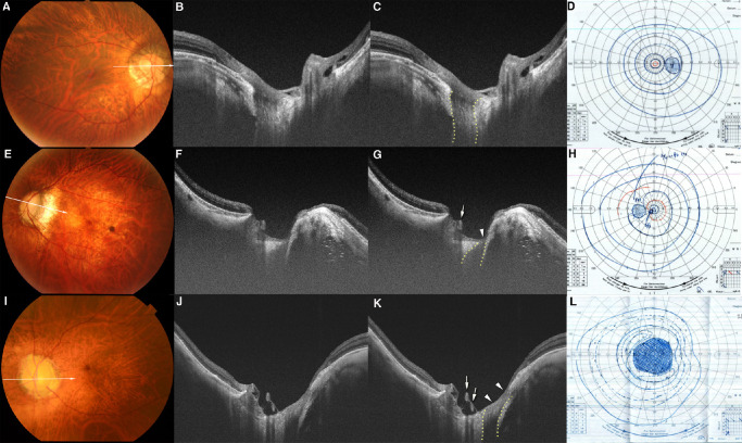
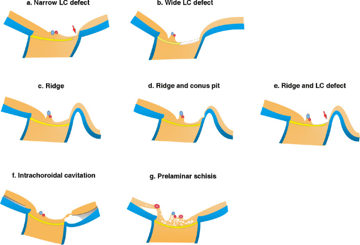
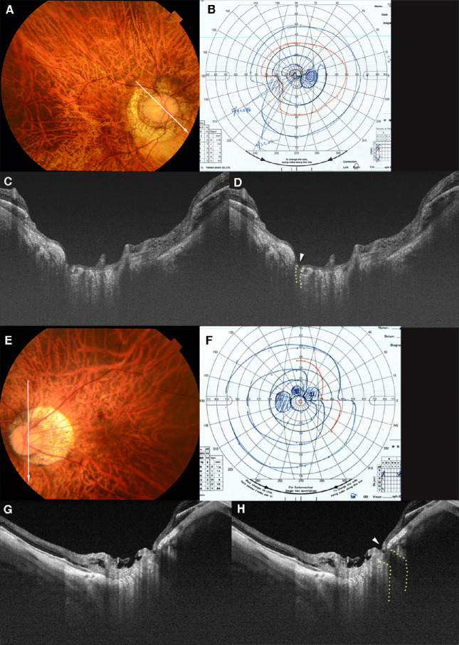
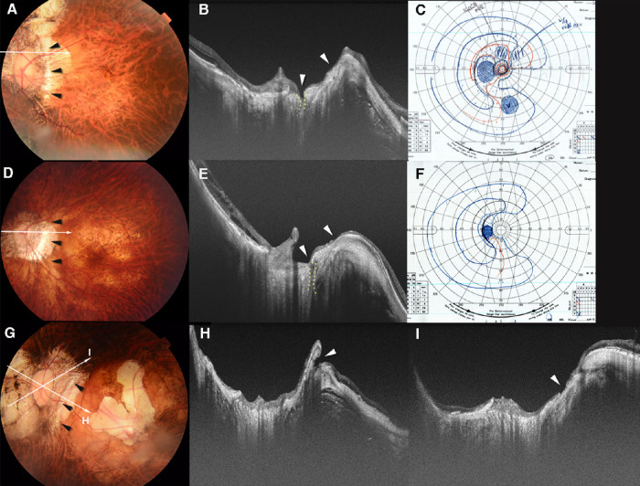
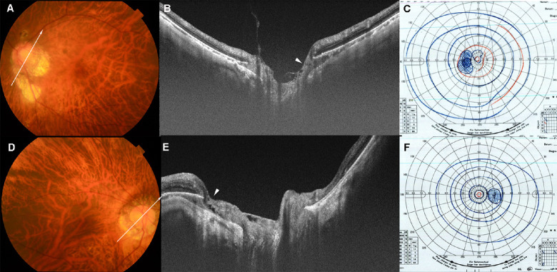
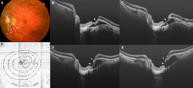
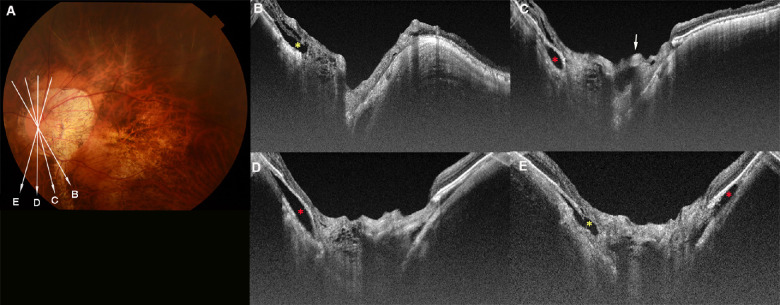
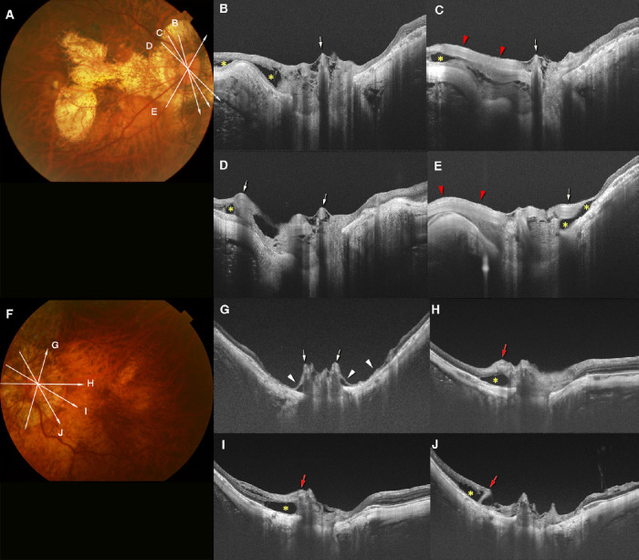
One hundred eight eyes of 70 patients with PM were retrospectively studied. The disc-centered swept source optical coherence tomographic images and the Goldmann VF recorded within 1 year of the optical coherence tomographic examination were analyzed. Four structural abnormalities were identified: lamina cribrosa (LC) defects, ridge protrusions, intrachoroidal cavitations (ICC), and prelaminar schisis. The correspondence of the VF defects with the structural abnormalities was assessed.

RESULTS

The mean age, axial length, and optic disc area of the 108 eyes were 58.7 ± 10.0 years, 31.1 ± 2.4 mm, and 4.7 ± 2.2 mm2, respectively. Eighty-five of the 108 eyes (78.7%) had at least one abnormality and 49.4% (42/85) had two or more abnormalities. LC defects, ridge protrusions, ICC, and prelaminar schisis were detected in 47.2%, 33.3%, 21.3%, and 30.6% of the eyes, respectively. VF defects at the corresponding areas of these structural abnormalities were seen in 63% of the eyes with LC defects, 39% of the eyes with ridge protrusions, and 21% of the eyes with ICC.

CONCLUSIONS

Four kinds of structural abnormalities with corresponding VF defects are commonly observed in the papillary and peripapillary region of eyes with PM. The presence of these abnormalities suggests a possibility of functional damage.

摘要

目的

在眼压正常的病理性近视(PM)眼的乳头和视盘周围区域识别结构异常,并确定其与视野(VF)缺损的关系。

方法

回顾性研究了 70 例 PM 患者的 108 只眼。对以视盘为中心的扫频源光学相干断层扫描图像和光学相干断层扫描检查后 1 年内记录的 Goldmann VF 进行了分析。共发现 4 种结构异常:筛板缺陷、脊突、脉络膜内陷和视盘前裂。评估了 VF 缺损与结构异常的对应关系。

结果

108 只眼中的平均年龄、眼轴长度和视盘面积分别为 58.7±10.0 岁、31.1±2.4mm 和 4.7±2.2mm²。108 只眼中 85 只(78.7%)至少存在 1 种异常,49.4%(42/85)存在 2 种或更多异常。47.2%、33.3%、21.3%和 30.6%的眼中分别发现了筛板缺陷、脊突、脉络膜内陷和视盘前裂。在 63%的存在筛板缺陷的眼中、39%的存在脊突的眼中和 21%的存在脉络膜内陷的眼中,相应区域可见 VF 缺损。

结论

在 PM 眼的乳头和视盘周围区域,常见到 4 种伴有相应 VF 缺损的结构异常。这些异常的存在提示存在功能损伤的可能性。

https://cdn.ncbi.nlm.nih.gov/pmc/blobs/6f84/9034715/02b836f1cc04/iovs-63-4-13-f008.jpg
https://cdn.ncbi.nlm.nih.gov/pmc/blobs/6f84/9034715/ff4589b29c8a/iovs-63-4-13-f001.jpg
https://cdn.ncbi.nlm.nih.gov/pmc/blobs/6f84/9034715/9b360566f05f/iovs-63-4-13-f002.jpg
https://cdn.ncbi.nlm.nih.gov/pmc/blobs/6f84/9034715/dbf8bc833776/iovs-63-4-13-f003.jpg
https://cdn.ncbi.nlm.nih.gov/pmc/blobs/6f84/9034715/dc9474221e33/iovs-63-4-13-f004.jpg
https://cdn.ncbi.nlm.nih.gov/pmc/blobs/6f84/9034715/23b3cdb48e28/iovs-63-4-13-f005.jpg
https://cdn.ncbi.nlm.nih.gov/pmc/blobs/6f84/9034715/ded1c32fe604/iovs-63-4-13-f006.jpg
https://cdn.ncbi.nlm.nih.gov/pmc/blobs/6f84/9034715/9e5a6b36c919/iovs-63-4-13-f007.jpg
https://cdn.ncbi.nlm.nih.gov/pmc/blobs/6f84/9034715/02b836f1cc04/iovs-63-4-13-f008.jpg

相似文献

1
Structural Abnormalities in the Papillary and Peripapillary Areas and Corresponding Visual Field Defects in Eyes With Pathologic Myopia.病理性近视眼中的乳头和视盘周围区域的结构异常及其对应的视野缺损。
Invest Ophthalmol Vis Sci. 2022 Apr 1;63(4):13. doi: 10.1167/iovs.63.4.13.
2
Optic Nerve Head Abnormalities in Nonpathologic High Myopia and the Relationship With Visual Field.非病理性高度近视的视神经乳头异常及其与视野的关系
Asia Pac J Ophthalmol (Phila). 2023;12(5):460-467. doi: 10.1097/APO.0000000000000636. Epub 2023 Sep 22.
3
Effect of focal lamina cribrosa defect on glaucomatous visual field progression.视盘局限性筛板缺损对青光眼视野进展的影响。
Ophthalmology. 2014 Aug;121(8):1524-30. doi: 10.1016/j.ophtha.2014.02.017. Epub 2014 Mar 31.
4
Lamina cribrosa defects and optic disc morphology in primary open angle glaucoma with high myopia.高度近视原发性开角型青光眼中的筛板缺损与视盘形态
PLoS One. 2014 Dec 22;9(12):e115313. doi: 10.1371/journal.pone.0115313. eCollection 2014.
5
Multiple Temporal Lamina Cribrosa Defects in Myopic Eyes with Glaucoma and Their Association with Visual Field Defects.近视性青光眼眼中多个时间性板层筛状缺损及其与视野缺损的关系。
Ophthalmology. 2017 Nov;124(11):1600-1611. doi: 10.1016/j.ophtha.2017.04.027. Epub 2017 May 24.
6
Optic disc torsion presenting as unilateral glaucomatous-appearing visual field defect in young myopic Korean eyes.视盘扭转导致年轻高度近视韩国人单侧青光眼样视野缺损。
Ophthalmology. 2014 May;121(5):1013-9. doi: 10.1016/j.ophtha.2013.11.014. Epub 2014 Feb 5.
7
Microstructure of Peripapillary Atrophy and Subsequent Visual Field Progression in Treated Primary Open-Angle Glaucoma.治疗原发性开角型青光眼后患区萎缩的微观结构与随后的视野进展。
Ophthalmology. 2016 Mar;123(3):542-51. doi: 10.1016/j.ophtha.2015.10.061. Epub 2015 Dec 12.
8
Optic Disc Characteristics and Visual Field Progression in Normal Tension Glaucoma Patients With Tilted Optic Discs.正常眼压性青光眼伴视盘倾斜患者的视盘特征与视野进展
J Glaucoma. 2016 Nov;25(11):901-907. doi: 10.1097/IJG.0000000000000565.
9
Effect of peripapillary tilt direction and magnitude on central visual field defects in primary open-angle glaucoma with high myopia.高度近视性原发性开角型青光眼的视盘倾斜方向和程度对中心视野缺损的影响。
Jpn J Ophthalmol. 2020 Jul;64(4):414-422. doi: 10.1007/s10384-020-00747-y. Epub 2020 Jun 22.
10
Association between peripheral visual field defects and focal lamina cribrosa defects in highly myopic eyes.高度近视眼中周边视野缺损与局限性筛板缺损之间的关联
Jpn J Ophthalmol. 2022 May;66(3):285-295. doi: 10.1007/s10384-022-00909-0. Epub 2022 Mar 20.

引用本文的文献

1
Detection of optic disc abnormalities using retro-mode scanning laser ophthalmoscopy.使用后向模式扫描激光检眼镜检测视盘异常。
Jpn J Ophthalmol. 2025 Sep 12. doi: 10.1007/s10384-025-01260-w.
2
Clinical features of high myopia with optic nerve head prelaminar schisis: Wuhan high myopia study.伴有视神经乳头层前劈裂的高度近视的临床特征:武汉高度近视研究
Eye (Lond). 2025 Sep 4. doi: 10.1038/s41433-025-03939-3.
3
Interplay Between γ-Zone Peripapillary Atrophy and Optic Disc Parameters in Central Visual Field Impairment in Highly Myopic Eyes.

本文引用的文献

1
The Effect of Ametropia on Glaucomatous Visual Field Loss.屈光不正对青光眼性视野缺损的影响。
J Clin Med. 2021 Jun 25;10(13):2796. doi: 10.3390/jcm10132796.
2
Clinical Features of Advanced Glaucoma With Optic Nerve Head Prelaminar Schisis.伴有视神经乳头板层前劈裂的晚期青光眼的临床特征
Am J Ophthalmol. 2021 Dec;232:17-29. doi: 10.1016/j.ajo.2021.06.007. Epub 2021 Jun 18.
3
IMI Pathologic Myopia.病理性近视
高度近视眼中央视野损害中γ区视乳头周围萎缩与视盘参数之间的相互作用
Invest Ophthalmol Vis Sci. 2025 Jun 2;66(6):74. doi: 10.1167/iovs.66.6.74.
4
Enlarged Blind Spot Linked to Gamma Zone and Peripapillary Hyperreflective Ovoid Mass-Like Structures in Non-Pathological Highly Myopic Eyes.非病理性高度近视眼中扩大的盲点与γ区及视乳头周围高反射卵圆体样结构相关。
Invest Ophthalmol Vis Sci. 2025 Apr 1;66(4):5. doi: 10.1167/iovs.66.4.5.
5
Multimodal imaging of optic nerve head abnormalities in high myopia.高度近视性视神经乳头异常的多模态成像
Front Neurol. 2024 Apr 23;15:1366593. doi: 10.3389/fneur.2024.1366593. eCollection 2024.
6
Papillary and Peripapillary Hemorrhages in Eyes With Pathologic Myopia.病理性近视眼中的视盘及视盘周围出血。
Invest Ophthalmol Vis Sci. 2022 Nov 1;63(12):28. doi: 10.1167/iovs.63.12.28.
7
Optic nerve cavitations in glaucoma suspect and glaucoma patients.可疑青光眼和青光眼患者的视神经空洞形成
Am J Ophthalmol Case Rep. 2022 Oct 20;28:101733. doi: 10.1016/j.ajoc.2022.101733. eCollection 2022 Dec.
8
Three-Dimensional Volume Calculation of Intrachoroidal Cavitation Using Deep-Learning-Based Noise Reduction of Optical Coherence Tomography.基于深度学习的光相干断层扫描降噪技术对脉络膜内空泡的三维容积计算。
Transl Vis Sci Technol. 2022 Jul 8;11(7):1. doi: 10.1167/tvst.11.7.1.
Invest Ophthalmol Vis Sci. 2021 Apr 28;62(5):5. doi: 10.1167/iovs.62.5.5.
4
Association of Optic Nerve Head Prelaminar Schisis With Glaucoma.视乳头前板层裂与青光眼的相关性。
Am J Ophthalmol. 2021 Mar;223:246-258. doi: 10.1016/j.ajo.2020.10.021. Epub 2020 Nov 6.
5
Border Tissue Morphology Is Spatially Associated with Focal Lamina Cribrosa Defect and Deep-Layer Microvasculature Dropout in Open-Angle Glaucoma.眼前节组织形态与开角型青光眼局灶性筛板缺陷和深层微血管丢失呈空间相关。
Am J Ophthalmol. 2019 Jul;203:89-102. doi: 10.1016/j.ajo.2019.02.023. Epub 2019 Feb 28.
6
IMI - Defining and Classifying Myopia: A Proposed Set of Standards for Clinical and Epidemiologic Studies.IMI - 定义和分类近视:临床和流行病学研究的一套标准建议。
Invest Ophthalmol Vis Sci. 2019 Feb 28;60(3):M20-M30. doi: 10.1167/iovs.18-25957.
7
Pulling and Tugging on the Retina: Mechanical Impact of Glaucoma Beyond the Optic Nerve Head.牵拉视网膜:视神经头以外青光眼的机械性影响。
Invest Ophthalmol Vis Sci. 2019 Jan 2;60(1):26-35. doi: 10.1167/iovs.18-25837.
8
Posterior staphyloma in pathologic myopia.病理性近视的后葡萄肿。
Prog Retin Eye Res. 2019 May;70:99-109. doi: 10.1016/j.preteyeres.2018.12.001. Epub 2018 Dec 8.
9
Ametropia, retinal anatomy, and OCT abnormality patterns in glaucoma. 1. Impacts of refractive error and interartery angle.屈光不正、视网膜解剖和青光眼的 OCT 异常模式。1. 屈光不正和动脉间夹角的影响。
J Biomed Opt. 2017 Dec;22(12):1-11. doi: 10.1117/1.JBO.22.12.121713.
10
Peripapillary retinal splitting visualized on OCT in glaucoma and glaucoma suspect patients.青光眼和疑似青光眼患者经光学相干断层扫描(OCT)显示的视乳头周围视网膜劈裂。
PLoS One. 2017 Aug 23;12(8):e0182816. doi: 10.1371/journal.pone.0182816. eCollection 2017.