Department of Orthopedic Surgery, Kindai University Hospital, Osaka-Sayama City, Osaka.
Eur J Histochem. 2022 Apr 22;66(2):3393. doi: 10.4081/ejh.2022.3393.
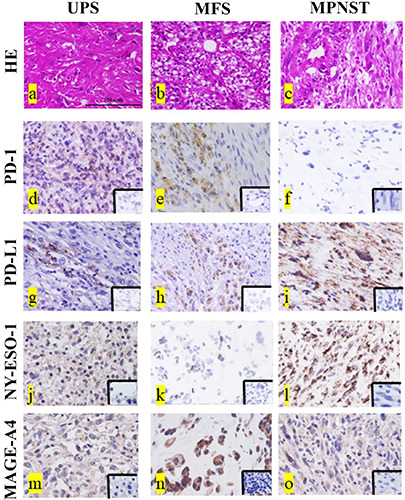
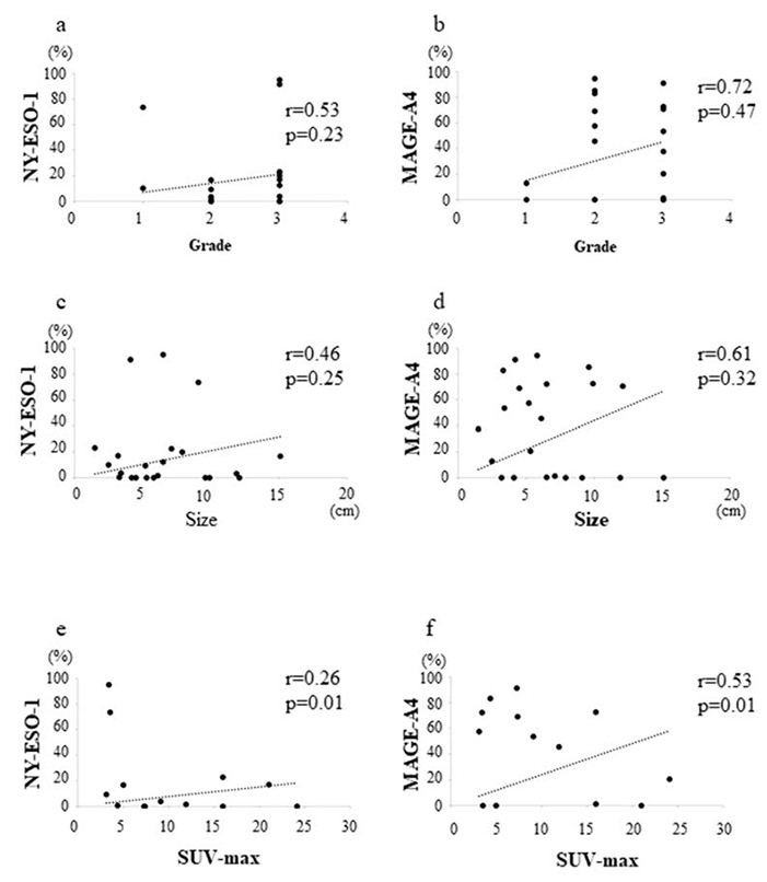
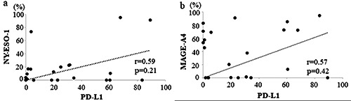
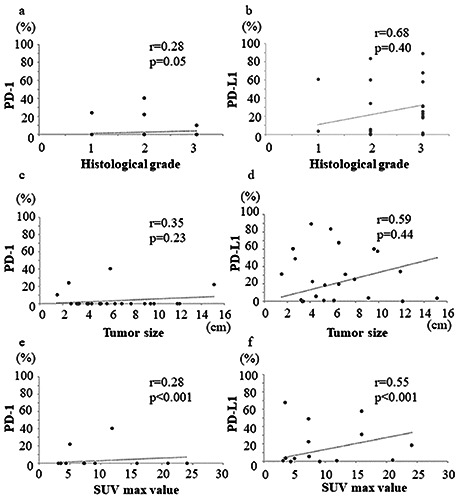
Immunotherapy has altered the treatment paradigm for soft tissue sarcomas (STSs). Considering the limited information regarding the clinical significance of immunohistochemical markers in STS, the purpose of this study was to determine the clinical significance of programmed cell death-1 (PD-1), PD ligand-1 (PD-L1), New York esophageal squamous cell carcinoma-1 (NY-ESO-1), and melanoma-associated antigen-A4 (MAGE-A4) expression in STSs. Twenty-two patients (median age, 72.5 years) with STSs treated at our hospital were included in this study. The specimens obtained at the time of biopsy were used to perform immunostaining for PD-1, PD-L1, NY-ESO, and MAGE-A4. The rates of PD-1-, PD-L1-, NY-ESO-, and MAGE-A4-positive cells and cases were calculated. The correlations among the positive cell rates of the immunohistochemical markers as well as their correlations with the histological grade, tumor size, or maximum standardized uptake (SUVmax) value were also determined. The average rates of PD-1-, PD-L1-, NY-ESO-, and MAGE-A4-positive cells were 4.39%, 28.0%, 18.2%, and 39.4%, respectively. Although the PD-1-positive cell rate showed no correlation with the rates of NY-ESO-1- and MAGE-A4-positive cells, the PD-L1-positive cell rates showed strong positive correlations with the rates of NY-ESO-1- and MAGE-A4-positive cells. PD-1-, PD-L1-, NY-ESO-1-, and MAGE-A4-positive cell rates showed weak to moderate correlations with histological grade or tumor size, while the PD-1-, PD-L1-, and MAGE-A4-positive cell rates showed strong to very strong positive correlations with the SUVmax value. Thus, PD-1, PD-L1, NY-ESO, and MAGE-A4 expressions are correlated and may be involved in the aggressive elements of STSs.
免疫疗法改变了软组织肉瘤(STS)的治疗模式。鉴于免疫组化标志物在 STS 中的临床意义信息有限,本研究旨在确定程序性细胞死亡蛋白-1(PD-1)、PD 配体-1(PD-L1)、纽约食管鳞状细胞癌-1(NY-ESO-1)和黑色素瘤相关抗原-A4(MAGE-A4)在 STS 中的表达的临床意义。本研究纳入了在我院治疗的 22 名 STS 患者(中位年龄 72.5 岁)。活检时获得的标本用于进行 PD-1、PD-L1、NY-ESO 和 MAGE-A4 的免疫组化染色。计算 PD-1、PD-L1、NY-ESO 和 MAGE-A4 阳性细胞和病例的比例。还确定了免疫组化标志物阳性细胞率之间的相关性及其与组织学分级、肿瘤大小或最大标准化摄取(SUVmax)值之间的相关性。PD-1、PD-L1、NY-ESO 和 MAGE-A4 阳性细胞的平均比例分别为 4.39%、28.0%、18.2%和 39.4%。尽管 PD-1 阳性细胞率与 NY-ESO-1 和 MAGE-A4 阳性细胞率无相关性,但 PD-L1 阳性细胞率与 NY-ESO-1 和 MAGE-A4 阳性细胞率呈强正相关。PD-1、PD-L1、NY-ESO-1 和 MAGE-A4 阳性细胞率与组织学分级或肿瘤大小呈弱至中度相关,而 PD-1、PD-L1 和 MAGE-A4 阳性细胞率与 SUVmax 值呈强至非常强的正相关。因此,PD-1、PD-L1、NY-ESO 和 MAGE-A4 的表达相关,可能参与 STS 的侵袭性成分。