Department of Anatomy, Brain Health Research Centre, Brain Research New Zealand, University of Otago, Dunedin, New Zealand; Neurophysiological Pharmacology Section, National Institute of Neurological Disorders and Stroke, National Institutes of Health, 35 Convent Drive, Building 35 Room 1C 903, Bethesda, MD 20892-3702, USA.
Neurophysiological Pharmacology Section, National Institute of Neurological Disorders and Stroke, National Institutes of Health, 35 Convent Drive, Building 35 Room 1C 903, Bethesda, MD 20892-3702, USA.
Exp Neurol. 2022 Aug;354:114089. doi: 10.1016/j.expneurol.2022.114089. Epub 2022 Apr 22.
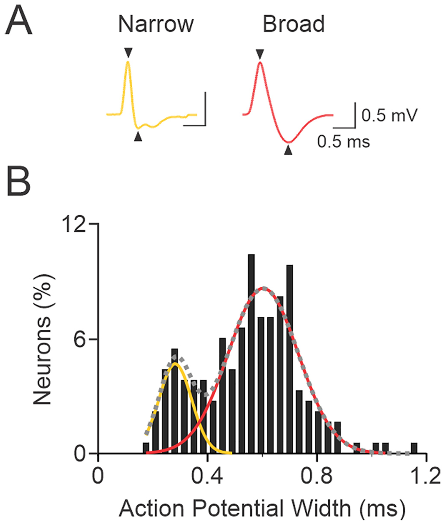
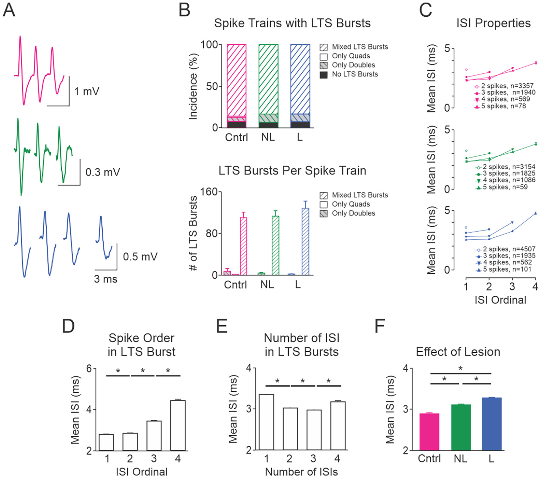
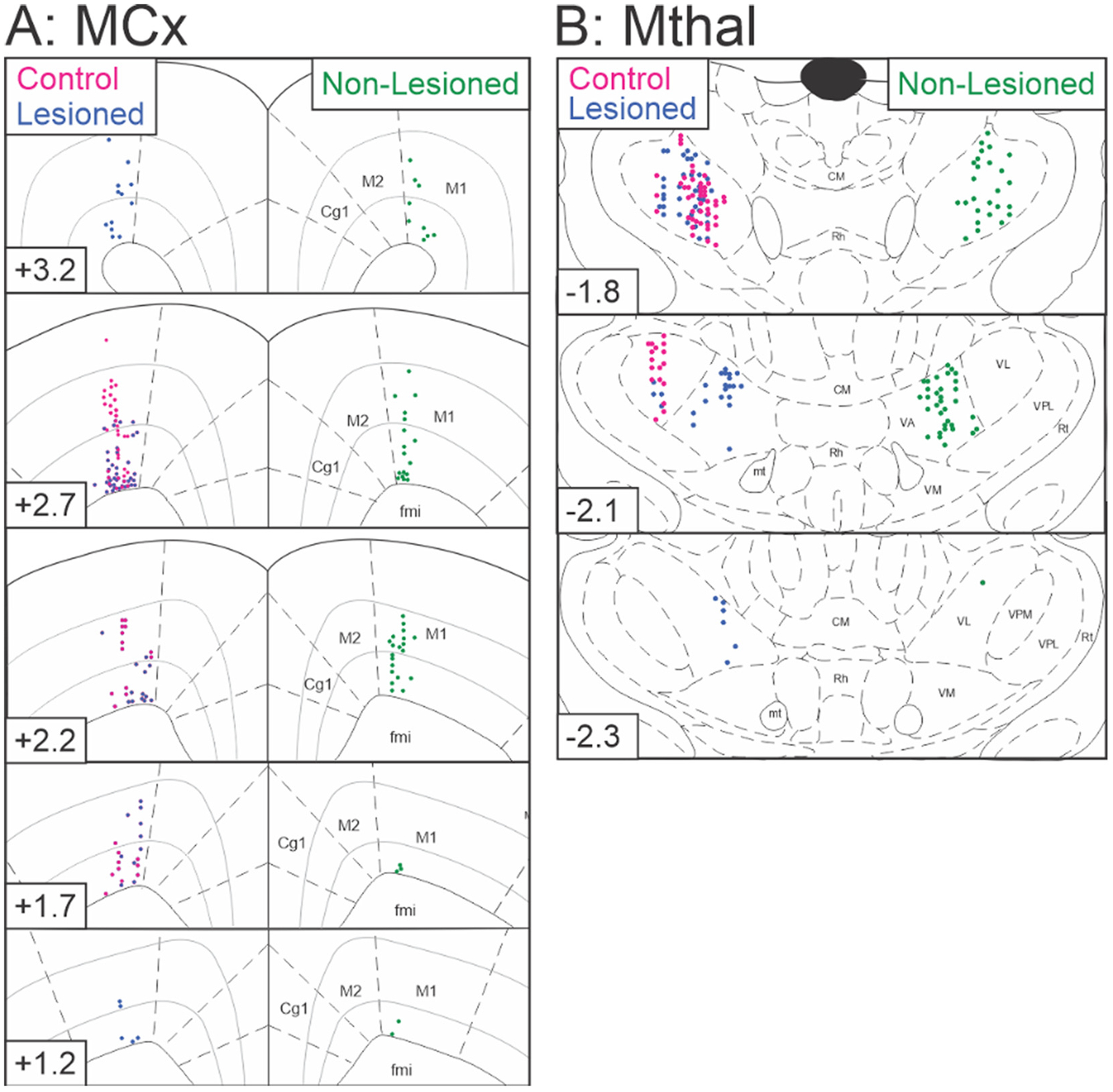
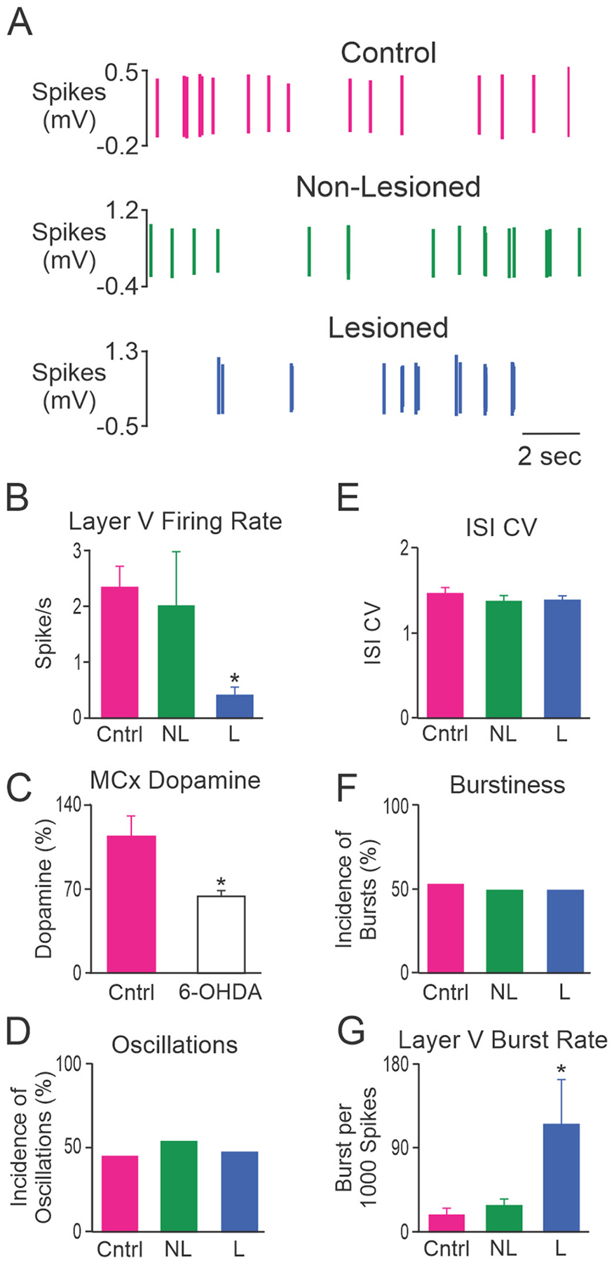
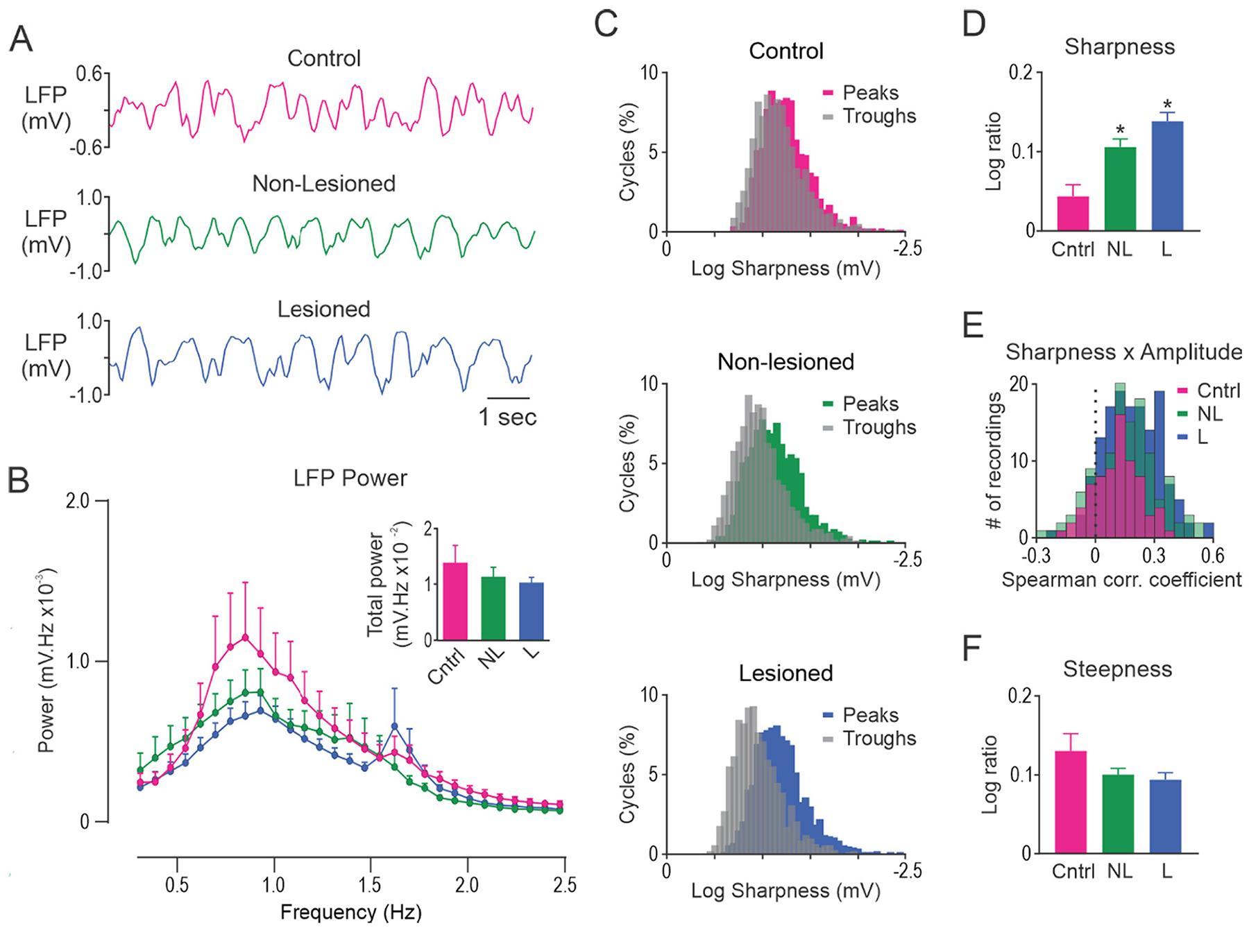
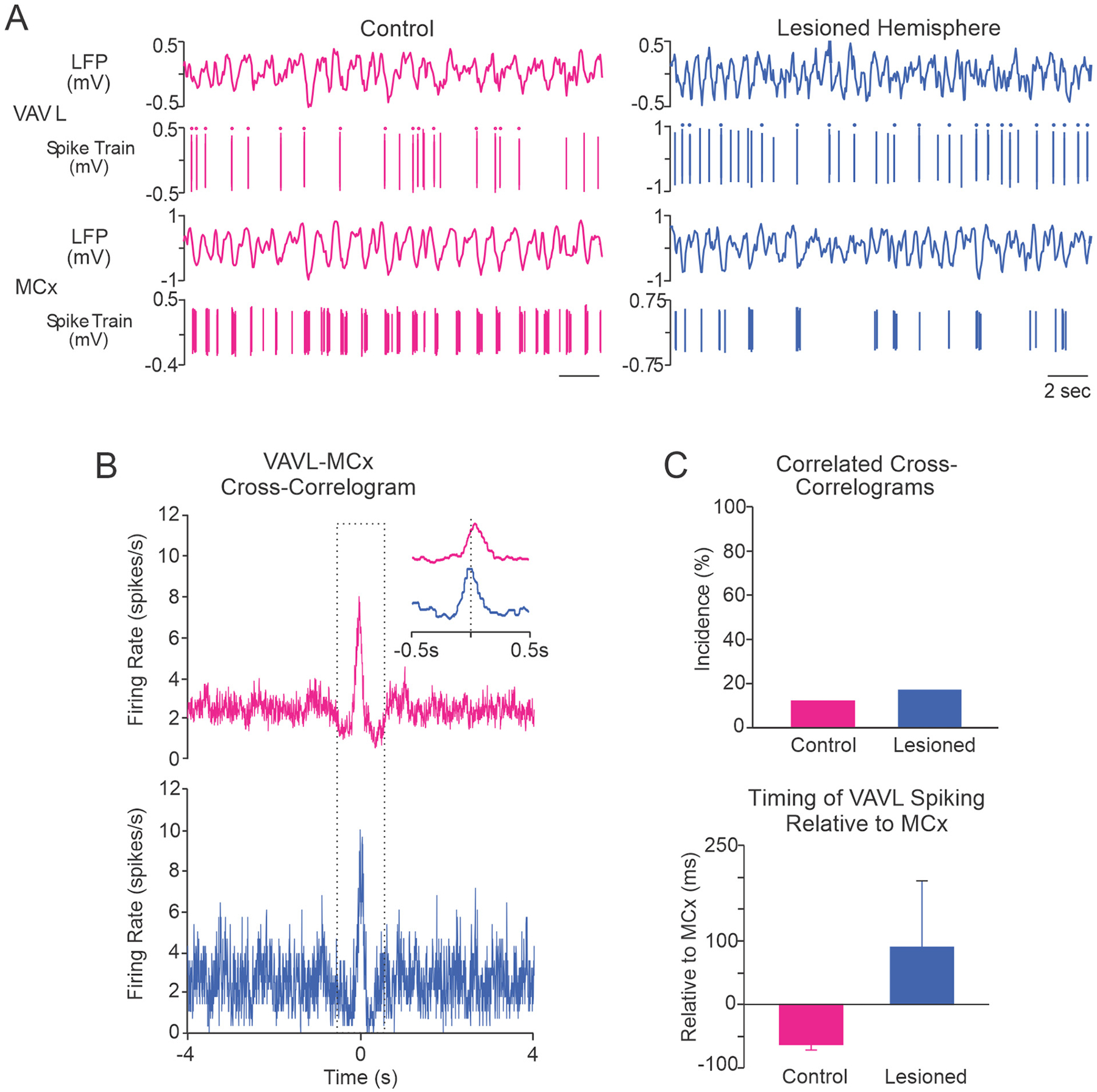
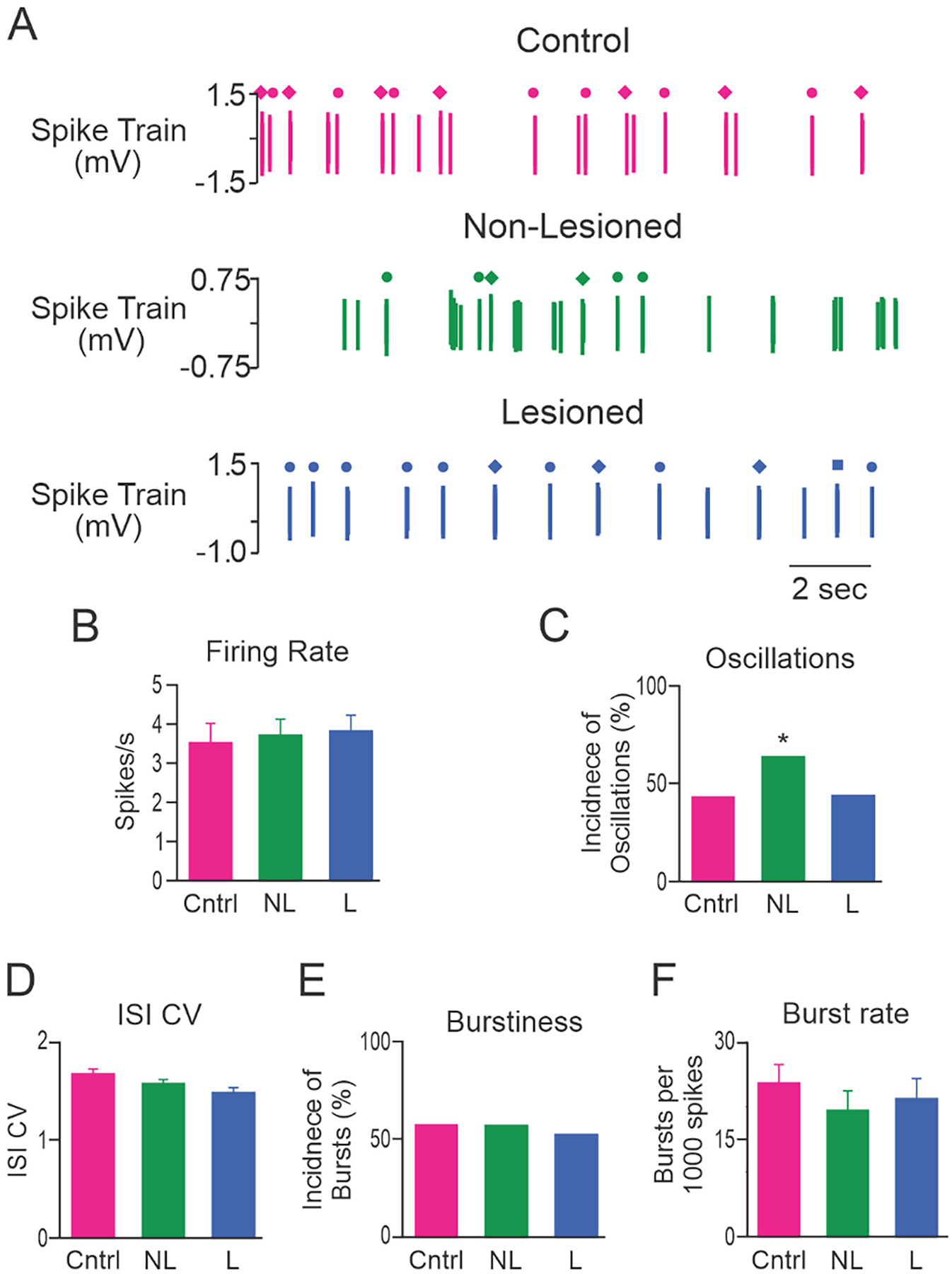
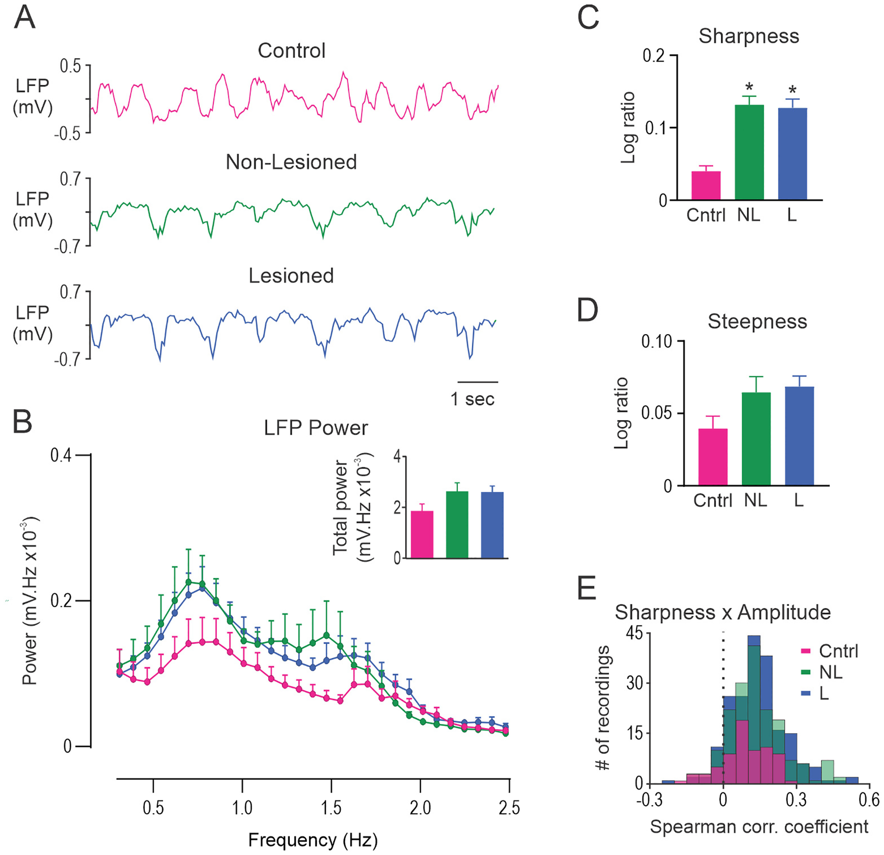
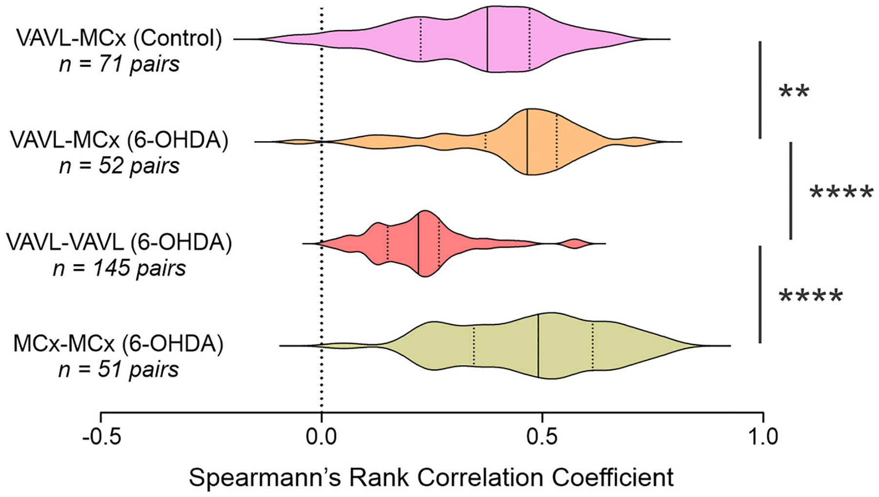
Parkinson's disease (PD) causes bursty and oscillatory activity in basal ganglia output that is thought to contribute to movement deficits through impact on motor thalamus and motor cortex (MCx). We examined the effect of dopamine loss on motor thalamus and motor cortex activity by recording neuronal and LFP activities in ventroanterior-ventrolateral (VAVL) thalamus and MCx in urethane-anesthetised control and parkinsonian rats. Dopamine lesion decreased the firing rate and increased the bursting of putative pyramidal neurons in layer V, but not layer VI, of the MCx without changing other aspects of firing pattern. In contrast, dopamine lesion did not affect VAVL firing rate, pattern or low threshold calcium spike bursts. Slow-wave (~1 Hz) oscillations in LFP recordings were analyzed with conventional power and waveform shape analyses. While dopamine lesion did not influence total power, it was consistently associated with an increase in oscillatory waveform sharpness asymmetry (i.e., sharper troughs vs. peaks) in both motor thalamus and MCx. Furthermore, we found that measures of sharpness asymmetry were positively correlated in paired motor thalamus-MCx recordings, and that correlation coefficients were larger in dopamine lesioned rats. These data support the idea that dysfunctional MCx activity in parkinsonism emerges from subsets of cell groups (e.g. layer V pyramidal neurons) and is evident in the shape but not absolute power of slow-wave oscillations. Hypoactive layer V pyramidal neuron firing in dopamine lesioned rats is unlikely to be driven by VAVL thalamus and may, therefore, reflect the loss of mesocortical dopaminergic afferents and/or changes in intrinsic excitability.
帕金森病(PD)导致基底神经节输出的爆发和振荡活动,据认为这种活动通过对运动丘脑和运动皮层(MCx)的影响导致运动缺陷。我们通过在麻醉的对照和帕金森大鼠的腹侧前腹侧-腹侧外侧(VAVL)丘脑和 MCx 中记录神经元和 LFP 活动,研究了多巴胺丧失对运动丘脑和运动皮层活动的影响。多巴胺损伤降低了 MCx 中第 V 层(但不是第 VI 层)的推测性锥体神经元的放电率并增加了其爆发率,而没有改变其他放电模式方面。相比之下,多巴胺损伤不会影响 VAVL 的放电率、模式或低阈值钙脉冲爆发。使用传统的功率和波形形状分析对 LFP 记录中的慢波(约 1 Hz)振荡进行了分析。虽然多巴胺损伤不影响总功率,但它始终与运动丘脑和 MCx 中的振荡波形锐度不对称性增加相关(即,波谷比波峰更尖锐)。此外,我们发现,在配对的运动丘脑-MCx 记录中,锐度不对称性的测量值呈正相关,并且在多巴胺损伤大鼠中,相关系数更大。这些数据支持运动皮层功能障碍的帕金森病中出现的想法,即源于细胞群亚群(例如,V 层锥体神经元)的活动,并且在慢波振荡的形状而不是绝对功率中明显。多巴胺损伤大鼠中低活性的 V 层锥体神经元放电不太可能由 VAVL 丘脑驱动,因此可能反映了中皮质多巴胺能传入的丧失和/或内在兴奋性的变化。