Silvestri Erica, Moretto Manuela, Facchini Silvia, Castellaro Marco, Anglani Mariagiulia, Monai Elena, D'Avella Domenico, Della Puppa Alessandro, Cecchin Diego, Bertoldo Alessandra, Corbetta Maurizio
Department of Information Engineering, University of Padova, 35131 Padova, Italy.
Padova Neuroscience Center, University of Padova, 35129 Padova, Italy.
Brain Commun. 2022 Apr 8;4(2):fcac082. doi: 10.1093/braincomms/fcac082. eCollection 2022.
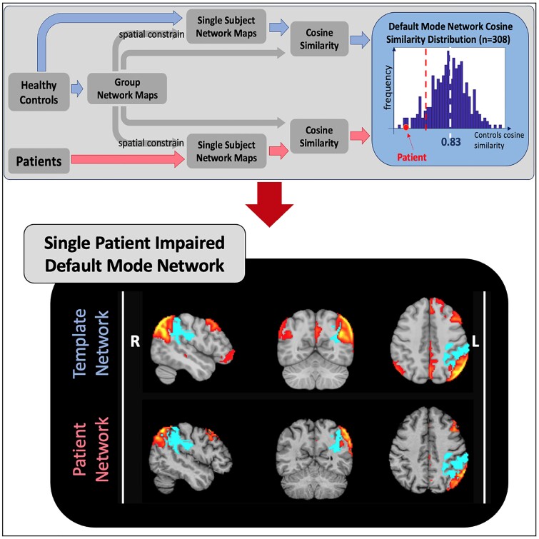
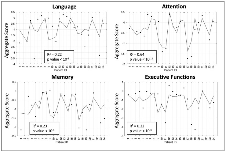
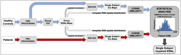
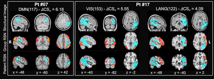
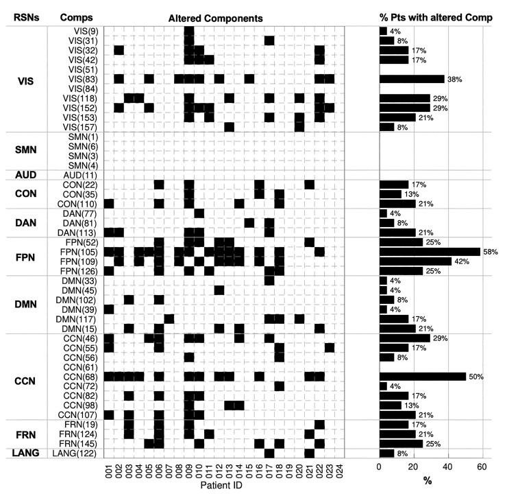
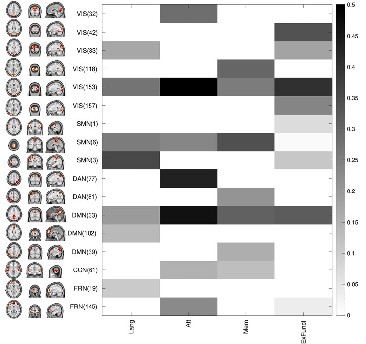
Assessment of impaired/preserved cortical regions in brain tumours is typically performed via intraoperative direct brain stimulation of eloquent areas or task-based functional MRI. One main limitation is that they overlook distal brain regions or networks that could be functionally impaired by the tumour. This study aims (i) to investigate the impact of brain tumours on the cortical synchronization of brain networks measured with resting-state functional magnetic resonance imaging (resting-state networks) both near the lesion and remotely and (ii) to test whether potential changes in resting-state networks correlate with cognitive status. The sample included 24 glioma patients (mean age: 58.1 ± 16.4 years) with different pathological staging. We developed a new method for single subject localization of resting-state networks abnormalities. First, we derived the spatial pattern of the main resting-state networks by means of the group-guided independent component analysis. This was informed by a high-resolution resting-state networks template derived from an independent sample of healthy controls. Second, we developed a spatial similarity index to measure differences in network topography and strength between healthy controls and individual brain tumour patients. Next, we investigated the spatial relationship between altered networks and tumour location. Finally, multivariate analyses related cognitive scores across multiple cognitive domains (attention, language, memory, decision making) with patterns of multi-network abnormality. We found that brain gliomas cause broad alterations of resting-state networks topography that occurred mainly in structurally normal regions outside the tumour and oedema region. Cortical regions near the tumour often showed normal synchronization. Finally, multi-network abnormalities predicted attention deficits. Overall, we present a novel method for the functional localization of resting-state networks abnormalities in individual glioma patients. These abnormalities partially explain cognitive disabilities and shall be carefully navigated during surgery.
评估脑肿瘤中受损/保留的皮质区域通常通过术中对明确区域进行直接脑刺激或基于任务的功能磁共振成像来进行。一个主要限制是它们忽略了可能因肿瘤而功能受损的远端脑区或网络。本研究旨在:(i)研究脑肿瘤对通过静息态功能磁共振成像测量的脑网络皮质同步性的影响(静息态网络),包括病变附近和远处的网络;(ii)测试静息态网络的潜在变化是否与认知状态相关。样本包括24名不同病理分期的神经胶质瘤患者(平均年龄:58.1±16.4岁)。我们开发了一种用于单受试者静息态网络异常定位的新方法。首先,我们通过组引导独立成分分析得出主要静息态网络的空间模式。这是由从健康对照的独立样本中得出的高分辨率静息态网络模板提供信息的。其次,我们开发了一种空间相似性指数来测量健康对照与个体脑肿瘤患者之间网络拓扑和强度的差异。接下来,我们研究了改变的网络与肿瘤位置之间的空间关系。最后,多变量分析将多个认知领域(注意力、语言、记忆、决策)的认知分数与多网络异常模式相关联。我们发现脑胶质瘤会导致静息态网络拓扑结构的广泛改变,主要发生在肿瘤和水肿区域之外的结构正常区域。肿瘤附近的皮质区域通常显示出正常的同步性。最后,多网络异常预示着注意力缺陷。总体而言,我们提出了一种用于个体神经胶质瘤患者静息态网络异常功能定位的新方法。这些异常部分解释了认知障碍,在手术过程中应仔细考虑。