Department of Psychiatry, Vagelos College of Physicians and Surgeons, Columbia University, New York, NY 10032, USA; Division of Molecular Therapeutics, New York State Psychiatric Institute, New York, NY 10032, USA.
Department of Structural Biology, St. Jude Children's Research Hospital, Memphis, TN 38105, USA.
Cell. 2022 May 12;185(10):1661-1675.e16. doi: 10.1016/j.cell.2022.03.042. Epub 2022 Apr 27.
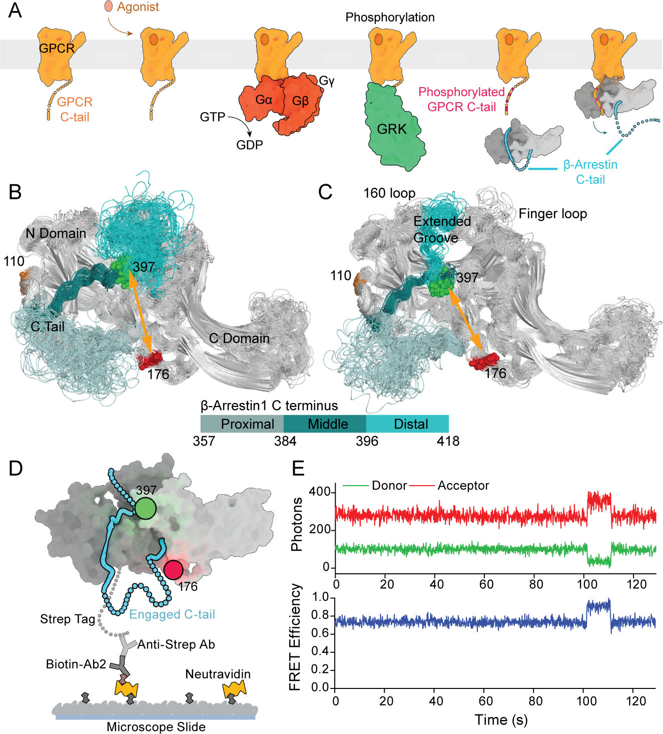
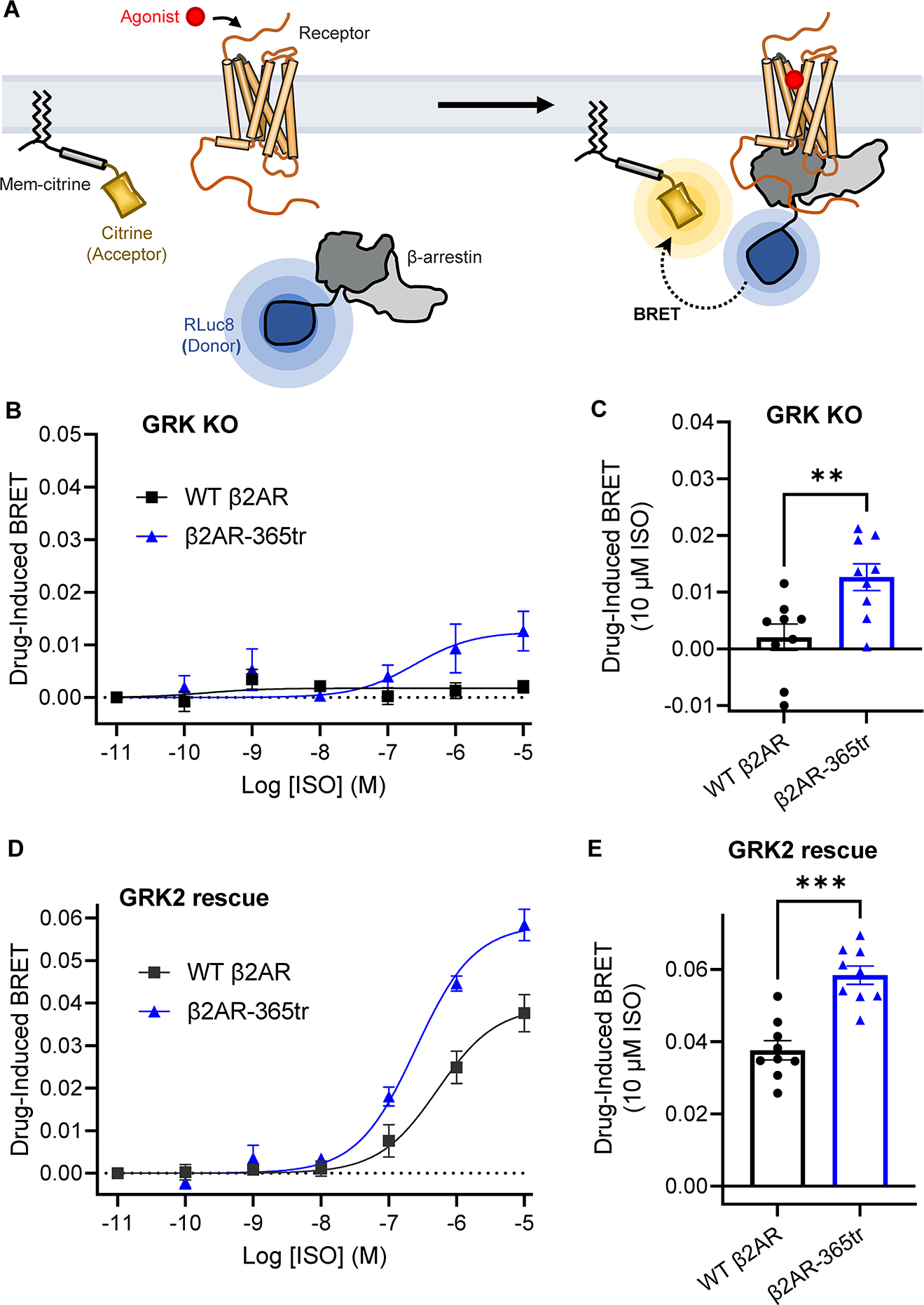
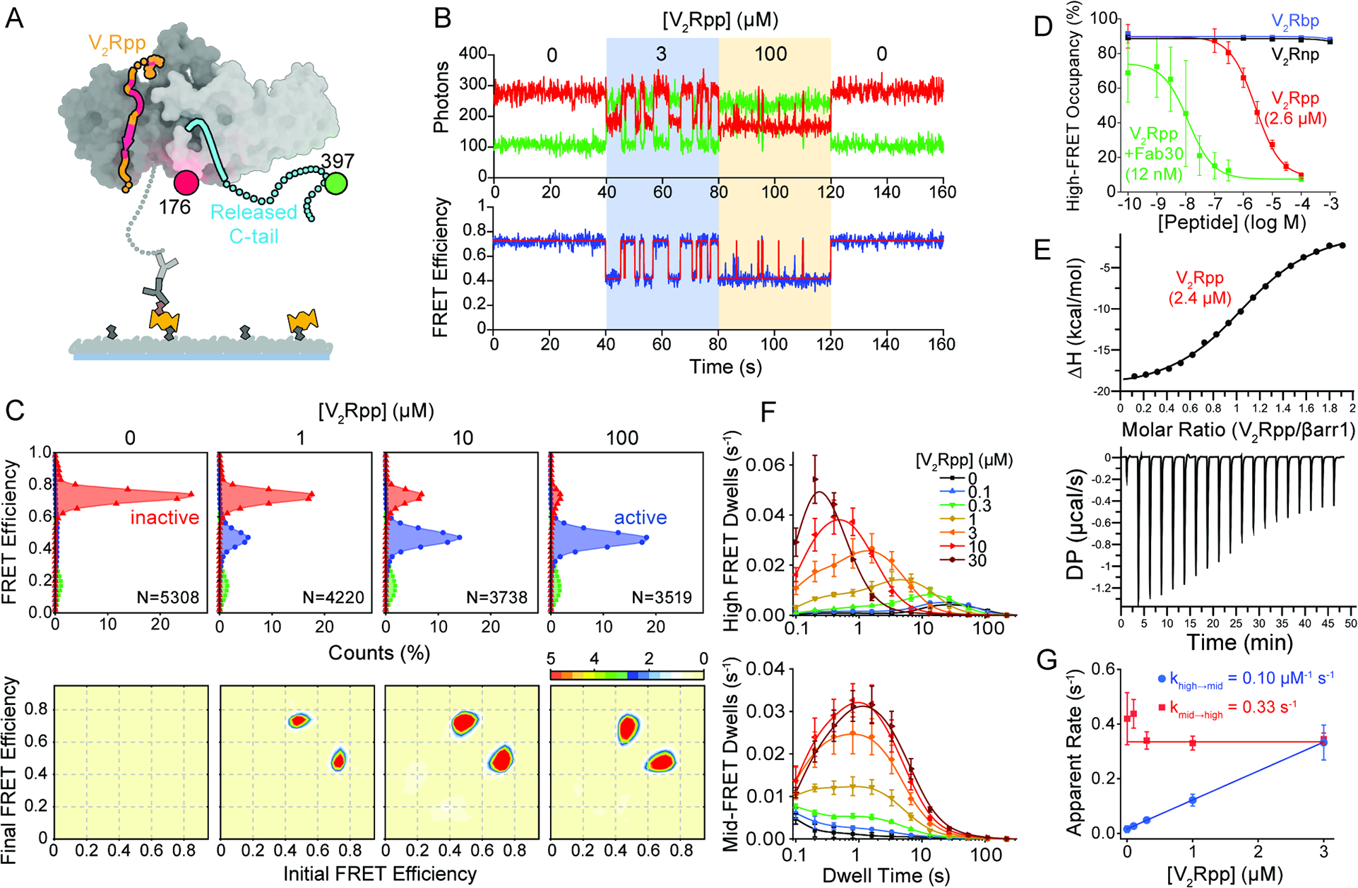
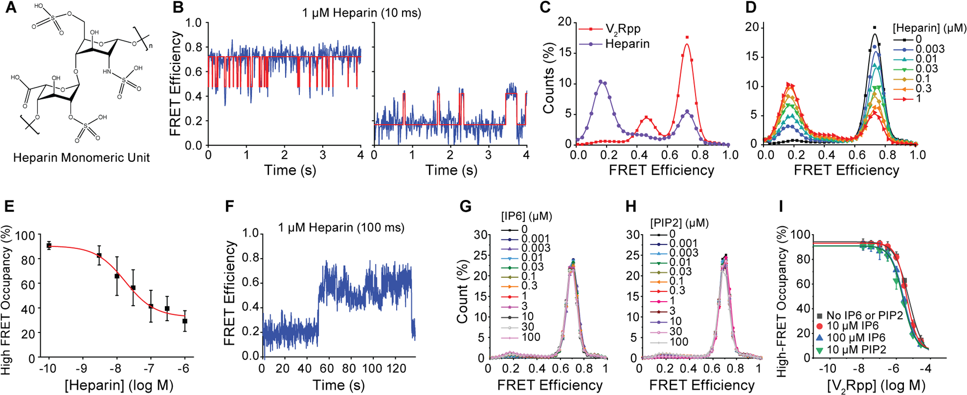
β-arrestins bind G protein-coupled receptors to terminate G protein signaling and to facilitate other downstream signaling pathways. Using single-molecule fluorescence resonance energy transfer imaging, we show that β-arrestin is strongly autoinhibited in its basal state. Its engagement with a phosphopeptide mimicking phosphorylated receptor tail efficiently releases the β-arrestin tail from its N domain to assume distinct conformations. Unexpectedly, we find that β-arrestin binding to phosphorylated receptor, with a phosphorylation barcode identical to the isolated phosphopeptide, is highly inefficient and that agonist-promoted receptor activation is required for β-arrestin activation, consistent with the release of a sequestered receptor C tail. These findings, together with focused cellular investigations, reveal that agonism and receptor C-tail release are specific determinants of the rate and efficiency of β-arrestin activation by phosphorylated receptor. We infer that receptor phosphorylation patterns, in combination with receptor agonism, synergistically establish the strength and specificity with which diverse, downstream β-arrestin-mediated events are directed.
β-arrestins 与 G 蛋白偶联受体结合,从而终止 G 蛋白信号转导,并促进其他下游信号通路。通过单分子荧光共振能量转移成像,我们发现 β-arrestin 在其基础状态下受到强烈的自身抑制。它与模拟磷酸化受体尾部的磷酸肽结合,可有效地将β-arrestin 尾部从其 N 结构域释放出来,从而形成不同的构象。出乎意料的是,我们发现与磷酸化受体的结合,即使其磷酸化条码与分离的磷酸肽完全相同,β-arrestin 的结合效率也非常低,而且需要激动剂促进受体激活,才能使 β-arrestin 激活,这与被隔离的受体 C 尾部的释放一致。这些发现,以及有针对性的细胞研究,揭示了激动剂和受体 C 尾部的释放是磷酸化受体激活β-arrestin 的速度和效率的特定决定因素。我们推断,受体的磷酸化模式与受体激动剂相结合,协同建立了不同的、下游的β-arrestin 介导的事件的强度和特异性。