Weill Cornell Medicine-Qatar, Doha, Qatar.
Department of Biology, University of Antwerp, Antwerp, Wilrijk, Belgium.
PLoS One. 2022 May 5;17(5):e0267837. doi: 10.1371/journal.pone.0267837. eCollection 2022.
Pial collateral blood flow is a major determinant of the outcomes of acute ischemic stroke. This study was undertaken to determine whether retinal vessel metrics can predict the pial collateral status and stroke outcomes in patients.
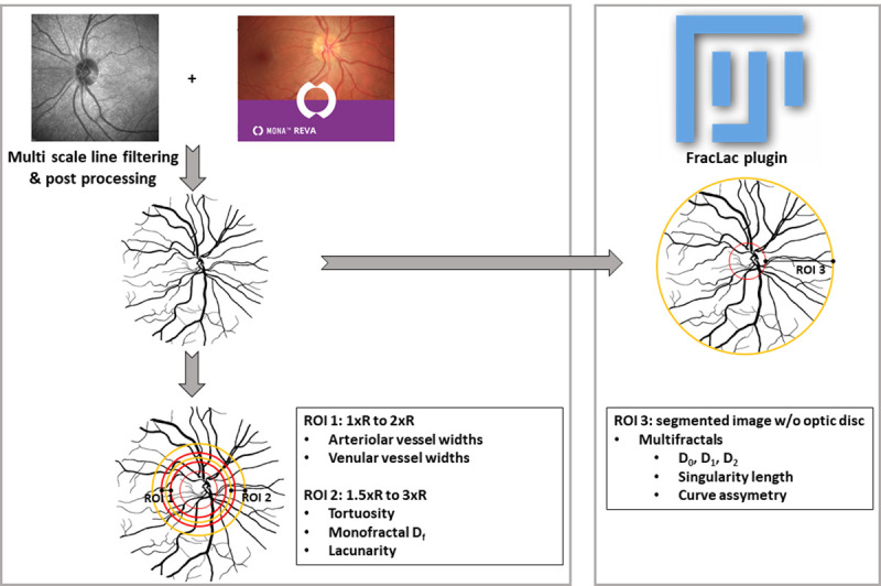
Thirty-five patients with acute stroke secondary to middle cerebral artery (MCA) occlusion underwent grading of their pial collateral status from computed tomography angiography and retinal vessel analysis from retinal fundus images.
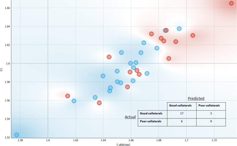
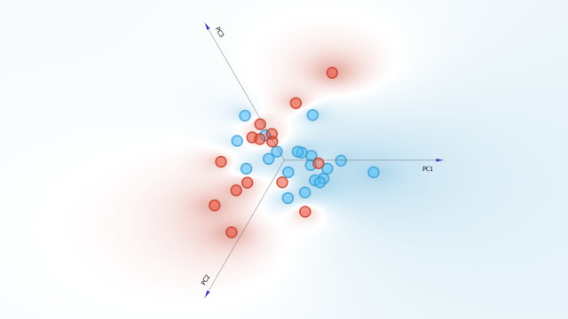
The NIHSS (14.7 ± 5.5 vs 10.1 ± 5.8, p = 0.026) and mRS (2.9 ± 1.6 vs 1.9 ± 1.3, p = 0.048) scores were higher at admission in patients with poor compared to good pial collaterals. Retinal vessel multifractals: D0 (1.673±0.028vs1.652±0.025, p = 0.028), D1 (1.609±0.027vs1.590±0.025, p = 0.044) and f(α)max (1.674±0.027vs1.652±0.024, p = 0.019) were higher in patients with poor compared to good pial collaterals. Furthermore, support vector machine learning achieved a fair sensitivity (0.743) and specificity (0.707) for differentiating patients with poor from good pial collaterals. Age (p = 0.702), BMI (p = 0.422), total cholesterol (p = 0.842), triglycerides (p = 0.673), LDL (p = 0.952), HDL (p = 0.366), systolic blood pressure (p = 0.727), HbA1c (p = 0.261) and standard retinal metrics including CRAE (p = 0.084), CRVE (p = 0.946), AVR (p = 0.148), tortuosity index (p = 0.790), monofractal Df (p = 0.576), lacunarity (p = 0.531), curve asymmetry (p = 0.679) and singularity length (p = 0.937) did not differ between patients with poor compared to good pial collaterals.
This is the first translational study to show increased retinal vessel multifractal dimensions in patients with acute ischemic stroke and poor pial collaterals. A retinal vessel classifier was developed to differentiate between patients with poor and good pial collaterals and may allow rapid non-invasive identification of patients with poor pial collaterals.
软脑膜侧支循环血流是急性缺血性脑卒中结局的主要决定因素。本研究旨在确定视网膜血管指标是否可以预测患者的软脑膜侧支循环状态和卒中结局。
35 例因大脑中动脉(MCA)闭塞导致急性卒中的患者接受了计算机断层血管造影和视网膜眼底图像的视网膜血管分析,以对软脑膜侧支循环状态进行分级。
与软脑膜侧支循环良好的患者相比,软脑膜侧支循环不良的患者入院时 NIHSS(14.7 ± 5.5 与 10.1 ± 5.8,p = 0.026)和 mRS(2.9 ± 1.6 与 1.9 ± 1.3,p = 0.048)评分更高。视网膜血管多重分形:D0(1.673±0.028 与 1.652±0.025,p = 0.028)、D1(1.609±0.027 与 1.590±0.025,p = 0.044)和 f(α)max(1.674±0.027 与 1.652±0.024,p = 0.019)在软脑膜侧支循环不良的患者中更高。此外,支持向量机学习对区分软脑膜侧支循环不良和良好的患者具有良好的敏感性(0.743)和特异性(0.707)。年龄(p = 0.702)、BMI(p = 0.422)、总胆固醇(p = 0.842)、甘油三酯(p = 0.673)、LDL(p = 0.952)、HDL(p = 0.366)、收缩压(p = 0.727)、HbA1c(p = 0.261)和标准视网膜指标,包括 CRAE(p = 0.084)、CRVE(p = 0.946)、AVR(p = 0.148)、扭曲指数(p = 0.790)、单分形 Df(p = 0.576)、空隙度(p = 0.531)、曲线不对称性(p = 0.679)和奇点长度(p = 0.937)在软脑膜侧支循环不良和良好的患者之间没有差异。
这是第一项表明急性缺血性脑卒中患者软脑膜侧支循环不良时视网膜血管多重分形尺寸增加的转化研究。开发了一种视网膜血管分类器来区分软脑膜侧支循环不良和良好的患者,可能允许快速非侵入性地识别软脑膜侧支循环不良的患者。